Description of Pediatric Tuberculosis Evaluated in a Referral Center in Istanbul Turkey
Seda Geylani Gulec,
1Leyla Telhan,
2Tanyel Koçkaya,
1Ela Erdem,
1Banu Bayraktar,
3and Ayse Palanduz
4Departments of 1Pediatrics, 2Pediatric Infectious Diseases, and 3Microbiology, Sisli Etfal Training and Research Hospital, Istanbul;
4Department of Family Medicine, Istanbul Faculty of Medicine, Istanbul University, Istanbul, Turkey.
Received: May 3, 2011 Revised: January 3, 2012 Accepted: January 5, 2012
Corresponding author: Dr. Seda Geylani Gulec, Department of Pediatrics,
Sisli Etfal Training and Research Hospital, Adnan Saygun Cad. Ilgın Sk. Yeni Ulus Sitesi A1 Blok, Daire: 8 Ulus/Besiktas Istanbul, Turkey.
Tel: 90-212-2810566, Fax: 90-216-4188752 E-mail: [email protected]
This study has been presented at 14th annual meeting of Turkish Thoracic Society.
∙ The authors have no financial conflicts of interest.
© Copyright:
Yonsei University College of Medicine 2012 This is an Open Access article distributed under the terms of the Creative Commons Attribution Non- Commercial License (http://creativecommons.org/
licenses/by-nc/3.0) which permits unrestricted non- commercial use, distribution, and reproduction in any medium, provided the original work is properly cited.
Purpose: Diagnosis of tuberculosis (TB) in children is more challenging than in adults. This study aimed to describe demographical, clinical and laboratory findings of children diagnosed with tuberculosis in Turkey, including the issues of contact tracing, culture positivity and forms of the disease. Materials and Methods: Clini- cal and laboratory data of 51 children with a mean age of 8.0±4.6 years who were diagnosed with TB were retrospectively reviewed. Main diagnostic tools included tuberculin skin test, chest X-ray, sputum/gastric aspirate culture with sensitivity test- ing, and direct microscopy for acid-fast bacilli on available samples. Clinical char- acteristics and outcomes of the patients were examined. Results: Thirty-six (70.6%) children were diagnosed with intra-thoracic and 15 (29.4%) with extra-tho- racic tuberculosis. Twenty-eight of the patients had a positive Bacillus Calmette- Guérin vaccine scar (28/51, 54.9%) and 23/51 (45.1%) had a positive tuberculin skin test. An adult TB contact was identified in 27 (52.9%) of the cases. On direct microscopy, acid-fast bacilli were found in nine (17.6%) patients and positive cul- ture for Mycobacterium tuberculosis was found in 19 (37.3%). Drug resistance to isoniazid was detected in four (7.8%). One patient with nephrotic syndrome and miliary tuberculosis died during follow-up. All other patients responded well to the treatment. Conclusion: Focusing on active contact tracing among all household contacts of tuberculous cases may be helpful in early identification and controlling childhood disease, even in regions with low disease prevalence. Adopting a suspi- cious and proactive approach in this particular age group is warranted.
Key Words: Childhood tuberculosis, extra-thoracic tuberculosis, Mycobacterium tuberculosis, intra-thoracic tuberculosis, tuberculin skin test
INTRODUCTION
Tuberculosis is still a global health problem, particularly in high-burden areas, namely Africa, South East Asia, Western Pacific and Eastern Mediterranean re- gions, with respective incidences of 363, 181, 108, and 105 cases per 100000 pop- ulation per year.1 Turkey on the other hand has a lower disease burden with 30/100000 cases per year incidence, a rate close to America (32/100000) and Eu-
ter where further investigations such as computerized to- mography or bronchoscopy were performed, irrespective of negative microscopic findings. Diagnosis of the screened individuals was established using both clinical and labora- tory evidence.
Diagnostic procedures
For each case, TST was performed by intradermal injection of 0.1 mL (5 TU) PPD (5 tuberculin units of purified protein derivative) solution. After 48 to 72 hours, induration was measured in millimeters on the horizontal axis. In children with no risk factors, no history of disease and no Bacille Calmette-Guérin vaccine (BCG) scar, a reactive area of ≥10 mm was classified as a positive result. In children with a BCG scar, an induration of ≥15 mm was considered a posi- tive reaction.9
All cases had chest X-ray. Induced sputum sample could only be obtained in five patients and gastric aspirates were used for diagnostic purposes in the remaining 46 patients.
Sputum/gastric aspirate samples were subjected to direct mi- croscopic examination and culture in Mycobacteria Growth Indicator Tube/Lowenstein-Jensen medium. The detection of acid-fast bacilli (AFB) by direct microscopy was done using Erlich Ziehl Nielsen, Auromine Rhodamin and fluo- roscopic examinations.
The diagnosis of TB was made through clinical findings, history of exposure to a positive source case, a positive TST, microbiological results, radiological findings, and histopath- ological findings. Cases with pulmonary, intra-thoracic nod- al, pleural and pericardial involvement were classified as having intra-thoracic tuberculosis whereas cases with pe- ripheral nodal, central nervous system, abdominal and other organ involvement including miliary tuberculosis were clas- sified as extra-thoracic tuberculosis.10
Treatment protocol
The treatment of pulmonary disease, TB peritonitis, and lymph node disease included a regimen of three drugs: iso- niazid (10-15 mg/kg/day po, max 300 mg/day); rifampin (10-20 mg/kg/day po, max 600 mg/day); and pyrazinamide (20-40 mg/kg/day po, max 2 g/day during the first two months of treatment). A fourth drug, ethambutol (15-25 mg/
kg/day po, max 2.5 g/day) or streptomycin (20-40 mg/kg/
day, max 1 g/day, intramuscular), was added in cases with TB meningitis and intestinal TB. Parenteral steroids were added (1-2 mg/kg/day) in cases of endobronchial pulmo- nary TB, TB meningitis, pericarditis, and miliary disease.
rope (49/100000).1 This is most probably a result of the country’s effective tuberculosis control program, which has been in effect for decades.
Childhood tuberculosis actually reflects uncontrolled adult tuberculosis since it develops as a result of dissemina- tion from adults and adolescents with cavitary lung disease, and it represents a major unrecognized cause of disease and death during childhood in endemic countries.2 HIV infected infants and children in particular are at high risk to develop tuberculosis infection with a disseminated and severe pat- tern.3,4 Given the importance of the condition, tuberculosis control programs focus on strict reporting of children with the disease as well as diagnosis and treatment of smear pos- itive cases.3,5
Data on the incidence and clinical course of pediatric tu- berculosis are limited and mostly reported from low-burden countries.6 Recently, this condition has stimulated interest since 15 to 20% of all tuberculosis cases are children and this figure may reach up to 40% in countries where the dis- ease is endemic.3,6-8 Moreover, diagnosis of tuberculosis (TB) is particularly challenging in children.
The aim of this study is to describe pediatric tuberculosis cases in Turkey, including the issues of contact tracing, cul- ture positivity and forms of the disease.
MATERIALS AND METHODS
Study population
Fifty-one children who were diagnosed with tuberculosis between January 2007 and May 2008 at the Department of Infectious Disease, Sisli Etfal Research and Training Hos- pital, Istanbul, Turkey were included in this study. All pa- tients had signs and symptoms suggestive of tuberculosis, and they had been referred for further investigation and treatment. The institution is a 1000-bed hospital providing tertiary healthcare services in metropolitan area of Istanbul.
Medical records of the patients were reviewed for clinical, demographic and laboratory data.
An investigation for a source case/contact in families was conducted by local TB control officials. Chest X-ray (CXR) was obtained from all family members whereas tuberculin skin test (TST) was done in the subjects younger than 35 years of age. In case where these investigations reveal sus- picious findings, direct microscopy and culture of sputum samples were done. Patients with clinical or radiological evidence of tuberculosis were referred to a specialized cen-
(29/51, 56.9%), and increased C-reactive protein (28/51, 54.9%). The mean value for leukocyte count, erythrocyte sedimentation rate, and C-reactive protein was 10601±
3864/mm3, 39.3±32.0 mm/h, and 38.6±63.5 mg/L, respec- tively. Other specific laboratory investigations performed are shown in Table 2.
Special investigations
According to CXR and computerized CT scans, the most common radiological findings were mediastinal lymphade- nopathy (37/51, 72.5%), pneumonic infiltration and consol- idation (28/51, 54.9%), pleural effusion (7/51, 13.7%), and miliary pattern (5/51, 9.8%).
Description of extra pulmonary TB cases
There were four cases with central nervous system TB. Ce- rebrospinal fluid analyses of these patients revealed low chloride and glucose levels, turbid appearance, elevated pressure and presence of protein. All of them had pleocyto- sis with lymphocytic predominance. Acid-fast stain of cere- brospinal fluid for tuberculosis bacilli was negative for all samples, and bacterial cultures yielded no growth. In one case, AFB was negative but Mycobacterium tuberculosis was found in the gastric aspirate sample. In another case, both acid-fast stain and mycobacterial culture of gastric as- pirate sample yielded positive results. In three patients, con- solidation and infiltration were observed on CXR. Menin- geal involvement, hydrocephalus, and tuberculoma were identified by cranial MRI. None of the patients had enhance- ment of basal cisterns.11 A shunt was created in three patients with hydrocephalus. Cerebrospinal fluid examination find- ings were pathologic in only one out of five patients with miliary tuberculosis. In that case, in addition to lymphocyto- sis, cerebrospinal fluid pressure and protein level were in- creased, and glucose and chloride were decreased.
Treatment was given for nine months for pulmonary tuber- culosis and tuberculous lymphadenitis, whereas one year for tuberculous meningitis, pericarditis, peritonitis, intesti- nal tuberculosis, miliary tuberculosis, and endobronchial tuberculosis.
RESULTS
Clinical findings
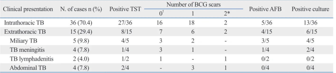
The mean age of the patients was 8±4.6 years (range: 5 months to 15 years). Twenty-eight (55%) patients were male. Most common clinical symptoms were high fever (28/51, 54.9%), cough (26/51, 51%) and weight loss (15/51, 29.4%). Thirty-six (36/51, 70.6%) children had intra-tho- racic and 15/51 (29.4%) had extra-thoracic tuberculosis.
Distribution of the cases by clinical form and microbiologi- cal data are shown in Table 1. Histopathological evidence of granulomatous inflammation and caseous necrosis con- sistent with TB were found in the patients with pericarditis, abdominal TB, and lymphadenitis.
Laboratory findings
The most common laboratory findings were increased eryth- rocyte sedimentation rate (31/51, 60.8%), leukocytosis
Table 1. Distribution of the Cases and Diagnostic Findings by Clinical Form of the Disease
Clinical presentation N. of cases n (%) Positive TST Number of BCG scars Positive AFB Positive culture
0† 1 2*
Intrathoracic TB 36 (70.4) 27/36 16 18 2 5/36 13/36
Extrathoracic TB 15 (29.4) 8/15 7 6 2 4/15 6/15
Miliary TB 5 (9.8) 4/5 3 2 - 3/5 4/5
TB meningitis 4 (7.8) 1/4 3 1 - 1/4 2/4
TB lymphadenitis 2 (4.0) 1/2 1 - 1 0/2 0/2
Abdominal TB 4 (7.8) 2/4 - 3 1 0/4 0/4
TB, tuberculosis; AFB, acid-fast bacilli; BCG, Bacillus Calmette-Guérin vaccine; TST, tuberculin skin test.
n/n denotes number of patients with positive result/number of patients with available data.
*Multiple application was possible in the past.
†No scar, but documentation not available.
Table 2. Specific Laboratory Investigations
Investigation N. of patients
Lumbar puncture for CSF sampling 9
Pleural fluid sampling 2
Colonoscopy and biopsy 1
Bronchoscopy and lavage 4
Fine needle aspiration biopsy 3
Computerized tomography 44
Cranial MRI 4
CSF, cerebrospinal fluid.
with miliary TB. They all were treated with four anti-tuber- culosis drugs without discontinuing isoniazid. As the addi- tional drug, three patients received streptomycin and one received ethambutol. Three of the cases were also given steroid therapy. In three of these patients, isoniazid resis- tance was also identified in the source cases. All patients were followed once a week during the first month and once a month thereafter. One patient with nephrotic syndrome and miliary tuberculosis died during follow-up. All other patients responded well to the treatment. One patient devel- oped severe hepatotoxicity, which improved following tran- sient interruption of the drug treatment.
Source identification
None of the patients had a positive history of exposure to adult TB; however, a source case was detected in 52.9% of children by contact tracing. Thus, 27 adult contacts were treated for TB as a result of active contact tracing after the pediatric cases were diagnosed; however, details of how the diagnosis was made are unknown.
DISCUSSION
Tuberculosis is a major health problem, particularly in the developing world. Although it geographically neighbors a high-prevalence region, Turkey has a low TB prevalence, most probably owing to its policies on vaccination, treat- ment and surveillance that have been in effect for decades.
There is a nation-wide network of specialized healthcare fa- cilities to combat against the disease. All infants routinely receive BCG vaccination two months after birth. After the age of 3 months, TST positive infants are vaccinated. How- ever, only about half of the children in this study had BCG scar, which is similar to the rate reported by Aycicek, et al.13 (66%) in another series of pediatric tuberculosis patients.
Despite strict procedures for BCG vaccination, infants oc- casionally skip vaccination due to sociocultural issues in this country. This low rate of vaccination among diseased children suggests that lack of vaccination may be a contrib- uting factor for the development of the disease, although protection rate of BCG vaccination is controversial.
Diagnosis of TB in children is difficult and poses prob- lems that are different from adults. Clinical and radiological findings of childhood tuberculosis are nonspecific and quite variable. In addition, sampling, confirmation and final diag- nosis pose a challenge.5,7 Sputum samples are difficult to Bronchoscopy was performed in four patients for persis-
tent radiological findings and worsening clinical symptoms (wheezing and hemoptysis), and endobronchial TB was documented in those patients. However, bronchoalveolar fluid examination did not reveal AFB nor cultural growth was observed, although gastric lavage fluid direct micros- copy and/or culture had been positive among those patients at the initial examination.
Two patients had TB lymphadenitis. In one of them, it was localized in the cervical region and along the axillary line in the other. In the case of axillary adenitis, BCG reac- tion was excluded since the patient did not have a history of recent BCG vaccination. Histopathological findings of fine needle aspiration biopsy samples confirmed the diagnosis of TB.
Four patients had abdominal TB. Three of them were ad- mitted with acute abdominal pain and they underwent sur- gery. One patient had colonoscopy. In these patients, exam- ination of biopsy samples revealed granulomatous reaction and pathological findings consistent with tuberculosis.
One TB pericarditis case presented with constrictive peri- carditis. Although TST, AFB and culture were negative in this patient, protein level was high, and glucose and chloride levels were low in pericardial fluid samples. Diagnosis of TB was confirmed by histological findings of fine needle as- piration biopsy samples.
Immunocompromised children
Three patients were immunocompromised, which presented with miliary TB, TB peritonitis and TB meningitis, respec- tively. The patient with miliary tuberculosis was receiving steroid for focal segmental glomerulosclerosis and died dur- ing treatment. The second patient with tuberculous peritoni- tis was receiving steroid for nephritic syndrome. The third patient with tuberculous meningitis had also Hodgkin’s lym- phoma. The latter two patients completed their tuberculosis treatments and they are currently under remission.
Treatment
Nineteen cases were treated with four drugs; fifteen re- ceived streptomycin and four received ethambutol as the additional agent. All four patients who received ethambutol as the fourth drug had abdominal involvement and all pa- tients who received this agent were older than 5 years.12 Iso- niazid resistance was reported in four patients (7.8%): a 12-year-old girl with a cavitary lesion; a patient with TB meningitis; a patient with endobronchial TB; and a child
somewhat similar rates for these laboratory parameters with corresponding figures of 60.8% and 56.9% for increased erityrocyte sedimentation rate and leukocyte count, respec- tively.
Radiological findings support the diagnosis of TB. In the study of Cosar, et al.,22 radiological findings were noted as lymphadenopathy (63.6%), primary focus-calcification (56.8%), miliary pattern (20.4%), pneumonic infiltration (15.9%) and cavitary lesion (9%) among pediatric patients.
In this study, cavitary lesion was observed in only one case.
However, neither primary foci nor calcification were ob- served in any of the cases, and typical miliary patterns were observed in five patients (9.8%). Since hematogenous spread of TB is common in infants and children younger than four years of age, these patients are particularly at greater risk of developing TB meningitis. In this series, a relatively high rate of TB meningitis was observed (four cases, 7.8%), and two of the cases were below the age of four. Thus, prompt diagnosis and immediate treatment of childhood TB before the development of potentially fatal complications is im- portant.26
TST is not specific and sensitive enough, since BCG im- munization can cause false positive results. In addition, mal- nutrition and miliary tuberculosis may result in false negative results.27-29 In this study, similar to previous reports, positive TST rate was higher in intra-thoracic TB than in extra-tho- racic disease, although the incidences of BCG scar were sim- ilar.30 TST was negative in 31.5% of culture positive cases, suggesting a low sensitivity for TST. In contrast, however, another study found higher positive TST rate among culture positive patients with chest X-ray findings of pulmonary tu- berculosis and concluded that the test may be a useful diag- nostic tool particularly in regions where the disease preva- lence is high.29 Thus, diagnostic value of this test seems to be variable, depending on patient groups and different preva- lence rates.5 Although several tests including IFN-γ release assays (IGRAs) have so far been developed for the diagnosis of LTBI, a substantial advance in TB diagnostics will require more study in children, in order to improve the diagnosis in this age group.7,28,31 Polymerase chain reaction (PCR) also has a limited place in the diagnosis of childhood TB.5,32,33
Accurate diagnosis and isolation of drug-resistant strains as well as identification of antimicrobial susceptibility are be- coming more and more important in childhood tuberculosis.3 When microbiological and molecular techniques are used to- gether, the diagnosis of TB becomes more definite. Myco- bacterial culture is more sensitive than direct microscopy. In collect from children3,14,15 and sputum-smear microscopy/
culture yields are lower.7,16 Diagnostic scoring systems show high variability in case yield and their agreement is usually poor.17 Moreover, acute tuberculosis may present in the form of acute pneumonia in children, which is quite dif- ficult to differentiate clinically and radiologically from con- ditions caused by other pathogens.3 TST on the other hand can be used only in the diagnosis of latent tuberculosis in- fection (LTBI); and it is not able to differentiate from active disease. Children with the appropriate clinical presentation, positive TST results, tuberculosis specific radiological find- ings, and a history of no clinical improvement after appro- priate antibiotic treatment are diagnosed with tuberculosis, even if AFB are not found in the sputum and/or gastric aspi- rates.18,19
The high prevalence of childhood TB is an indication of the continued dissemination of the disease. The most im- portant approach in the fight against the disease is determi- nation, identification and treatment of the source cases, as well as BCG vaccination soon after birth.20,21 In our coun- try, chest X-ray examination and TST are done in all mem- bers of the household of an adult index case. Isoniazid pro- phylaxis for 6 months is initiated in household members younger than 35 years of age (<16 years of age in some provinces). However, in certain regions follow-up and ac- cess to the patients are problematic, and complying with strict procedures is occasionally not possible. Although es- sential, finding the source case may sometimes be difficult.
In this study, 27 of the 51 patients (52.9%) had contact with an adult case of TB. High TB contact rate found on active screening in this study suggests that adults even in low HIV prevalence areas may not present with typical TB symptoms.
Corresponding figures for contact rates ranged from 22.6 to 59% in previous studies.13,22-25 Thus, many adults would have been missed if not traced. However, it is of note to mention that this study included only the children admitted with a clinical presentation suggestive of tuberculosis infection since our institution is a referral center; thus, none of them was identified as a result of contact tracing.
Clinical manifestations and physical findings of childhood TB may vary. In this study, the most common clinical signs were high fever (54.9%), cough (51%) and weight loss (29.4%). Cosar, et al.22 reported that fever (63.6%), weight loss (61.3%) and cough (61.3%) were the most frequent clinical signs. In that study by Cosar, et al., the rate of in- creased erythrocyte sedimentation rate and leukocyte count was 86.3% and 65.9%, respectively. In this study, we found
TB/2009411; 2009.
2. Marais BJ. Childhood tuberculosis: epidemiology and natural his- tory of disease. Indian J Pediatr 2011;78:321-7.
3. Zar HJ, Pai M. Childhood tuberculosis-a new era. Paediatr Respir Rev 2011;12:1-2.
4. Marais BJ, Gie RP, Schaaf HS, Hesseling AC, Obihara CC, Starke JJ, et al. The natural history of childhood intra-thoracic tuberculo- sis: a critical review of literature from the pre-chemotherapy era.
Int J Tuberc Lung Dis 2004;8:392-402.
5. Nicol MP, Zar HJ. New specimens and laboratory diagnostics for childhood pulmonary TB: progress and prospects. Paediatr Respir Rev 2011;12:16-21.
6. Moyo S, Verver S, Mahomed H, Hawkridge A, Kibel M, Hatherill M, et al. Age-related tuberculosis incidence and severity in chil- dren under 5 years of age in Cape Town, South Africa. Int J Tu- berc Lung Dis 2010;14:149-54.
7. Zar HJ, Connell TG, Nicol M. Diagnosis of pulmonary tuberculo- sis in children: new advances. Expert Rev Anti Infect Ther 2010;8:277-88.
8. Marais BJ, Schaaf HS. Childhood tuberculosis: an emerging and previously neglected problem. Infect Dis Clin North Am 2010;
24:727-49.
9. World Health Organization. A research agenda for childhood tu- berculosis. WHO/HTM/TB:381; 2007.
10. Hesseling AC, Marais BJ, Kirchner HL, Mandalakas AM, Brittle W, Victor TC, et al. Mycobacterial genotype is associated with disease phenotype in children. Int J Tuberc Lung Dis 2010;14:
1252-8.
11. Bharath RD, Sinha S, Vasudev MK, Ravishankar S, Chandrashek- ar N. Tuberculous meningitis presenting as isolated interhemi- spheric exudates. J Med Imaging Radiat Oncol 2010;54:129-33.
12. Graham SM. Treatment of paediatric TB: revised WHO guide- lines. Paediatr Respir Rev 2011;12:22-6.
13. Aycicek A, Aktas GS, Celen OF. [Clinical, radiological and epide- miological characteristics of 69 pediatric tuberculosis cases from Sanliurfa district]. Turkish Pediatr J 2006;49:205-12.
14. Newton SM, Brent AJ, Anderson S, Whittaker E, Kampmann B.
Paediatric tuberculosis. Lancet Infect Dis 2008;8:498-510.
15. Tuberculosis fact sheet. American Lung Association; 2006.
16. Marais BJ, Pai M. New approaches and emerging technologies in the diagnosis of childhood tuberculosis. Paediatr Respir Rev 2007;8:124-33.
17. Hatherill M, Hanslo M, Hawkridge T, Little F, Workman L, Ma- homed H, et al. Structured approaches for the screening and diag- nosis of childhood tuberculosis in a high prevalence region of South Africa. Bull World Health Organ 2010;88:312-20.
18. Lobato MN, Sun SJ, Moonan PK, Weis SE, Saiman L, Reichard AA, et al. Underuse of effective measures to prevent and manage pediatric tuberculosis in the United States. Arch Pediatr Adolesc Med 2008;162:426-31.
19. Cruz AT, Starke JR. Clinical manifestations of tuberculosis in chil- dren. Paediatr Respir Rev 2007;8:107-17.
20. Magdorf K, Detjen AK. Proposed management of childhood tu- berculosis in low-incidence countries. Eur J Pediatr 2008;167:927- 21. Guthmann JP, de La Rocque F, Boucherat M, van Cauteren D, 38.
Fonteneau L, Lécuyer A, et al. [BCG vaccine coverage in private medical practice: first data in children below two years old, seven months after the end of compulsory vaccination in France]. Arch Pediatr 2009;16:489-95.
this study, culture was positive only in 37.2% of patients. In most previous studies from other geographical regions, posi- tive culture rates ranging between 30 and 40% were reported, similar to the figure obtained in this study,27,34,35 although rates below this range have also been reported. For example, mi- crobial growth was possible in 25% of the cases in a pediat- ric referral hospital setting34 and a 10% rate was obtained in a community-based study among patients with contact his- tory or among suspicious cases.35 Patients referred to our institution that turned out to have LTBI after investigations were not included in this study, therefore, our study popula- tion might have been consisted of patients with relatively se- vere disease, which may account for the slightly high rate of culture positivity when compared to the latter studies. In summary, culture results seem to show variability across dif- ferent patient populations and disease severities.
Although multi-drug resistant tuberculosis constitutes less than 1% of the cases in the United States, a rate as high as 15% have been reported from Kazakistan.30 In the present study, INH resistance was found in four cases (7.8%), which were successfully treated by using four-drug regimens in- cluding streptomycin and ethambutol. None of these four cases had been exposed to INH previously. Considering this relatively high drug resistance rate, we suggest that drug sus- ceptibility testing should be done and history of previous drug treatments should be questioned in every index case.
Otherwise, delay in recognizing drug-resistant tuberculosis may increase mortality and morbidity in children.36
The main limitations of this study include retrospective design and inaccessibility of novel diagnostic techniques such as IGRA and PCR. Further studies on the diagnosis and management of tuberculosis with more robust design are warranted in this age group.
In summary, childhood tuberculosis is still an important health problem even in regions with relatively low tubercu- losis prevalence. Focusing on active contact tracing among all household contacts of index cases may be helpful in identification and controlling the disease. Adopting a more suspicious and proactive approach in this particular age group would prevent delay in diagnosis and disease related complications.
REFERENCES
1. World Health Organization. Global tuberculosis control: epidemi- ology, strategy, financing: WHO report 2009. WHO/HTM/
31. Mazurek GH, Jereb J, Lobue P, Iademarco MF, Metchock B, Ver- non A; Division of Tuberculosis Elimination, National Center for HIV, STD, and TB Prevention, Centers for Disease Control and Prevention (CDC). Guidelines for using the QuantiFERON-TB Gold test for detecting Mycobacterium tuberculosis infection, United States. MMWR Recomm Rep 2005;54:49-55.
32. Cordova J, Shiloh R, Gilman RH, Sheen P, Martin L, Arenas F, et al. Evaluation of molecular tools for detection and drug suscepti- bility testing of Mycobacterium tuberculosis in stool specimens from patients with pulmonary tuberculosis. J Clin Microbiol 2010;48:1820-6.
33. Lima JF, Montenegro LM, Montenegro Rde A, Cabral MM, Lima AS, Abath FG, et al. Performance of nested PCR in the specific detection of Mycobacterium tuberculosis complex in blood sam- ples of pediatric patients. J Bras Pneumol 2009;35:690-7.
34. Zar HJ, Hanslo D, Apolles P, Swingler G, Hussey G. Induced spu- tum versus gastric lavage for microbiological confirmation of pul- monary tuberculosis in infants and young children: a prospective study. Lancet 2005;365:130-4.
35. Hatherill M, Hawkridge T, Zar HJ, Whitelaw A, Tameris M, Workman L, et al. Induced sputum or gastric lavage for communi- ty-based diagnosis of childhood pulmonary tuberculosis? Arch Dis Child 2009;94:195-201.
36. Al-Dabbagh M, Lapphra K, McGloin R, Inrig K, Schaaf HS, Marais BJ, et al. Drug-resistant tuberculosis: pediatric guidelines.
Pediatr Infect Dis J 2011;30:501-5.
22. Cosar H, Onay H, Bayram N, Ozkınay F. The evaluation of the epidemiological and clinical findings and the prognosis of the 44 pediatric tuberculosis patients. J Pediatr Infect 2008;2:1-6.
23. Vellema SC, Durrheim DN, Smith JE. Diagnosing childhood tu- berculosis in rural clinics in Mpumalanga Province, South Africa.
Curationis 2008;31:52-8.
24. Schaaf HS, Marais BJ, Whitelaw A, Hesseling AC, Eley B, Hussey GD, et al. Culture-confirmed childhood tuberculosis in Cape Town, South Africa: a review of 596 cases. BMC Infect Dis 2007;7:140.
25. Tanrikulu AC, Suner A, Dagli CE, Hosoglu S, Gurkan F. [Clinical and laboratory features of disseminated tuberculosis cases with lung involvement]. Klimik Journal 2004;17:200-4.
26. Brent AJ, Anderson ST, Kampmann B. Childhood tuberculosis:
out of sight, out of mind? Trans R Soc Trop Med Hyg 2008;102:
217-8.
27. Marais BJ. Advances in the clinical diagnosis of TB in children.
Pediatr Res 2008;63:116.
28. Lewinsohn DA, Lewinsohn DM. Immunologic susceptibility of young children to Mycobacterium tuberculosis. Pediatr Res 2008;63:115.
29. Pan W, Matizirofa L, Workman L, Hawkridge T, Hanekom W, Mahomed H, et al. Comparison of mantoux and tine tuberculin skin tests in BCG-vaccinated children investigated for tuberculo- sis. PLoS One 2009;4:e8085.
30. Cruz AT, Starke JR. Pediatric tuberculosis. Pediatr Rev 2010;
31:13-25.