764 Original Article
Korean Circulation J 2006;36:764-766
ISSN 1738-5520
ⓒ 2006, The Korean Society of Circulation CASE REPORT
Primary Cardiac Osteosarcoma
Jun Ho Bae, MD1, Geu-Ru Hong, MD2, Sang-Hee Lee, MD2, Woong Kim, MD2,
Jong-Seon Park, MD2, Dong-Gu Shin, MD2, Young-Jo Kim, MD2 and Bong-Sub Shim, MD2
1Division of Cardiology, Dongguk University Hospital, Gyeongju, 2Division of Cardiology, Departments of Internal Medicine, Yeungnam University Hospital, Daegu, Korea
ABSTRACT
A 52-year-old woman, who was suffering from aplastic anemia, presented with the clinical features of severe heart failure. The transthoracic echocardiogram showed a heterogeneous, huge mass on the base of the posterior mitral valve. We guessed that the mass would be a benign neoplasm and probably myxoma, and we decided upon surgical resection. After tumor resection, an unexpected result of the histopathology was a high grade osteosarcoma. The other studies that were done after we had the diagnosis could not reveal any evidence of metastatic malignancy.
(Korean Circulation J 2006;36:764-766)
KEY WORDS:Cardiac neoplasms;Osteosarcoma.
Introduction
Primary cardiac neoplasms are rare tumors. About 25% of primary cardiac neoplasms are considered as malignancy, and the majority of these malignancies are angiosarcoma and undifferentiated sarcoma. Cardiac os- teosarcomas account for only 3-9% of all cardiac sarco- mas,1) and few cases of cardiac osteosarcoma have been reported. We report here on an extremely rare case that proved to be a primary cardiac osteosarcoma.
Case
A 52-year-old woman, who had been receiving treat- ment for 5 years due to aplastic anemia, was referred to our cardiologic department because of her aggravated dyspnea on exertion for several weeks. At the time of admission, her vital signs were relatively stable and the physical examination was unremarkable exclusive for soft crackle sounds on both lower lung fields. The laboratory findings were pancytopenia(white blood cells: 2,740×
103/μL, hemoglobin: 3.7 gm/dl, platelets: 84,000×
103/μL), serum iron: 139 μg/dL, serum ferritin: 11336 ng/mL and serum TIBC: 192 μg/dL. The cardiac enzy-
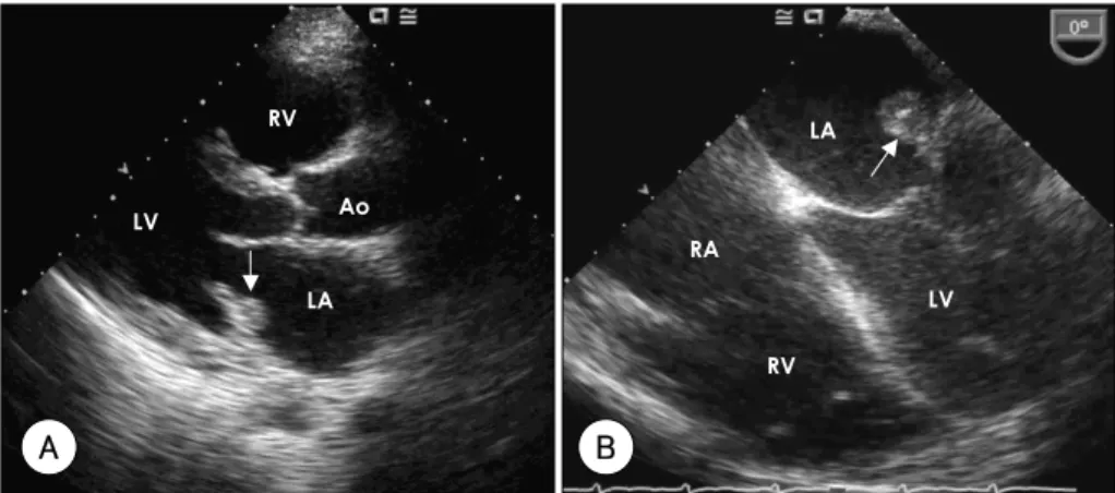
mes and serum electrolytes were normal. Chest radio- graphy demonstrated cardiomegaly and pulmonary con- gestion. A transthoracic echocardiogram(TTE) showed a small sized echogenic mass in the left atrium(LA), a dilated left ventricular dimension(left ventricular end diastolic dimension: 58 mm), a decreased left ventricu- lar systolic function(left ventricular ejection fraction:
34%) and significant mitral regurgitation(Fig. 1A). The transesophageal echocardiogram demonstrated about a 1 cm sized heterogeneous mass on the base of the posterior mitral valve, which extended to the mid portion of it (Fig. 1B). At that time, our diagnosis was nonbacterial, thrombotic endocarditis and dilated cardiomyopathy due to hemochromatosis. After conventional therapy for con- gestive heart failure and anticoagulation, she became much better and was discharged.
Alas, she arrived at the emergency department about 4 months later because of severe dyspnea and orthopnea.
The physical examination revealed an apical diastolic murmur and coarse crackle sounds on the mid to lower lung fields. On TTE, the mass shown by the previous study had rapidly increased to about 5×4 cm in size with broad base, and this mass occupied most of the left atrial chamber. Further, the echogenicity of the mass was more inhomogeneous. It had relatively low echogenicity within the center and it was attached to the base of the posterior mitral valve(Fig. 2). We guessed that the mass would be a benign neoplasm and probably myxoma, and we deci- ded upon surgical resection. During the operation, we saw the palpable mass in the LA and it had invaded the base of the posterior mitral leaflet with extension into
Received:July 6, 2006
Revision Received:September 7, 2006 Accepted:September 11, 2006
Correspondence:Geu-Ru Hong, MD,Division of Cardiology, Departments of Internal Medicine, Yeungnam University Hospital, 317-1 Daemyeong- dong, Nam-gu, Daegu 705-717, Korea
Tel: 82-53-620-3830, Fax: 82-53-620-8386 E-mail: grhong@med.yu.ac.kr
Jun Ho Bae, et al:Osteosarcoma of the Heart·765
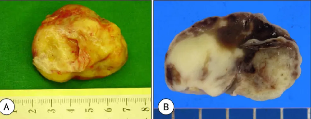
the posterior wall of the LA. After resection, it seemed to be a myxoma in appearance(Fig. 3), but the histopatho- logy was that of a high grade osteosarcoma composed of polygonal to spindle cells, patchy areas of necrosis and cal- cification with cartilage and osteoid formation(Fig. 4).2) The bone marrow biopsy and a 131-Iodine whole body bone scan along with the other studies we conducted re- vealed there was no evidence of metastatic malignancy.
We finally came to a conclusion that the mass was a rapid growing, primary cardiac osteosarcoma.
Discussion
Primary cardiac neoplasms are infrequent tumors, with metastases to the heart being about 20-40 times more common than primary tumors. An autopsy series repor- ted a prevalence of 0.1% to 0.3% for primary cardiac tumors.1) About 25% of primary cardiac tumors are con- sidered as malignancy; the majority(95%) are sarcomas, primarily angiosarcomas and undifferentiated sarcomas.
Cardiac osteosarcoma accounts for only 3-9% of all car- diac sarcomas. They may be predominantly osteoblastic or they may show chondroblastic or fibroblastic differen- tiation.3) Unlike metastatic osteosarcoma, primary cardiac
osteosarcomas occur predominantly in the LA and they frequently involve the pulmonary veins.4)5) Therefore, they are usually accompanied by the signs and symptoms of congestive heart failure. Histologically, they contain malignant bone-producing cells. The heterogeneous tis- sue contains spindle cells, mature osteoid with acellular lacunae and calcification. Calcification is often noted in the gross surgical specimens. Although cardiac neoplasms can arise from any cardiac structure, most exhibit a pre- dilection for a specific chamber. Mainly because of their left atrial location, primary cardiac osteosarcomas are often confused upon radiology with left atrial myxomas.
Echocardiography has been the modality of choice for making the diagnosis of intracardiac disease.6) It allows real-time imaging, and it can show tumor mobility and those features that are typically seen for atrial myxomas and less often for sarcoma.
Computed tomography(CT) and Magnetic Reso- nance Imaging(MRI) provide better soft-tissue contrast and visualization of extracardiac structures than echo- cardiography. CT can depict calcification and fat, and it may allow tissue diagnosis of some masses such as lipo- mas. CT may show dense calcifications within a low- attenuation mass. However, calcification may also be
Fig. 1. Photograph of the transthoracic echocardiogram showing the parasternal long axis view of a 1 cm sized, round shaped, echogenic mass in the left atrium (arrow) (A). Transesophageal echocardiogram showing the heterogeneous mass on the base of the posterior mitral valve (arrow) (B). LA: left atrium, LV: left ventricle, RA: right atrium, RV: right ventricle, Ao: aorta.
LV Ao
RV
LA
A
RV RA
LA
LV
B
Fig. 2. Photograph of the follow up transthoracic echocardiogram showing the parasternal long axis view of the 5×4 cm sized mass (arrow). It had relatively low echogenicity within the center and it was attached to the base of the posterior mitral valve (A). The apical four chamber view showed the mass (arrow) occupying most of the left atrial chamber and protruding into the left ventricle during diastole (B). LA: left atrium, LV: left ventricle, RA: right atrium, RV: right ventricle, Ao: aorta.
LV
LA RV
Ao
A
RV
RA LA
LV
B
766·Korean Circulation J 2006;36:764-766
minimal and it can in may be mistaken in the early stages for benign, dystrophic calcification.3) The sugges- tive image features of osteosarcomas are a broad base of attachment, an aggressive growth pattern such as exten- sion into the pulmonary veins, invasion of the atrial septum or infiltrative growth along the epicardium, and a tumor location within the left atrium. Not only are
they unusual, but they are also often asymptomatic until they become large, and even then they produce non- specific symptoms. Complete excision has been associa- ted with increased survival. Therefore, an early diagnosis may permit more complete debulking and improve the outcome. However, patient survival is poor for this ma- lady. Despite resection, chemotherapy and even cardiac transplantation, these patients generally succumb to their tumor burden or metastases.
REFERENCES
1) Lurito KJ, Martin T, Cordes T. Right atrial primary cardiac os- teosarcoma. Pediatr Cardiol 2002;23:462-5.
2) Yamagishi M, Bando K, Furuichi S, Ishibashi-Ueda H, Yutani C, Miyatake K. Primary cardiac osteosarcoma in right ventricular outflow tract. Circulartion 2000;101:2220-1.
3) Araoz PA, Eklund HE, Welch TJ, Breen JF. CT and MR imaging of primary cardiac malignancies. Radiographics 1999;19:1421-34.
4) Burke AP, Virmani R. Osteosarcomas of the heart. Am J Surg Pathol 1991;15:289-95.
5) Burke AP, Cowan D, Virmani R. Primary sarcomas of the heart.
Cancer 1992;69:387-95.
6) Chang BI, Lee SJ, Sung HD, et al. A case of primary malignant mesenchymoma of the heart. Korean Circ J 2003;33:625-8.
Fig. 3. The left atrium mass measured 31.5 g in weight and 5.1×4.2×3.0 cm in size. The outer surface was pale yellow to grayish brown with a slightly lobulated contour (A). On sectioning, the cut surface was homegeneously pale, rubbery to firm and it showed a somewhat myxomatous appearance and focal hemorrhage (arrows) (B).
A B
Fig. 4. The histopathology showed a high grade osteosarcoma compo- sed of polygonal to spindle cells, patchy areas of necrosis and calcifica- tion with cartilage and osteoid formation.