J Korean Surg Soc 2013;84:267-272 http://dx.doi.org/10.4174/jkss.2013.84.5.267
ORIGINAL ARTICLE
JKSS JKSS JKSS
Journal of the Korean Surgical Society pISSN 2233-7903ㆍeISSN 2093-0488
Received October 26, 2012, Revised February 11, 2013, Accepted March 8, 2013 Correspondence to: Jeong Eon Lee
Division of Breast and Endocrine Surgery, Department of Surgery, Samsung Medical Center, Sungkyunkwan University School of Medicine, 81 Irwon-ro, Gangnam-gu, Seoul 135-710, Korea
Tel: +82-2-3410-0260, Fax: +82-2-3410-6982, E-mail: [email protected]
cc Journal of the Korean Surgical Society is an Open Access Journal. All articles are distributed under the terms of the Creative Commons Attribution Non-Commercial License (http://creativecommons.org/licenses/by-nc/3.0/) which permits unrestricted non-commercial use, distribution, and reproduction in any medium, provided the original work is properly cited.
Primary breast lymphoma: a single institution’s experience
Seung Pil Jung
1,3, Minkuk Kim
1, Kang Min Han
2, Jung-Han Kim
1, Jee Soo Kim
1, Seok Jin Nam
1, Jeoung Won Bae
3, Jeong Eon Lee
11Division of Breast and Endocrine Surgery, Department of Surgery, Samsung Medical Center, Sungkyunkwan University School of Medicine, Seoul, 2Department of Pathology, Samsung Medical Center, Sungkyunkwan University School of Medicine, Seoul,
3Division of Breast and Endocrine Surgery, Department of Surgery, Korea University Hospital, Korea University College of Medicine, Seoul, Korea
Purpose: Primary breast lymphoma is a very rare disease, accounting for 0.4–0.5% of all breast malignancies. Due to the rar- ity, there are only limited reports of this disease in Korean women. In this reason, we report the experience of a single in- stitution in Korea with primary breast lymphoma (PBL). Methods: We retrospectively reviewed the medical records of 9 pa- tients with PBL and evaluated the clinicopathologic characteristics and treatment outcomes. Results: All nine patients were female and had diffuse large B-cell lymphoma (DLBL). The median age at diagnosis was 47.9 years and the median tumor size was 3.8 cm in diameter. The most common symptom was a painless palpable mass. Five patients were classified as stage IEA and four patients were IIEA according to the Ann Arbor staging system. Four patients underwent excisional biopsy and one patient underwent a lumpectomy with sentinel lymph node biopsy due to uncertain histology of the preoperative core needle biopsy. Nine patients received anthracycline containing combined chemotherapy; among them, five patients were treated with a rituximab containing regimen. Four patients received radiotherapy combined with chemotherapy. A complete response was achieved in eight patients. During the 44 months of the median follow-up period, three cases of relapse oc- curred, and among them, two patients died due to disease progression. Conclusion: Most PBLs are B-cell origin, with DLBL being the most common histologic type. A combined treatment modality has been known to have positive effects on prog- nosis, and surgery should be limited to a diagnostic purpose.
Key Words: Breast, Diffuse large B-cell lymphoma, Combined modality therapy, Treatment outcome
INTRODUCTION
Breast lymphoma is rare clinical entity. The disease may arise in both sexes, while it occurs almost exclusively in women. About 25–40% of non-Hodgkin’s lymphoma
(NHL) patients present with a primary extranodal origin [1,2], and the extranodal lymphoma could arise in almost every organ in the body [2,3]. However, because of paucity of the lymphoid tissue in the breast [4], primary breast lymphoma (PBL) and secondary involvement of the breast
by lymphoma are rare [5,6].
PBL is diagnosed when the breast is the first site or ma- jor manifestation of the lymphoma, and there is no doc- umentation of lymphoma elsewhere, except the ipsilateral axillary node [7]. PBL accounts for more than 40% of cases of breast lymphoma [8]. PBL has been rarely reported, and accounts for less than 1% of all NHL and 1.7–2.2% of extra- nodal NHL [1,9]. Diffuse large B-cell lymphoma (DLBL) is the most common histologic type of PBL, whereas low grade lymphomas, including mucosal-associated lym- phoid tissue (MALT) lymphoma, marginal zone B-cell lymphoma and follicular lymphoma (FL), are the majority of disseminated lymphomas involving the breast [8].
Surgery, chemotherapy and/or radiotherapy, as either monotherapy or combined treatment have been reported as treatment modalities for PBL.
Due to the rarity of PBL, limited information about this disease in Korean women is available. Here, we report our experience of the clinicopathologic characteristics and treatment outcomes of this rare disease in our institution.
METHODS
We retrospectively reviewed the electronic database of the Samsung Medical Center, Seoul, Korea, for the years between 1997 and 2009. Twenty three patients with in- filtration of lymphoid malignant cells in the mammary tis- sue were identified. We adapted the original criteria of PBL by Wiseman and Liao [7], PBL was diagnosed when the patients fulfilled the following criteria: 1) technically adequate pathologic specimen, 2) close association of breast tissue and lymphomatous infiltration, 3) absence of previous extramammary lymphoma and 4) no evidence of widespread documentation of a similar histologic type of the lymphoma except in the ipsilateral axillary lymph nodes. Among the 23 patients, 9 women met the eligibility criteria for PBL, and a retrospective review was con- ducted.
All patients were diagnosed histologically by excisional biopsy or core needle biopsy. All pathologic specimens were reviewed by a pathologist, and the histologic type of lymphoma was classified according to the World Health
Organization classification [10] based on morphologic ex- amination of hematoxylin & eosin stain coupled with im- munohistochemical stains. Patients were staged accord- ing to the Ann Arbor system [11], and the prognostic index was evaluated for all patients according to the Interna- tional Prognostic Index (IPI) score [12]. Initial staging pro- cedures included a complete blood count, chemistry, chest X-ray, mammography, breast sonography and computed tomography of the thorax, abdomen and pelvis. Aspirate and bone marrow biopsy was performed in all cases.
Fluorodeoxyglucose-positron emission tomography and computed tomography (FDG-PET CT) was performed from 2005 for staging work-up.
Treatment response was assessed after initial treatment, and response criteria was determined following the guidelines published by the National Cancer Institute [13].
Complete remission (CR) was defined as the complete dis- appearance or no evidence of all detectable clinical and ra- diological evidence of disease and disease-related symp- toms after systemic therapy or local therapy. Partial re- mission (PR) was defined as at least 50% decrease in the sum of the products of the greatest diameters (SPD). Stable disease (SD) was defined as less than a PR but is not pro- gressive disease. Relapsed or progressive disease was con- sidered when new lesions appeared or at least 50% in- crease from nadir in the SPD of any previously involved lesions after CR, or after PR or SD. Relapse-free survival (RFS) was defined as the time from diagnosis until disease relapse after CR or last follow-up, and disease-specific sur- vival (DSS) was defined as the time from diagnosis until death as a result of the disease or last follow-up.
We used the Kaplan-Meier method to estimate the RFS and DFS. Statistical analyses were performed using PASW ver. 18.0 (IBM Co., Armonk, NY, USA).
RESULTS
Clinicopathologic characteristics
A total of 9 patients were identified. Table 1 lists the baseline characteristics of patients. All patients were female. All specimen was diagnosed with DLBL (Fig. 1).
All The median age at diagnosis was 47.9 years (range, 28
Fig. 1. Microscopic findings of primary diffuse large B-cell lymphoma (DLBL) of the breast (A: H&E, ×12.5). Note the large cells with no particular arrangement. Most cells are very large with vesicular and prominent nuclei, and have significant nuclear pleomorphism (B: H&E, ×200). The specimen shows immuno- reactivity for CD20 (C, ×200) and BCL-2 (D, ×100).
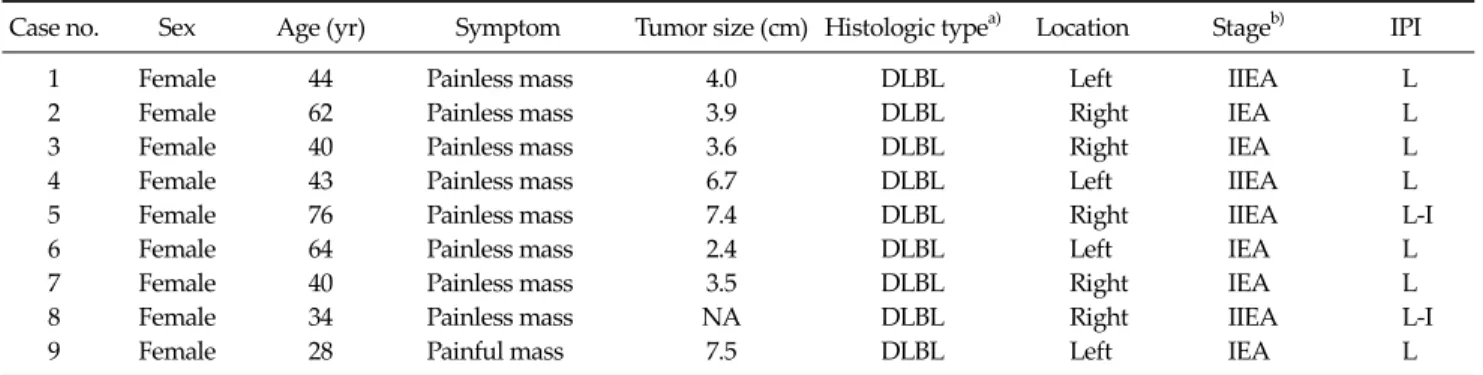
Table 1. Clinicopathologic characteristics of patients
Case no. Sex Age (yr) Symptom Tumor size (cm) Histologic typea) Location Stageb) IPI
1 Female 44 Painless mass 4.0 DLBL Left IIEA L
2 Female 62 Painless mass 3.9 DLBL Right IEA L
3 Female 40 Painless mass 3.6 DLBL Right IEA L
4 Female 43 Painless mass 6.7 DLBL Left IIEA L
5 Female 76 Painless mass 7.4 DLBL Right IIEA L-I
6 Female 64 Painless mass 2.4 DLBL Left IEA L
7 Female 40 Painless mass 3.5 DLBL Right IEA L
8 Female 34 Painless mass NA DLBL Right IIEA L-I
9 Female 28 Painful mass 7.5 DLBL Left IEA L
IPI, International Prognostic Index; DLBL, diffuse large B-cell tumor; L, low risk; L-I, low-intermediate risk; NA, not available.
a)Histologic type according to the World Health Organization classification. b)Stage according to the Ann Arbor staging system.
to 76 years). Eight patients initially visited the hospital with complaints of palpable breast mass without pain, and one patient presented with painful mass in the involved
breast. No patients had B symptoms, including fever, night sweats or weight loss. No patients showed evidence of human immunodeficiency virus, previous organ trans- plantation or breast tumor. The median tumor size was 3.8 cm (range, 1.7 to 7.5 cm) in diameter. Five cases presented in the right breast and four cases in the left breast. Five pa- tients were classified as IEA and four patients were classi- fied as IIEA as based on the Ann Arbor staging system.
Seven patients were classified as low risk group and the others were classified as low-intermediate risk group ac- cording to the IPI. Serum lactate dehydrogenase (LDH) was measured in all patients, and no patient had an ele- vated serum LDH.
Treatment and clinical outcomes
The median follow-up time was 44 months (range, 18 to 90 months). First-line therapy and clinical outcomes are documented in Table 2. Five patients underwent surgery;
one case of lumpectomy with sentinel lymph node biopsy because of uncertain diagnosis and four cases of excisional biopsy for diagnostic purposes. All patients received sys- temic chemotherapy with an anthracycline containing regimen (CHOP, cyclophosphamide, adriamycin, vincris- tine and prednisolone). Of those who received chemo- therapy, five patients received rituximab-containing che- motherapy (R-CHOP) and one patient received bleomy- cine-containing chemotherapy (CHOP-Bleo). Four pa- tients were treated with radiotherapy combined systemic chemotherapy at a total radiation therapy dose of 5,000 cGy in 25 fractions.
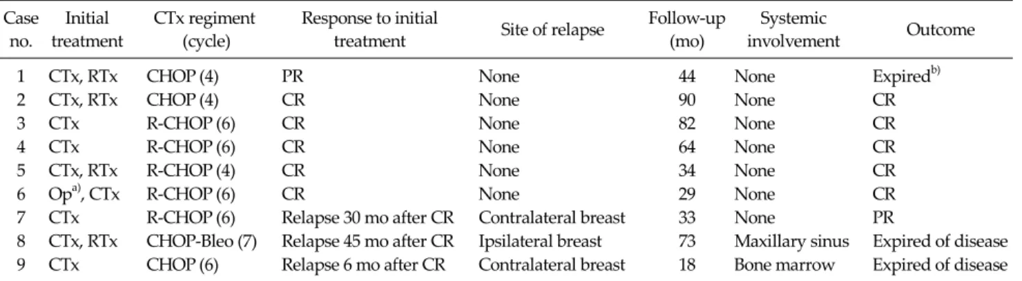
Table 2. Initial treatment and clinical outcomes of patients Case
no.
Initial treatment
CTx regiment (cycle)
Response to initial
treatment Site of relapse Follow-up (mo)
Systemic
involvement Outcome
1 CTx, RTx CHOP (4) PR None 44 None Expiredb)
2 CTx, RTx CHOP (4) CR None 90 None CR
3 CTx R-CHOP (6) CR None 82 None CR
4 CTx R-CHOP (6) CR None 64 None CR
5 CTx, RTx R-CHOP (4) CR None 34 None CR
6 Opa), CTx R-CHOP (6) CR None 29 None CR
7 CTx R-CHOP (6) Relapse 30 mo after CR Contralateral breast 33 None PR
8 CTx, RTx CHOP-Bleo (7) Relapse 45 mo after CR Ipsilateral breast 73 Maxillary sinus Expired of disease 9 CTx CHOP (6) Relapse 6 mo after CR Contralateral breast 18 Bone marrow Expired of disease CTx, chemotherapy; RTx, radiation therapy; CHOP, cyclophosphamide, doxorubicin, vincristine and prednisone; PR, partial remission; CR, complete remission; R-CHOP, rituximab-CHOP; Op, operation; Bleo, bleomycin.
a)Lumpectomy with sentinel lymph node biopsy. b)Death from accident.
A CR was achieved in eight patients after first-line therapy. However, among the CR patients, three patients experienced disease relapse. One patient relapsed on the ipsilateral breast and two patients on the contralateral breast. Systemic relapse occurred in two patients, includ- ing one case in the maxillary sinus and one case in the bone marrow. The estimated mean relapse free period was 64 months (95% confidence interval [CI], 42 to 87 months).
There was one case of SD (case no. 1); she expired 44 months after diagnosis because of an accident. In this study, there were two cases of disease-related death (cases no. 9 and 10), 73 months and 18 months after diagnosis, respectively. The estimated mean DSS was 77 months (96%
CI, 61 to 93 months).
DISCUSSION
PBL is a rare disease entity, likely due to the paucity of lymphoid tissue in breast. By the dedicated efforts of sev- eral investigators, a number of studies have been under- taken regarding PBL, including a report of more than 200 patients from the International Extranodal Lymphoma Study Group [14] and a prospective study of 96 patients [15]. However, most reports regarding Korean PBL pa- tients are retrospective and included small numbers of pa- tients [16-19]. For these lesions, we report our experience of PBL in a single institution. To the best of our knowledge, this is one of the largest series in Korea women.
According to the definition set out by Wiseman and Liao [7], PBL is diagnosed when the breast is the first or major manifestation site of lymphoma without the in- volvement of lymphoma in any other organ, except the ip- silateral axillary lymph node. Patients with concurrent widespread disease or preceding extramammary lympho- ma are also not defined as having PBL. Therefore, this defi- nition encompasses only tumors classified as stage I or II disease according to the Ann Arbor staging system, and lymphomas involving the breast but not meeting these cri- teria are diagnosed as secondary breast lymphoma.
The most common clinical symptom in 60–100% of pa- tients is a painless breast mass [9,20,21]. Other symptoms have been reported, including palpable lymph node, pain- ful mass, local inflammation, or breast swelling. Similar to previous reports, all of our patients visited the hospital with the complaint of a palpable breast mass, with only one of them experiencing a localized pain. B symptoms are uncommon. The incidental mammographic detection rate has been previously noted to be about 12% [20]. Mammo- graphic findings are nonspecific, and most lesions are oval-shaped and high-density lesions. There is no specific mammographic finding that could differentiate PBL from other invasive carcinomas of the breast. On ultrasound ex- amination, lymphomas are detected as single, oval, and hypoechoic lesion without speculated margins or calcifi- cation [8,22,23].
Fine needle aspiration, core needle biopsy, and exci- sional biopsy are effective methods for evaluating breast
mass and axillary lymph nodes. However, immunohisto- chemical or genetic studies are sometimes needed for ex- act diagnosis. In one case in our study (case no. 7), the pathologic department could not exactly diagnose lym- phoma with the specimen from the core needle biopsy.
Because the core needle biopsy was performed in outside institution, and the specimens were contaminated with the artifact. We performed a lumpectomy with sentinel lymph node biopsy based on the pre-operative diagnosis of “poorly differentiated invasive carcinoma”. After the operation, immunohistochemical staining revealed that antibodies against the CD20, BCL-2 and BCL-6 were pos- itive, and CD3 was negative on the surgically resected specimen. The specimen was diagnosed as DLBL of the breast based on microscopic morphological features and immunohistochemistry. Histologically, most PBLs are B-cell subtype. The majority of PLB cases in both our result and many published articles are DLBL, which account for 40–100% of PLB [20,24]. Low-grade subtypes of PBL, such as FL, MALT lymphoma, and marginal zone lymphoma are less commonly reported [8,25]. In our study, all cases were DLBL. In the pathologic laboratories, all specimens were showed no-particular arranged large lymphoid cells with vesicular and prominent nuclei. And the tissues were stained positively for the B-cell markers, including CD20 and Bcl-6, and negatively for the CD3, CD5 and CD10.
The prognosis of PBL has been reported that the behav- ior and clinical outcomes are similar to that of systemic lymphomas of the same histological types and stages.
Therefore, prognostic factors for patient with PBL have been reported as histologic subtype, IPI, the use of an- thracycline containing chemotherapy and/or radiation therapy [14,20], stage according to the Ann Arbor staging system [20], LDH level and age [9,26]. In a report by Ryan et al. [14] of 204 patients with primary DLBL of the breast, the median overall survival period was 8.0 years, and the median progression free period was 5.5 years. Our result showed similar results that estimated DSS and RFS were 6.4 years and 5.4 years, respectively. These results are im- proved over those of previous reports of Korean women with PBL. In a reports by Park et al. [18], the median overall survival and disease-free survival were calculated as 12 months and 6.5 months, respectively. We do not fully ex-
plain the exact cause of the differences in prognosis be- tween our results and those of Park et al. [18], but the dif- ferent prognosis may arise from differences in patients’
characteristics and from improvements in treatment modalities.
There have been controversies regarding the treatment of PBL. In general, PBL treatment is similar to that of pa- tients with systemic lymphoma of similar histologic type.
Combination chemotherapy with or without radiation therapy is accepted as the main treatment method. A pro- spective study by Aviles et al. [15], found that the 10 year rate of event-free survival was 50%, 57%, and 83% for ra- diotherapy, chemotherapy, and combined therapy, re- spectively. Rituximab, a monoclonal antibody targeting the CD20 antigen, is known to have high efficacy for DLBL, and R-CHOP has been the mainstay for treatment of B-cell lymphoma [27]. However, due to the low in- cidence of PBL, there are no useful data regarding the effi- cacy of R-CHOP compared with CHOP for the treatment of PBL. In our study, R-CHOP was used for all patients di- agnosed after July, 2005. All patients who received R-CHOP chemotherapy achieved CR, and among them, only one patient had a relapse in the contralateral breast.
The use of CHOP and R-CHOP could be one of the reasons that the prognosis of our patients was better than the re- sults of Park et al. [18]. Park et al. [18] which included pa- tients diagnosed between 1989 and 2001. Three out of their patients were received anthracycline-containing chemo- therapy, and no patients received a rituximab containing regimen.
Surgery is only recommended for diagnostic purposes, and is not recommended as a first line therapy, as ex- tensive surgery may delay the beginning of chemotherapy [14]. Furthermore, Misra et al. [28] and Park et al. [18] sug- gested that surgery should be performed only after the lo- cal failure of systemic treatment. In our study, one patients received operation not for diagnostic purpose and the pa- tient underwent a lumpectomy with sentinel lymph node biopsy because of an unsatisfactory diagnosis before the operation. Due to the small numbers in our series, no con- clusions can be drawn regarding the prognosis according to stage and treatment methods.
In conclusion, due to the rarity of the PBL, there have
been a few reports about PBL in Korean patients. In our study, all PBL cases are B-cell origin, with DLBL being the most common histologic type. A combined treatment mo- dality has been known to have positive effects on prog- nosis, and surgery should be limited to diagnostic purposes. Further studies about PBL in Korean women are necessary to improve understanding of this disease. And, we also hope that further studies could reveal the impact of treatment protocol for Korean women with PBL.
CONFLICTS OF INTEREST
No potential conflict of interest relevant to this article was reported.
REFERENCES
1. Freeman C, Berg JW, Cutler SJ. Occurrence and prognosis of extranodal lymphomas. Cancer 1972;29:252-60.
2. d'Amore F, Christensen BE, Brincker H, Pedersen NT, Thorling K, Hastrup J, et al. Clinicopathological features and prognostic factors in extranodal non-Hodgkin lymphomas.
Danish LYFO Study Group. Eur J Cancer 1991;27:1201-8.
3. Krol AD, le Cessie S, Snijder S, Kluin-Nelemans JC, Kluin PM, Noordijk EM. Primary extranodal non-Hodgkin's lymphoma (NHL): the impact of alternative definitions tested in the Comprehensive Cancer Centre West pop- ulation-based NHL registry. Ann Oncol 2003;14:131-9.
4. Ferguson DJ. Intraepithelial lymphocytes and macro- phages in the normal breast. Virchows Arch A Pathol Anat Histopathol 1985;407:369-78.
5. Topalovski M, Crisan D, Mattson JC. Lymphoma of the breast: a clinicopathologic study of primary and secondary cases. Arch Pathol Lab Med 1999;123:1208-18.
6. Gholam D, Bibeau F, El Weshi A, Bosq J, Ribrag V. Primary breast lymphoma. Leuk Lymphoma 2003;44:1173-8.
7. Wiseman C, Liao KT. Primary lymphoma of the breast.
Cancer 1972;29:1705-12.
8. Domchek SM, Hecht JL, Fleming MD, Pinkus GS, Canellos GP. Lymphomas of the breast: primary and secondary involvement. Cancer 2002;94:6-13.
9. Ha CS, Dubey P, Goyal LK, Hess M, Cabanillas F, Cox JD.
Localized primary non-Hodgkin lymphoma of the breast.
Am J Clin Oncol 1998;21:376-80.
10. International Agency for Research on Cancer, Swerdlow S, Campo E, Harris NL, Jaffe ES, editors. WHO classification of tumours of haematopoietic and lymphoid tissues. 4th ed. Lyon: World Health Organization; 2008. p.439.
11. Carbone PP, Kaplan HS, Musshoff K, Smithers DW,
Tubiana M. Report of the Committee on Hodgkin's Disease Staging Classification. Cancer Res 1971;31:1860-1.
12. A predictive model for aggressive non-Hodgkin's lym- phoma. The International Non-Hodgkin's Lymphoma Prognostic Factors Project. N Engl J Med 1993;329:987-94.
13. Cheson BD, Horning SJ, Coiffier B, Shipp MA, Fisher RI, Connors JM, et al. Report of an international workshop to standardize response criteria for non-Hodgkin's lym- phomas. NCI Sponsored International Working Group. J Clin Oncol 1999;17:1244.
14. Ryan G, Martinelli G, Kuper-Hommel M, Tsang R, Pruneri G, Yuen K, et al. Primary diffuse large B-cell lymphoma of the breast: prognostic factors and outcomes of a study by the International Extranodal Lymphoma Study Group.
Ann Oncol 2008;19:233-41.
15. Aviles A, Delgado S, Nambo MJ, Neri N, Murillo E, Cleto S.
Primary breast lymphoma: results of a controlled clinical trial. Oncology 2005;69:256-60.
16. Jung TH, Chung KS, Kim WM, Yu BJ, Chung CH, Lee MJ, et al. A case of primary breast lymphoma. Korean J Hematol 1992;27:409-13.
17. Jung SW, Kim BS, Park JH, Kim SK, Seo HE, Shin DK, et al.
A case of primary MALT lymphoma of the breast. Cancer Res Treat 2001;33:269-73.
18. Park YH, Kim SH, Choi SJ, Ryoo BY, Kang YK, Lee SS.
Primary malignant lymphoma of the breast: clinicopatho- logical study of nine cases. Leuk Lymphoma 2004;45:
327-30.
19. Lim HJ, Cho KR, Kim I, Hwang KW, Seo BK, Woo OH, et al.
Primary peripheral T-cell lymphoma of the breast: radiologic and pathologic findings. J Breast Cancer 2010;13:318-22.
20. Jeanneret-Sozzi W, Taghian A, Epelbaum R, Poortmans P, Zwahlen D, Amsler B, et al. Primary breast lymphoma: pa- tient profile, outcome and prognostic factors. A multi- centre Rare Cancer Network study. BMC Cancer 2008;8:86.
21. Brustein S, Filippa DA, Kimmel M, Lieberman PH, Rosen PP. Malignant lymphoma of the breast: a study of 53 patients. Ann Surg 1987;205:144-50.
22. Lyou CY, Yang SK, Choe DH, Lee BH, Kim KH.
Mammographic and sonographic findings of primary breast lymphoma. Clin Imaging 2007;31:234-8.
23. Yang WT, Lane DL, Le-Petross HT, Abruzzo LV, Macapin- lac HA. Breast lymphoma: imaging findings of 32 tumors in 27 patients. Radiology 2007;245:692-702.
24. Brogi E, Harris NL. Lymphomas of the breast: pathology and clinical behavior. Semin Oncol 1999;26:357-64.
25. Martinelli G, Ryan G, Seymour JF, Nassi L, Steffanoni S, Alietti A, et al. Primary follicular and marginal-zone lym- phoma of the breast: clinical features, prognostic factors and outcome: a study by the International Extranodal Lymphoma Study Group. Ann Oncol 2009;20:1993-9.
26. Hugh JC, Jackson FI, Hanson J, Poppema S. Primary breast lymphoma: an immunohistologic study of 20 new cases.
Cancer 1990;66:2602-11.
27. Coiffier B. Rituximab therapy in malignant lymphoma.
Oncogene 2007;26:3603-13.
28. Misra A, Kapur BM, Rath GK. Primary breast lymphoma. J Surg Oncol 1991;47:265-70.