© 2016 The Korean Ophthalmological Society
This is an Open Access article distributed under the terms of the Creative Commons Attribution Non-Commercial License (http://creativecommons.org/licenses /by-nc/3.0/) which permits unrestricted non-commercial use, distribution, and reproduction in any medium, provided the original work is properly cited.
Original Article
Comparison of Astigmatism Induced by Combined Inferior Oblique Anterior Transposition Procedure and Lateral Rectus Recession Alone
Sun Jung Eum, Bo Young Chun
Department of Ophthalmology, Kyungpook National University School of Medicine, Daegu, Korea
Purpose: The purpose of this study is to compare the magnitude and axis of astigmatism induced by a combined inferior oblique (IO) anterior transposition procedure with lateral rectus (LR) recession versus LR recession alone.
Methods: Forty-six patients were retrospectively analyzed. The subjects were divided into two groups: those having concurrent inferior oblique muscle overaction (IOOA) and intermittent exotropia (group 1, 20 patients) and those having only intermittent exotropia as a control (group 2, 26 patients). Group 1 underwent combined anterior transposition of IO with LR recession and group 2 underwent LR recession alone. Induced astigma- tism was defined as the difference between preoperative and postoperative astigmatism using double-angle vector analysis. Cylinder power, axis of induced astigmatism, and spherical equivalent were analyzed at 1 week, 1 month, and 3 months after surgery.
Results: Larger changes in the axis of induced astigmatism were observed in group 1, with 4.5° incyclotorsion, than in group 2 at 1 week after surgery (axis, 84.5° vs. 91°; p < 0.001). However, there was no statistically significant inter-group difference thereafter. Relaxation and rapid regression in the incyclotorsion of induced astigmatism were observed over-time. Spherical equivalent significantly decreased postoperatively at 1 month in both groups, indicating a myopic shift (p = 0.011 for group 1 and p = 0.019 for group 2) but did not show sig- nificant differences at 3 months after surgery (p = 0.107 for group 1 and p = 0.760 for group 2).
Conclusions: Combined IO anterior transposition procedures caused an increased change in the axis of in- duced astigmatism, including temporary incyclotorsion, during the first week after surgery. However, this signif- icant difference was not maintained thereafter. Thus, combined IO surgery with LR recession does not seem to produce a sustained astigmatic change, which can be a potential risk factor of postoperative amblyopia or diplopia compared with LR recession alone.
Key Words: Astigmatism, Exotropia, Inferior oblique muscle, Strabismus
The inferior oblique (IO) muscle acts as an excyclotorter, with secondary actions of elevation and adduction [1]. In-
ferior oblique muscle overaction (IOOA) is a common dis- order of ocular motility [2], and surgical correction is the main treatment modality. Previous clinical studies have re- ported that surgical weakening of the IO muscle produces incyclorotation of the axis of astigmatism by 10° [3,4]. Re- fractive changes after strabismus surgery have been stud- ied extensively, but the effect of surgery on refraction and astigmatism is controversial [5-13]. Several studies have re-
Received: February 1, 2016 Accepted: June 1, 2016
Corresponding Author: Bo Young Chun, MD, PhD. Department of Oph- thalmology, Kyungpook National University Hospital, #130 Dongdeok-ro, Jung-gu, Daegu 41944, Korea. Tel: 82-53-420-5818, Fax: 82-53-426-6552, E-mail: [email protected]
ported that recession of a single rectus muscle is correlated with an increase in power in the meridian of the recessed muscle [13-15]. This effect is related to a change in corneal curvature secondary to reduction in tension of the recessed extraocular muscle transmitted via the sclera to the cornea [12,16,17].
An investigation of the relationship between muscle overaction and astigmatism is beneficial because induced astigmatism after strabismus surgery can affect patient vi- sual acuity and may increase the potential risk of postop- erative amblyopia in children. We previously demonstrated that the larger is the amount of lateral rectus (LR) reces- sion, the greater is the cylinder power obtained postopera- tively [18]. Hainsworth et al. [17] reported that the amount of muscle resected or recessed may affect the amount of corneal curvature change after strabismus surgery. In their study, comparison of the calculated corneal power change from preoperative to postoperative demonstrated a statisti- cally significant difference between the recession proce- dure group and the recession-resection procedure group.
Therefore, it can be presumed that there may be some dif- ference in the degree of postoperative astigmatism accord- ing to the involved extraocular muscle. A variety of studies on the effect of strabismus surgery on refraction and astig- matism in patients with intermittent exotropia, X(T), have been reported [5-8,12]. However, there is relatively little in- formation on the objective changes in surgically-induced astigmatism after a combined IO weakening procedure with LR recession.
Thus, we analyzed surgically-induced astigmatism in patients who underwent concurrent anterior transposition of the IO muscle and LR recession and compared the find- ings with those in patients who underwent LR recession only. The postoperative changes in the axis and cylindrical power of induced astigmatism were calculated using dou- ble-angle vector analysis [18-20].
Materials and Methods
A retrospective review of medical records was conduct- ed on 322 patients who underwent strabismus surgery for X(T) and IOOA between March 2007 and June 2010. Ex- clusion criteria for this study were age younger than 5 years, a history of strabismus surgery, coexisting dissociat- ed vertical deviation, corneal opacity, craniofacial anoma-
lies, lack of cooperation during measurement for refrac- tion, ocular misalignment or visual acuity, and follow-up duration less than 3 months.
A total of 46 patients were enrolled in this study and were divided into two groups based on the type of surgery:
20 patients (group 1) with bilateral IOOA and X(T) who underwent combined bilateral anterior transposition of the IO with bilateral LR recession, and 26 patients (group 2) with only X(T) who underwent bilateral LR recession. The following parameters were analyzed: sex, age at surgery, distant and near deviation angles, preoperative and postop- erative non-cycloplegic autorefraction, calculated cylinder power and axis of induced astigmatism using double-angle vector analysis, spherical equivalent (SE), and best-correct- ed visual acuity (BCVA).
All patients received a comprehensive ophthalmological examination on their scheduled follow-up date. Visual acu- ities were measured with Snellen’s chart, and values were converted to the logarithm of the minimum angle of reso- lution (logMAR). Deviation angles were evaluated using the alternate prism cover test, and the degree of IOOA was graded as +1 to +4 based on evaluation of ocular misalign- ment in nine diagnostic gaze positions. All surgeries were performed by one surgeon (BYC). Under general anesthe- sia, the surgeon made a limbic conjunctival incision and recessed the LR muscle. The bilateral LR recession proce- dure was performed according to the deviation angle of the patient. Among the various surgical techniques for IO weakening procedures, all 20 patients in group 1 under- went full anterior transposition of the IO muscle regardless of the degree of IOOA. The surgical technique of anterior transposition was as follows: after the eyeball was retract- ed superiorly, the conjunctiva and Tenon’s capsule were in- cised 8 mm from the limbus in the inferotemporal direc- tion. The IO muscle was isolated with a muscle hook, and the muscle was clipped with two mosquitos and severed with Wescott scissors. One 7-0 Vicryl double-armed suture was placed through the insertion site of the muscle and re- attached to the sclera adjacent to the temporal side of the inferior rectus muscle insertion site. Refraction was mea- sured through non-cycloplegic refraction with an auto re- fractometer (KR-8100; Topcon, Tokyo, Japan) at 1 week before surgery and at 1 week, 1 month, and 3 months after surgery. Three consecutive refraction readings were evalu- ated to reach a median value of cylinder power, cylinder axis, and SE.
Induced astigmatism was defined as the difference be- tween postoperative and preoperative refraction [19,21]. To analyze the changes in the amount and axis of cylinder with statistical analysis, we calculated induced astigma- tism using the double-angle vector analysis described by Retzlaff et al. [19] and further developed by others [20-22].
Polar values (cylinder and axis) were converted to Carte- sian (x and y) values according to the method described by Holladay et al. [20] using formulas to determine the mean cylinder power and axis of induced astigmatism. On a double-angle plot, the x-axis is coincident with the axis of non-oblique (90°, 0°) astigmatism and the y-axis is coinci- dent with the axis of oblique (45°, 135°) astigmatism [20].
The surgically-induced astigmatism vector is determined by subtracting the preoperative vector from the postopera- tive vector. We cited an example of subtracting cylinders using the double-angle method [18,20]. In the following formula, the postoperative vector and preoperative vector are designated as numbers 3 and 1, respectively.
Example of subtracting cylinders using the double-angle mathematical method for subtraction of refraction
Postoperative cylinder (3) - preoperative cylinder (1) = induced cylinder (2)
For example, postoperative cylinder = +1.0, axis = 4°;
preoperative cylinder = +1.0, axis = 70°
Step 1: Determine the x and y components of the vectors representing the cylinders.
x1 = cylinder1 × cos(2 × axis1) x1 = -0.766 x3 = cylinder3 × cos(2 × axis3) x3 = 0.990 y1 = cylinder1 × sin(2 × axis1) y1 = 0.643 y3 = cylinder3 × sin(2 × axis3) y3 = 0.139 Step 2: Determine the induced cylinder axis (axis2).
Axis2 = (arctan[(y3 - y1) / (x3 - x1)]) / 2 = arctan(-0.504 / 1.756) / 2 = -8° = 172°
Step 3: Determine the induced cylinder power (cylinder power 2).
Cylinder power 2 = [cylinder3 × (cos(2 × (axis3 - axis2)))] - [cylinder1 × (cos(2 × (axis1 - axis2)))]
= 1.0 × cos(2 × (4 - 172)) - 1.0 × cos(2 × (70 - 172))
= 0.914 + 0.914 = 1.83 D
X = mean value of x, Y = mean value of y
Cylinder power of induced astigmatism = X2 + Y2, Axis of induced astigmatism = arctan (Y / X) / 2
The cylinder power (diopter, D) and axis (°) of the in- duced astigmatism were analyzed at 1 week, 1 month, and 3 months after surgery. All patients in this study under- went surgery on both eyes, although we chose only the right eyes of the patients for data analysis. The Mann-Whit- ney U-test was used for comparison of induced astigma- tism, SE, and BCVA between the two groups. A paired t-test was used to compare changes in parameters accord- ing to postoperative time points.
Statistical analysis was performed with PAWS ver. 18.0 (SPSS Inc., Chicago, IL, USA). The results are expressed as mean ± standard deviation. Statistical significance was defined as p-value <0.05 for all tests.
Results
Clinical findings of patients are shown in Table 1. A total of 46 eyes of from 46 patients were included from 26 male and 20 female patients. Patients were divided into two groups: group 1 (20 eyes of 20 patients) receiving concur- rent bilateral anterior transposition of the IO muscle and bilateral LR recession, and group 2 (26 eyes of 26 patients) with bilateral LR recession only. The mean age at surgery was 6.75 ± 2.22 years in group 1 and 7.42 ± 1.96 years in group 2. There were no significant differences in preopera- tive horizontal deviation angle at distance in the primary position or amount of LR recession between the two groups (p= 0.434 and p = 0.598, respectively). The median amount of oblique muscle overaction was +3.5 in group 1. The mean preoperative cylinder power of astigmatism was 0.72
± 0.61 D in group 1 and 0.80 ± 0.95 D in group 2, and the axis of astigmatism was 89.0° in group 1 and 89.2° in group 2. There was no statistically significant difference in cylin- der power or axis of preoperative astigmatism between the two groups (p = 0.735 and p = 0.581, respectively).
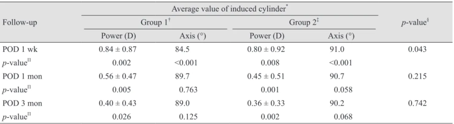
Table 2 presents the changes in induced astigmatism cal- culated with the double-angle mathematical method at postoperative 1 week, 1 month, and 3 months. We com-
pared the changes in induced astigmatism not only be- tween preoperative and postoperative times, but also be- tween the two subgroups. At 1 week after surgery, the mean cylinder power of induced astigmatism was 0.84 ± 0.87 D and the axis of induced astigmatism was 84.5° in group 1, while those in group 2 were 0.80 ± 0.92 D and 91.0°, respectively. An incyclotorsion of 4.5° in the axis of induced astigmatism in group 1 was observed with statisti- cally significant difference at 1 week after surgery (p
<0.001). However, this incyclotorsional change rapidly re- turned toward its preoperative value, and there was no sig- nificant difference in the cylinder axis of induced astigma- tism between preoperative and postoperative 1-month
follow-up (p = 0.763). Considering the changes in induced cylinder axis between the groups, there was a statistically significant difference in the axis of induced astigmatism between group 1 and group 2 at postoperative 1 week (p = 0.043). However, there was no significant inter-group dif- ference at 1 (p = 0.215) or 3 months (p = 0.742) postopera- tively. Comparing the cylinder power between preopera- tive and postoperative times, there was a statistically significant difference in the cylinder power of induced astigmatism compared with the preoperative value in both groups until the 3-month follow-up (p < 0.05). Considering the changes in cylinder power between the two groups, the average cylinder power of induced astigmatism for group
Table 1. Clinical characteristics of the patients
Group 1* Group 2† p-value
No. of patients 20 26
Sex (male : female) 12 : 8 14 : 12 0.676‡
Age at surgery (yr) 6.75 ± 2.22 7.42 ± 1.96 0.282§
Preop deviation of X(T) at distance (PD) 27.75 ± 5.73 29.04 ± 5.29 0.434§
Amount of LR recession (mm) 7.90 ± 0.93 8.04 ± 0.83 0.598§
Preop astigmatismΠ
Cylinder power (D) 0.72 ± 0.61 0.80 ± 0.95 0.735§
Axis (°) 89.0 89.2 0.581§
Values are presented as mean ± standard deviation.
Preop = preoperative; X(T) = exotropia; PD = prism diopter; LR = lateral rectus; D = diopter.
*Bilateral anterior transposition of the inferior oblique muscle and bilateral LR recession; †Bilateral LR recession; ‡Chi-square test;
§Mann-Whitney U-test; ΠPreoperative astigmatism is defined as the median value of three consecutive measurements.
Table 2. Changes in induced astigmatism Follow-up
Average value of induced cylinder*
p-value§
Group 1† Group 2‡
Power (D) Axis (°) Power (D) Axis (°)
POD 1 wk 0.84 ± 0.87 84.5 0.80 ± 0.92 91.0 0.043
p-valueΠ 0.002 <0.001 0.008 <0.001
POD 1 mon 0.56 ± 0.47 89.7 0.45 ± 0.51 90.7 0.215
p-valueΠ 0.005 0.763 0.001 0.058
POD 3 mon 0.40 ± 0.43 89.0 0.36 ± 0.33 90.2 0.742
p-valueΠ 0.026 0.125 0.002 0.068
Values are presented as mean ± standard deviation.
D = diopter; POD = postoperative day.
*The induced cylinder power and axis were calculated using the double-angle mathematical method; †Bilateral anterior transposition of the inferior oblique muscle and bilateral lateral rectus recession; ‡Bilateral lateral rectus recession; §Comparison of changes in the axis of induced astigmatism between the two groups (Mann-Whitney U-test); ΠComparison of cylinder power and axis between preoperative value and the value at each follow-up visit.
1 was slightly larger than that in group 2, but there was no significant inter-group difference at any postoperative time point after 1 week (p > 0.05).
The values of mean preoperative and postoperative SE over time are shown in Table 3. SE significantly decreased at postoperative 1 week and 1 month in both group 1 (p = 0.001, p = 0.011, respectively) and group 2 (p = 0.002, p = 0.019, respectively), indicating a shift in the myopic direc- tion. However, there was no significant difference at post- operative 3 months (p = 0.107 in group 1 and p = 0.760 in group 2). We also compared the SE between the groups at each follow-up date and found no significant difference (all p > 0.05). Comparing the overall mean change in SE be- tween preoperative and 3 month postoperative values in the two groups, there was no statistically significant differ- ence (-0.174 ± 0.487 in group 1, -0.073 ± 0.732 in group 2; p
= 0.165).
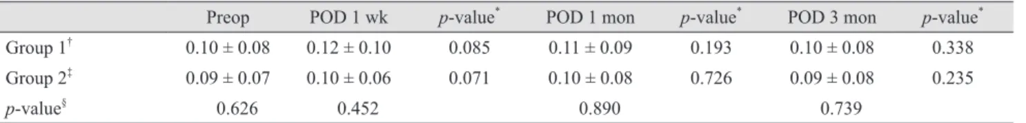
Table 4 presents the preoperative and postoperative BCVA (logMAR) of the two groups. No statistically sig- nificant difference was found between preoperative and postoperative BCVA (logMAR) either in group 1 or group 2 (all p > 0.05 in both groups). Moreover, there was no in- ter-group difference at any follow-up visit (all p > 0.05).
Discussion
The changes in refractive status and visual acuity fol- lowing uncomplicated strabismus surgery are generally short-lived and are customarily attributed to lid edema, use of eye drop medication, or photophobia [16]. If the astig- matic changes present without any of these factors, the true cause of changes in surgically-induced astigmatism and visual deficits must be determined. Refractive error changes after uncomplicated strabismus surgery have been documented in several studies [12,16]. Although the effect of extraocular muscle surgery on refractive and astigmatic changes is debatable [5-8], it is speculated that this effect is related to a change in corneal curvature secondary to the alterations in muscle tension transmitted via the sclera to the cornea [12,16,17]. Preslan et al. [12] previously reported a prospective study of preoperative and postoperative cy- cloplegic refractions in patients who had strabismus sur- gery, showing a statistically significant increase in with- the-rule astigmatism that persisted for at least 4 months.
Our previous study [18] demonstrated that LR recession induced an increase in surgically-induced astigmatism in the with-the-rule direction. The increased magnitude of
Table 3. Mean preoperative and postoperative spherical equivalents
Preop POD 1 wk p-value* POD 1 mon p-value* POD 3 mon p-value*
Group 1† (D) 0.07 ± 1.38 -0.61 ± 1.36 0.001 -0.22 ± 1.48 0.011 -0.10 ± 1.36 0.107 Group 2‡ (D) -0.66 ± 1.11 -0.98 ± 1.04 0.002 -0.70 ± 0.94 0.019 -0.74 ± 0.93 0.760
p-value§ 0.058 0.297 0.183 0.066
Values are presented as mean ± standard deviation.
Preop = preoperative; POD = postoperative day; D = diopter.
*Comparison of spherical equivalents between preoperative and 1 week, between 1 week and 1 month, and between 1 month and 3 months after surgery (paired t-test); †Bilateral anterior transposition of the inferior oblique muscle and bilateral lateral rectus recession;
‡Bilateral lateral rectus recession; §Comparison of spherical equivalents between the two groups at each follow-up visit (Mann-Whitney U-test).
Table 4. Preoperative and postoperative best-corrected visual acuity values (logMAR)
Preop POD 1 wk p-value* POD 1 mon p-value* POD 3 mon p-value*
Group 1† 0.10 ± 0.08 0.12 ± 0.10 0.085 0.11 ± 0.09 0.193 0.10 ± 0.08 0.338
Group 2‡ 0.09 ± 0.07 0.10 ± 0.06 0.071 0.10 ± 0.08 0.726 0.09 ± 0.08 0.235
p-value§ 0.626 0.452 0.890 0.739
Values are presented as mean ± standard deviation.
logMAR = logarithm of the minimum angle of resolution; Preop = preoperative; POD = postoperative day.
*Comparison of best-corrected visual acuity between preoperative and 1 week, between 1 week and 1 month, and between 1 month and 3 months after surgery (paired t-test); †Bilateral anterior transposition of the inferior oblique muscle and bilateral lateral rectus re- cession; ‡Bilateral lateral rectus recession; §Comparison of best-corrected visual acuity between the two groups at each follow-up visit (Mann-Whitney U-test).
surgically-induced astigmatism was proportional to the amount of LR recession, and this statistically significant change was observable at the 1 week postoperative exam- ination. However, this significant difference between the moderate- and large-recession groups was not maintained thereafter. Relaxation of astigmatism trended toward the preoperative value over time in both groups; at 1 month and 3 month follow-up, there was no statistically signifi- cant difference in the magnitude of induced astigmatism between subgroups [18].
Although the effect of strabismus surgery on refractive error has been studied extensively in previous reports [5- 13], there was little information about the effect on astig- matic change after oblique muscle surgery. According to the study of Kushner [3], it was assumed that surgery of an oblique muscle was expected to produce a torsional rotation of the globe; if this is true, patients with substantial astig- matic refractive errors should be expected to show a rota- tion of their axis of astigmatism after undergoing a surgical weakening or tightening procedure of an oblique muscle.
We agreed with Kushner’s assumption; therefore, we de- signed our study assuming it to be true. Factors that could worsen refractive errors after oblique muscle surgery should be sought and corrected, particularly important in preverbal children who have been previously amblyopic or are at high risk for amblyopia. Therefore, we aimed to evaluate and compare the magnitude and axis of surgical- ly-induced astigmatism by LR recession alone versus in combination with anterior transposition of the IO muscle.
In this study, the amount of surgically-induced astigma- tism was analyzed and compared in patients who had un- dergone either combined anterior transposition of IO mus- cle with LR recession (group 1) or LR recession alone (group 2) during the 3-month follow-up period. At 1 week after surgery, postoperative statistically significant incy- clotorsion of 4.5° in the axis of induced astigmatism in group 1 was observed compared with the preoperative val- ue (p < 0.001). However, this temporary incyclotorsion rapidly returned toward the preoperative value at postop- erative 1 month (p = 0.763) and was maintained until the 3 month follow-up (p = 0.125). In addition, comparing the postoperative induced cylinder axis between the two groups, there was a statistically significant difference in the axis of induced astigmatism at 1 week after surgery (84.5° in group 1 and 91.0° in group 2, p = 0.043). However, there was no significant inter-group difference at the post-
operative 1 month (p = 0.215) or 3 months follow-up (p = 0.742).
There have been various reports on the duration of tor- sional effect after IO weakening surgeries [3,23]. Kushner [3] reported that weakening of the IO muscle produced a long-term 10° incyclorotation of the astigmatic axis for at least 6 months, and there was no return of the axis toward the preoperative orientation. On the contrary, Santiago et al. [23] had reported that anterior transposition of the IO muscle initially decreased objective excyclotorsion, but the effect weakened after 10 weeks. The results of our study correspond with the above studies that have reported that IO weakening procedures caused an incyclotorsion of the globe. It has been reported that postoperative refractive er- ror stabilizes at 3 to 6 months after eye muscle surgery [3,5,12,13,23]. Therefore, we collected postoperative data at three time points (1 week, 1 month, and 3 months fol- low-up) to analyze the changes in refractive error. Notably, we found an unexpected difference in the recovery rates of surgically-induced incyclotorsion. In our study, the dura- tion of a temporary incyclotorsion after anterior transposi- tion of IO muscle combined with LR recession was rela- tively shorter than that in a previous study [3], and this abnormality rapidly recovered toward the preoperative value after 1 month. Furthermore, when comparing in- duced cylinder axes between the two groups, a statistically significant difference was observed only at postoperative 1 week. Thereafter, the axis of induced astigmatism after combined anterior transposition of the IO muscle with LR recession changed toward the axis of the control groups.
We postulate the reason for this rapid regression of chang- es in the induced astigmatic axis to be due to compensa- tion for cyclodeviation by the strong fusional power of tor- sional disparities of children. Ruttum and von Noorden [24] and von Noorden [25] reported the existence of an adaptive mechanism compensating for cyclodeviation by cyclofusion, which prevented awareness of image tilting and overcame cyclotropia based on empirical factors re- garding sensory reorientation. The existence of such sen- sorial adaptation to torsion eradicated the effect of IO weakening surgery.
The cylinder power of induced astigmatism did not vary significantly when comparing patients with solely bilateral LR recession to those with concurrent bilateral anterior transposition of the IO muscle and bilateral LR recession.
Al-Haddad et al. [26] also reported that no significant dif-
ference in the occurrence of astigmatism was observed be- tween a horizontal strabismus group and a horizontal stra- bismus combined with IOOA group. They explained this result with the hypothesis that tension of the overacting IO muscle per se did not cause a sufficient mechanical advan- tage on the globe to produce more pronounced astigma- tism in the presence of horizontal strabismus. As a result, rectus muscle weakening procedures contributed more to the postoperative change than any added effect from the IO muscle weakening surgery. Our findings correspond with the study of Al-Haddad et al. [26]. Although the two groups in our study showed comparable amounts of cylin- der power of induced astigmatism after surgery, slightly higher cylinder power in group 1 was observed until the 3 month follow-up. It seemed that a two-muscle surgery per eye in group 1 (IO and LR muscle) resulted in greater changes in the location of extraocular muscle insertion than that in group 2 (LR muscle only). Thus, the combined procedure might induce a longer inflammatory change, thus influencing an increased cylinder power of induced astigmatism in group 1 [1].
For statistical data analysis, we used the double-angle vector analysis method to subtract cylinders for calculating changes in the magnitude and axis of surgically-induced astigmatism. Most previous studies reporting on changes in refractive power after strabismus surgery evaluated mean changes in astigmatism using a keratometer or cor- neal topography [14,15,17]. These methods can generate discrepancies when representing magnitude and axis of in- duced astigmatism with angular data. Otherwise, dou- ble-angle vector analysis has the benefit of accounting for the directional effect of astigmatism. Therefore, we ana- lyzed periodic changes in astigmatism in terms of axis and cylinder power after combined anterior transposition of the IO muscle in patients who had concurrent X(T) using vector analysis of postoperative medical records.
The SE of the refraction in our study changed with a significant myopic shift at 1 week and 1 month after sur- gery, but it did not show significant difference between the groups at 3 months postoperative. The overall mean change in SE between preoperative and 3 months postop- erative values was -0.174 ± 0.487 in group 1 and -0.073 ± 0.732 in group 2, and there was no significant difference between the two groups ( p = 0.165). In our study, SE changed in agreement with previous studies of change in myopic direction [5,21]. Hong and Kang [5] reported that
the SE change represented a significant myopic shift after horizontal rectus muscle surgery at the first week. There- after, the myopic shift persisted until 6 months postopera- tively, though there was no statistically significant differ- ence. Furthermore, the study reported that the mean change in SE after surgery showed no significant in- ter-group difference. The same authors reported that the myopic shift might be caused by strabismus surgery itself considering that age did not show a significant correlation with SE; the results of our study closely corresponded with the above study. However, further studies to determine the exact mechanism between strabismus surgery and SE should be performed. Furthermore, future investigation about a relationship between degree of SE and type of strabismus surgery should be evaluated.
A statistically significant difference in the cylinder pow- er of induced astigmatism compared with the preoperative value was observed in both groups until the 3-month fol- low-up. Also, the cylinder axis of induced astigmatism compared with the preoperative value was significantly different at 1 week after surgery in both groups. These changes in astigmatism may induce vision changes, so we compared the visual acuity of the two groups before and after surgery. No statistically significant difference was found between preoperative and postoperative BCVA (log- MAR) either in group 1 or group 2 (all p > 0.05 in both groups), and there was also no inter-group difference (all p
> 0.05). Rajavi et al. [27] reported that LR recession in- duced significant change only in the astigmatic axis toward with-the-rule astigmatism, and not in astigmatic power or SE at 1 month or 3 months after surgery. Although the au- thors did not directly evaluate preoperative or postopera- tive visual acuity, they reported that shift of axis would not be practically notable, considering insignificant alter- ation in astigmatic power. Hong and Kang [5] also reported that small statistical astigmatic changes did not seem to be clinically important. In our study, cylinder power and axis of induced astigmatism showed statistically significant dif- ferences compared with the preoperative values, but these astigmatic changes did not affect vision changes in either group, in agreement with the previous studies [5,27].
Limitations of this study include analyses from retro- spective design, a small number of patients, and a relative- ly short follow-up period. In this study, all patients who underwent combined IO weakening surgery had only an- terior transposition of the IO muscle, so the differential ef-
fects of other various types of IO weakening surgeries were not studied. In addition, this study lacks measure- ments with subjective degrees of torsion such as the double Maddox rod test and objective degrees of torsion such as fundal photography. Kushner [3] attempted to correlate changes in the astigmatic axis with changes in subjective cyclorotation as measured with the double Maddox rod;
however, he encountered some problems; he found that measurement of cyclotropia with the double Maddox rod was often not reproducible to less than 5° in the same pa- tients. The amount of cyclotropia observed in the fixed eye seemed to vary considerably [3]. Also, patients with long-standing cyclotropia, often associated with primary oblique dysfunction, undergo a sensory reorientation of their cyclotropic eye and may not appear to have a cyclo- tropia on the double Maddox rod test [3,24]. However, he found that, in patients with subjective symptoms of cyclo- tropia, who did undergo double Maddox rod testing, the change in subjective cyclotropia postoperatively was simi- lar to the change in astigmatic axis [3]. Therefore, we also did not measure subjective cyclotropia by double Maddox rod; further, most of our patients were very young and were not able to understand the test. Future studies with more patients and a longer follow-up with different sur- gery types of horizontal and cyclovertical strabismus are highly recommended to confirm true statistically signifi- cant difference.
In conclusion, combined IO anterior transposition proce- dure with LR recession caused more changes in the axis of induced astigmatism than did LR recession alone during the first week after surgery. However, this incyclotorsion was rapidly restored toward the preoperative value and was maintained during postoperative follow-up, with no significant inter-group difference. Our results suggest that combined IO anterior transposition procedure with LR re- cession does not produce sustained astigmatic changes compared with LR recession alone, and these temporary changes may not interfere with the vision of patients through a longer postoperative period.
Conflict of Interest
No potential conflict of interest relevant to this article was reported.
References
1. von Noorden GK. Clinical observations in cyclodeviations.
Ophthalmology 1979;86:1451-61.
2. Parks MM. The overacting inferior oblique muscle. Am J Ophthalmol 1974;77:787-97.
3. Kushner BJ. The effect of oblique muscle surgery on the axis of astigmatism. J Pediatr Ophthalmol Strabismus 1986;23:277-80.
4. Sim JH, Lee SY. The effect of inferior oblique weakening procedures on the correction of ocular torsion. J Korean Ophthalmol Soc 2005;46:1020-6.
5. Hong SW, Kang NY. Astigmatic changes after horizontal rectus muscle surgery in intermittent exotropia. Korean J Ophthalmol 2012;26:438-45.
6. Noh JH, Park KH, Lee JY, et al. Changes in refractive er- ror and anterior segment parameters after isolated lateral rectus muscle recession. J AAPOS 2013;17:291-5.
7. Emre S, Cankaya C, Demirel S, Doganay S. Comparison of preoperative and postoperative anterior segment measure- ments with Pentacam in horizontal muscle surgery. Eur J Ophthalmol 2008;18:7-12.
8. Mun GH, Heo H, Park SW, Park YG. The changes of cor- neal astigmatism and refraction after horizontal rectus muscle surgery in intermittent exotropia. J Korean Oph- thalmol Soc 2010;51:581-7.
9. Snir M, Nissenkorn I, Buckman G, et al. Postoperative re- fractive changes in children with congenital esotropia: a preliminary study. Ophthalmic Surg 1989;20:57-62.
10. Marshall D. Changes in refraction following operation for strabismus. Arch Ophthalmol 1936;15:1020-31.
11. Dottan SA, Hoffman P, Oliver MD. Astigmatism after stra- bismus surgery. Ophthalmic Surg Lasers Imaging Retina 1998;19:128-9.
12. Preslan MW, Cioffi G, Min YI. Refractive error changes following strabismus surgery. J Pediatr Ophthalmol Stra- bismus 1992;29:300-4.
13. Killer HE, Bahler A. Significant immediate and long-term re- duction of astigmatism after lateral rectus recession in diver- gent Duane’s syndrome. Ophthalmologica 1999;213:209-10.
14. Nardi M, Rizzo S, Pellegrini G, Lepri A. Effects of strabis- mus surgery on corneal topography. J Pediatr Ophthalmol Strabismus 1997;34:244-6.
15. Kwitko S, Feldon S, McDonnell PJ. Corneal topographic changes following strabismus surgery in Grave’s disease.
Cornea 1992;11:36-40.
16. Thompson WE, Reinecke RD. The changes in refractive status following routine strabismus surgery. J Pediatr Ophthalmol Strabismus 1980;17:372-4.
17. Hainsworth DP, Bierly JR, Schmeisser ET, Baker RS. Cor- neal topographic changes after extraocular muscle surgery.
J AAPOS 1999;3:80-6.
18. Chun BY, Kim HK, Kwon JY. Comparison of magnitude of astigmatism induced by lateral rectus recession. Optom Vis Sci 2010;87:61-5.
19. Retzlaff J, Paden PY, Ferrell L. Vector analysis of astigma- tism: adding and subtracting spherocylinders. J Cataract Refract Surg 1993;19:393-8.
20. Holladay JT, Moran JR, Kezirian GM. Analysis of aggre- gate surgically induced refractive change, prediction error, and intraocular astigmatism. J Cataract Refract Surg 2001;27:61-79.
21. Bagheri A, Farahi A, Guyton DL. Astigmatism induced by simultaneous recession of both horizontal rectus muscles. J
AAPOS 2003;7:42-6.
22. Bartier M, Putteman A. Changes in astigmatism following surgery for strabismus. Bull Soc Belge Ophtalmol 1988;229:87- 96.
23. Santiago AP, Isenberg SJ, Apt L, Roh YB. The effect of anterior transposition of the inferior oblique muscle on oc- ular torsion. J AAPOS 1997;1:191-6.
24. Ruttum M, von Noorden GK. Adaptation to tilting of the visual environment in cyclotropia. Am J Ophthalmol 1983;96:229-37.
25. von Noorden GK. Clinical and theoretical aspects of cyclo- tropia. J Pediatr Ophthalmol Strabismus 1984;21:126-32.
26. Al-Haddad C, Antonios R, Khatib L, et al. Is inferior oblique overaction associated with astigmatism? J Pediatr Ophthalmol Strabismus 2015;52:288-93.
27. Rajavi Z, Rabei HM, Ramezani A, et al. Refractive effect of the horizontal rectus muscle recession. Int Ophthalmol 2008;28:83-8.