http://dx.doi.org/10.5468/ogs.2015.58.3.223 pISSN 2287-8572 · eISSN 2287-8580
Introduction
Endometriosis is defined as the presence of endometrial tissue outside the uterine cavity. The ectopic (extra-uterine) endome- trial tissue is histologically characterized by the presence of en- dometrial stroma and glands [1]. Endometriosis is considered an enigmatic disease, the exact mechanism of which remains unknown. Consequently, clinical eradication of the disease is very difficult, and recurrence is common [1,2].
While endometriosis is usually confined to the pelvis, it is also known to occur in extra-pelvic organs and tissues. Extra- pelvic endometriosis, although rare (8.9%), may occur in the gastrointestinal tract (32.3%), the urinary tract (5.9%), and
other sites (61.8%), which include the lung, the umbilicus, abdominal scars, the liver, the gall bladder, the pancreas, the
Clinical features of thoracic endometriosis: A single center analysis
Sun Mi Hwang, Chung Won Lee, Byung Seok Lee, Joo Hyun Park
Department of Obstetrics and Gynecology, Gangnam Severance Hospital, Yonsei University College of Medicine, Seoul, Korea
Objective
To analyze the diagnostic profiles and treatment outcomes of patients with thoracic endometriosis at a university hospital.
Methods
A retrospective review of medical records was performed for patients diagnosed with thoracic endometriosis at Gangnam Severance Hospital, Yonsei University College of Medicine, between January 2007 and January 2014.
Results
Fifteen patients (median age, 35 years; range, 23–48 years) were evaluated. Patients presented with catamenial hemoptysis (n=8), or catamenial pneumothorax (n=7). Patients with catamenial pneumothorax were significantly older than those presenting with hemoptysis (P=0.0002). Only 3 patients (20%) had coexisting pelvic endometriosis.
All patients underwent chest computed tomography; lesions were shown to predominantly affect the right lung (right lung, n=13, 86.7%; left lung, n=2, 13.3%), and were mainly distributed on the right upper lobe (n=9, 60%). Ten patients underwent video-assisted thoracoscopic surgery, and 1 patient underwent a thoracotomy.
Intraoperatively, endometriosis-specific findings were observed in 8/11 patients (72.7%); a further 5/11 patients (45.4%) had histologically detectable endometriosis. Over the follow-up period (mean, 18.4 months; range, 2–65 months) 5/15 patients (33%) had clinical signs of recurrence. Recurrence was not detected in any of the 5 catamenial pneumothorax patients that received adjuvant hormonal therapy after surgery.
Conclusion
The diagnosis and management of thoracic endometriosis requires a multidisciplinary approach, based upon skillful differential diagnosis, and involving careful gynecologic evaluation and assessment of the cyclicity of pulmonary symptoms. Imaging findings are non-specific, though there may be laterality towards the right lung. Since symptom recurrence is more common in those with presenting with pneumothorax, post-operative adjuvant medical therapy is recommended.
Keywords: Catamenial hemoptysis; Catamenial pneumothorax; Thoracic endometriosis
Received: 2014.8.30. Revised: 2014.12.1. Accepted: 2014.12.10.
Corresponding author: Joo Hyun Park
Department of Obstetrics and Gynecology, Gangnam Severance Hospital, Yonsei University College of Medicine, 211 Eonju-ro, Gangnam-gu, Seoul 135-720, Korea
Tel: +82-2-2019-3430 Fax: +82-2-2019-8537 E-mail: [email protected]
Articles published in Obstet Gynecol Sci are open-access, distributed under the terms of the Creative Commons Attribution Non-Commercial License (http://creativecommons.
org/licenses/by-nc/3.0/) which permits unrestricted non-commercial use, distribution, and reproduction in any medium, provided the original work is properly cited.
Copyright © 2015 Korean Society of Obstetrics and Gynecology
breasts, and the extremities [3-7].
Thoracic endometriosis is defined as the presence of ectopic endometrial tissue inside the thoracic cavity [8]. It usually pres- ents with pneumothorax, hemothorax, hemoptysis, lung nod- ules, isolated chest pain or pneumomediastinum; symptoms are synchronized with the menstrual cycle [5].
A pneumothorax occurring between 24 hours before and 72 hours after the onset of menstruation is described as
“catamenial.” Although this may include primary spontane- ous pneumothorax, occurring coincidently during the peri- menstrual period, the majority of recurrent episodes of cata- menial pneumothorax are caused by thoracic endometriosis. It is encountered in 20% to 30% of women with spontaneous pneumothorax [9].
Catamenial hemoptysis is characterized by cyclic pulmonary hemorrhage, synchronized with menstruation, which is asso- ciated with the presence of parenchymal or intra-bronchial endometrial tissue. Despite interest in thoracic endometriosis, the clinical profiles and etiology remain undetermined and obscure. Comprehensive analyses of women with catamenial pneumothorax or hemoptysis in the literature are lacking, likely due to the rarity of these symptoms. In the literature there is very little consensus management plans, although it is known that the recurrence rate is very high with conservative management only.
To better understand this enigmatic disease, we performed a retrospective analysis of 15 patients with thoracic endome- triosis, to determine the demographics, clinical presentations, pathological findings, and effectiveness of treatment, within a cohort from a single university hospital.
Materials and methods
We performed a retrospective analysis of the medical records of women diagnosed with thoracic endometriosis (presenting with catamenial pneumothorax, hemothorax, hemoptysis, and/ or lung nodules) between January 2007 and January 2014, at Gangnam Severance Hospital, Yonsei University Col- lege of Medicine.
Inclusion criteria consisted of the following: 1) women of reproductive age, 2) sufficient identifiable clinical information through history taking, physical examination, and imaging studies, 3) diagnosis based on the characteristics of intraop- erative findings, 4) diagnosis based on histopathological con- firmation, and 5) patients with full gynecological evaluation.
Exclusion criteria consisted of the following: 1) presence of other active lung disease/ unable to exclude malignancy and 2) loss to follow-up while still symptomatic. A total of 15 pa- tients were appropriate for inclusion in our study. All patients were admitted to our institution for diagnosis or treatment.
The study was approved by our institutional review board.
Medical records, including out-patient and in-patient reports and radiology results, were thoroughly reviewed. The follow- up period included the time from diagnosis to the completion of all treatment or indefinitely, until they relapsed. The type and duration of adjuvant hormone treatment, if prescribed, and recurrence of disease were analyzed. All 15 women un- derwent a full gynecological evaluation after the diagnosis of lung lesions with pelvic ultrasonography and/or computed tomography (CT) scan, before the start of adjuvant therapy;
serum CA-125 was also measured in all patients.
Results
1. Patient characteristics
The clinical profiles of all patients diagnosed with and treated for thoracic endometriosis at our institution are described in Table 1.
A total of 21 patients were retrieved, 15 of which were eligible for inclusion. Of these 15, 8 patients were diagnosed with tho- racic endometriosis with hemoptysis as their chief complaint, and 7 patients presented with pneumothorax. The median age was 35 years (range, 23–48 years). All patients displayed some degree of catamenial symptoms, although patterns differed;
some patients reported symptoms with every cycle of menstrual bleeding (n=7), whereas others showed only occasional episodes during menstruation (n=8). Patients had experienced between 1 and 7 catamenial episodes before presenting for medical advice.
None of the 15 patients were smokers; 1 patient had a history of asthma, and 2 patients presenting with catamenial hemoptysis had histories of empirical tuberculosis medication-use prior to being diagnosed with thoracic endometriosis. Three patients had previously undergone video-assisted thoracoscopic surgery (VATS), undertaken at other centers, following which they had not been diagnosed with thoracic endometriosis. In 1 patient with a his- tory of pneumothorax (P12), the episode occurred on the con- tralateral side to the presence of endometriotic lesions and the underlying etiology was considered equivocal.
2. Gynecological evaluation
Only 3/15 (20%) patients had coexisting pelvic endometriosis
(P1, P9, and P10). Of these, P1 underwent laparoscopy-guided enucleation of an ovarian endometriotic cyst; P9 displayed the typical “ground-glass” echogenicity of an ovarian endometriotic cyst on ultrasonography, but declined surgical removal due to the absence of any symptoms; P10 had a history of total hyster- ectomy with left salpingo-oophorectomy 6 years previously, and had a histological diagnosis of pelvic endometriosis. Thirteen of the 15 patients had a history of at least 1 pregnancy (gravidity or parity); no patient reported a history of infertility. Two patients (P1 and P9), both with pelvic endometriosis, had elevated serum CA-125.
3. Imaging results
Preoperative imaging profiles are summarized in Table 2. All patients underwent CT imaging of the chest for diagnosis.
Locations of the lesions shown on the CT scan were analyzed;
lesions were identified in the right lung (n=13, 86.6%), and the left lung (n=2, 13.4%). The majority of lesions in the right upper lobe (n=9, 60%). No patients had bilateral lesions. In patients presenting with hemoptysis, CT findings were primar- ily of ground glass opacity, suggesting pulmonary hemorrhage,
nodular lesions, and air filled cavities. All the patients presenting with hemoptysis had normal chest radiographs. One patient (P2) in whom the initial thoracic CT scan was unremarkable was sub- sequently shown to have a focal ground glass opacity lesion in a later CT scan, undertaken during her menstrual period. In those patients presenting with pneumothorax, there were no CT find- ings suggestive of endometriosis (Table 2).
4. Treatment modalities
Management of the study patients is summarized in Table 3.
With regard to management, the majority of patients (11/15) un- derwent surgery, while three out of 15 received hormone treat- ment only, and one patient received bronchial artery emboliza- tion as the initial treatment. Of the patients that presented with catamenial hemoptysis, 4 underwent VATS, 1 (P3) received bron- chial artery embolization, and 3 underwent hormone therapy with GnRH agonist as the primary treatment. The various treat- ment modalities attempted reflect the lack of specific treatment guidelines for catamenial hemoptysis. All 7 catamenial pneumo- thorax patients received thoracic surgery as the initial mode of treatment; 6 underwent VATS, and 1 underwent a thoracotomy.
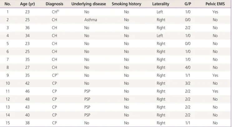
Table 1. Clinical characteristics of the patients with thoracic endometriosis
No. Age (yr) Diagnosis Underlying disease Smoking history Laterality G/P Pelvic EMS
1 23 CHb) No No Left 1/0 Yes
2 25 CH Asthma No Right 0/0 No
3 36 CH No No Right 2/2 No
4 34 CH No No Left 1/0 No
5 23 CH No No Right 0/0 No
6 25 CH No No Right 1/0 No
7 35 CH No No Right 1/0 No
8 27 CH No No Right 4/0 No
9 35 CPc) No No Right 1/1 Yes
10 42 CP No No Right 3/2 No
11 46 CP PSP No Right 2/2 Yes
12 48 CP PSP No Right 2/2 No
13 43 CP PSP No Right 2/2 No
14 40 CP PSP No Right 2/2 No
15 38 CP No No Right 1/1 No
G, gravidity; P, parity; EMS, endometriosis; PSP, primary spontaneous pneumothorax.
a)Hemoptysis or pneumothorax event that occurred with menstruation; b)Thoracic endometriosis with catamenial hemoptysis;
c)Thoracic endometriosis with catamenial pneumothorax.
Table 3. Treatments and prognosis in the patients with thoracic endometriosis
No. Diagnosis 1st line treatment Adjuvant treatment Date of diagnosis Follow- up (mo) Recurrencea)
1 CH VATS (segmentectomy) No Nov. 2008 35 Yes (1)
2 CH VATS (WR) No Dec. 2012 4 No
3 CH VATS (WR) No Nov. 2009 4 No
4 CH VATS (LUL lobectomy) No Nov. 2012 4 No
5 CH Hormone (GnRH a) No Nov. 2010 4 No
6 CH Hormone (GnRH a+dienogest) No June 2013 10 Yes (1)
7 CH Hormone (GnRH a+dienogest) No Apr. 2010 49 Yes (4)
8 CH BAE GnRH a×2 Jan. 2007 6 No
9 CP VATS (WR, DR) No June 2007 65 Yes (3)
10 CP VATS (DR) GnRH a×4 July 2010 10 No
11 CP Thoracotomy (DR) GnRH a×6 Nov. 2009 47 No
12 CP VATS (WR, DR) No Apr. 2010 14 Yes (3)
13 CP VATS (WR, DR) GnRH a×2 Sep. 2013 2 No
14 CP VATS (DR) GnRH a×6, MPA Sep. 2010 13 No
15 CP VATS (DR) GnRH a×6, OCb) Sep. 2010 9 No
CH, thoracic endometriosis with catamenial hemoptysis; VATS, video-assisted thoracoscopic surgery; WR, wedge resection; LUL, left upper lobe; BAE, bronchial artery embolization; DR, diaphragmatic resection; MPA, medroxyprogesterone acetate; OC, oral contraceptive.
a)Number of episode after initial treatment; b)Drospirenone 3 mg ethynyl estradiol 0.03 mg.
Table 2. Location of lesion and findings on chest CT at diagnosis
Patient Diagnosis Location CT Findings
GGO Nodule Air filled cavity Othersa)
1 CH LUL + + + -
2 CH RML + - - -
3 CH RUL + + - -
4 CH LUL + - - -
5 CH RUL + - - -
6 CH RML + - - -
7 CH RUL + - - -
8 CH RLL + + - -
9 CP RUL - - - +
10 CP RUL - - - +
11 CP RUL - - - +
12 CP RUL - - - +
13 CP RUL - - - +
14 CP RUL - - - +
15 CP RLL - - - +
CT, computed tomography; GGO, ground-glass opacity; CH, thoracic endometriosis with catamenial hemoptysis; LUL, left upper lobe;
RML, right middle lobe; CP, thoracic endometriosis with catamenial pneumothorax; RUL, right upper lobe.
a)Blebs, emphysema, subsegmental atelectasis, etc.
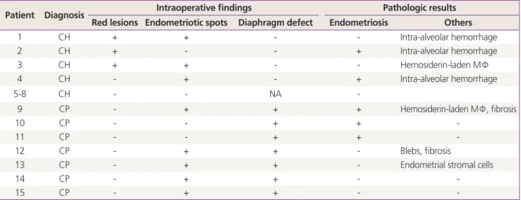
Table 4. Findings of intraoperative thoracic lesions and pathologic results
Patient Diagnosis Intraoperative findings Pathologic results
Red lesions Endometriotic spots Diaphragm defect Endometriosis Others
1 CH + + - - Intra-alveolar hemorrhage
2 CH + - - + Intra-alveolar hemorrhage
3 CH + + - - Hemosiderin-laden MΦ
4 CH - + - + Intra-alveolar hemorrhage
5-8 CH - - NA -
9 CP - + + + Hemosiderin-laden MΦ, fibrosis
10 CP - - + + -
11 CP - - + + -
12 CP - + + - Blebs, fibrosis
13 CP - + + - Endometrial stromal cells
14 CP - + + - -
15 CP - + + - -
CH, thoracic endometriosis with catamenial hemoptysis; MΦ, macrophage; NA, note applicable; CP, thoracic endometriosis with catamenial pneumothorax.
a)Patients 5 to 8 had not received surgery, treated by hormone therapy.
Fig. 1. Intraoperative photos from a patient presenting with catamenial hemoptysis (P4). (A) Multiple endometriotic spots and a hemorrhagic ap- pearance, observed on the surface of the left upper lobe. (B) After lobectomy, the left upper lobe (dimensions, 18×12×2.5 cm; weight, 160 g) shows significant hemorrhagic areas. Intraoperative photos from a patient presenting with catamenial pneumothorax (P13). (C) Multiple 1 to 5 mm dia- phragmatic holes are found on the central tendon of the diaphragm, with multiple endometriotic spots. (D) The specimen after diaphragmatic resec- tion (dimensions, 5×3×0.3 cm). Histology of the resected diaphragm showed the multiple nodules and endometrial stromal cells.
A B
C D
5. Intraoperative findings
Intraoperative findings are summarized in Table 4. Specific intraoperative findings characteristic of endometriosis (red le- sions, endometriotic spots) were observed in 9/11 (81%) of the patients who underwent thoracic surgery; 5 (45%) of these patients had endometriosis pathologically confirmed from a thoracic specimen. The dominant intraoperative findings in pa- tients with hemoptysis consisted of pleural endometriotic spots, hemorrhages, bullae, and blebs (Fig. 1A, B). In those with pneu- mothorax, ≥1 diaphragmatic defects, ranging from 1 to 5 mm were discovered, often coexisting with endometriotic spots in the adjacent tissues (Fig. 1C, D).
6. Histopathological results
Histopathological findings are summarized in Table 3. Specimens from 5/11 (45.4%) patients displayed the hallmark finding of
‘endometrial glands and stroma,’ in 1 patient the specimen dis- played only endometrial stromal cells. Another 2 patients had findings suggestive of endometriosis, including fibrosis without endometriotic glands. Hemosiderin laden macrophages, consid- ered compatible with or suggestive of endometriosis, were pres- ent in another 2 patients.
7. Postoperative management and outcomes
Table 4 summarizes the clinical course and outcomes of study patients. Mean follow-up time for the group was 18.4 months (range, 2–65 months). Of the patients that presented with he- moptysis, 3/4 (75%) patients in the surgical treatment group did not experience recurrence. In contrast, 2/3 (67%) patients expe- rienced recurrence in the hormone-only treatment group; these patients had been treated with dienogest (Visanne, a dosage of 2 mg/day) as maintenance, following 3 cycles of GnRH agonist (leuprolide acetate depot 3.75 mg every 4 weeks) induction.
In the surgical treatment group, 1 patient with coexisting pel- vic endometriosis experienced recurrence, and was treated with thoracic re-operation, without subsequent adjuvant medical therapy. Although the recurrence rate appears higher in the hor- mone treatment group, P6 had only one minor episode during 10 months follow-up, and P7 had a significant reduction in the amount of hemoptysis. The patient (P8) who received bronchial artery embolization and adjuvant hormone therapy with a GnRH agonist was successfully treated, without relapse.
Of the patients that presented with a pneumothorax, there were 3 episodes of recurrence in 2 patients (mean follow-up, 26.5 months; range, 10–65 months); neither had received post- operative adjuvant hormonal therapy. Following surgery, 5/7 pa-
tients received adjuvant hormonal therapy with a GnRH agonist;
this comprised 2 to 6 cycles of a GnRH agonist, followed by me- droxyprogesterone acetate (n=1) or an oral contraceptive (n=4), for 3 months. We recommended 6 cycles of the GnRH agonist, but 2 patients declined further cycles, after either 2 or 4 cycles, because of menopausal symptoms. However, in those who re- ceived serial adjuvant hormone therapy with a GnRH agonist, recurrence was not detected.
Discussion
While various hypotheses have been postulated to explain tho- racic endometriosis, none have been confirmed. Hypotheses include coelomic metaplasia of the pelvic or distant tissues into ectopic endometrial tissue; the physiologic hypothesis, where high levels of circulating prostaglandin F2 during menstruation causes vasoconstriction and bronchospasm, with subsequent alveolar rupture and pneumothorax; the migration theory, in which endometrial diaphragmatic implants may result from the migration of endometrial tissue from the uterus to the pelvis, and through the peritoneal fluid to the subdiaphragmatic area;
and metastatic or lymphovascular micro-embolization, which suggests a metastatic spread of endometrial cells to the lungs, through the venous or lymphatic vasculature. Finally, the trans- genital-transdiaphragmatic passage of air theory suggests that the passage of air through congenital or acquired (secondary to endometriosis) diaphragmatic defects may result in ectopic implants, has also been suggested [10,11].
According to a meta-analysis of articles published between 2001 and 2007, the clinical presentation of thoracic endo- metriosis includes pneumothorax (72%), hemoptysis (14%), hemothorax (12%), and lung nodules (2%) [12]. Notably, most studies included in this meta-analysis involved a thoracoscopic examination. In our hospital, patients presented with hemop- tysis in 53.3% of cases, and pneumothorax in 46.6%; a similar pattern was observed in the excluded patients.
Thoracic endometriosis is challenging in that diagnosis is ex- tremely difficult, unless it is strongly suspected by experienced clinicians with a multi-modality approach. First, symptoms that precede or occur concurrently with menstrual bleeding are typical; however, symptoms that occur in the inter-menstrual period will not exclude the diagnosis of thoracic endometriosis.
Thoracic endometriosis-related pneumothorax has even been reported during early pregnancy [13]. Second, patients com- monly complain of chest pain, hemoptysis, dyspnea, cough,
and scapular pain; these symptoms often accompany other more common pulmonary pathologies, particularly lung malig- nancies or tuberculosis, making differential diagnosis difficult.
Two patients in this series had received empirical tuberculosis medication, before receiving a confirmative diagnosis. Third, there are no specific diagnostic modalities or diagnostic crite- ria, and in cases involving hemoptysis in particular, radiological abnormalities are transient. The CT findings for thoracic endo- metriosis may include ground glass opacities, nodular lesions, thin-walled cavities, or bullae [14].
The mechanism of right lung dominance in thoracic endo- metriosis has yet to be explained. A meta-analysis of 74 cases of catamenial hemoptysis, with diagnoses dating from 1956, revealed that 37 cases (59.6%) occurred in the right lung, and 19 cases (30.6%) occurred in the left lung. Six patients (9.7%) had bilateral lesions [11]. In our series, the vast major- ity of cased had right lung lesions only. A possible explanation for this phenomenon is that endometrial tissue may circulate clockwise, with the flow of peritoneal fluid in the abdominal cavity [15].
Histological confirmation of ectopic endometrium is not always performed, nor is it always possible. Previous defini- tions of thoracic endometriosis have specified that histological
confirmation requires the presence of both endometrial stroma and glands; the presence of only stroma or pulmonary paren- chymal hemorrhages and/or hemosiderin laden macrophages was considered suggestive [16]. These different patterns of histology were also evident in our series, and the overall clini- cal picture, in conjunction with the biopsy findings, should be considered for appropriate diagnosis.
The prevalence of concurrent pelvic endometriosis in patients with catamenial pneumothorax is reported to be anywhere between 18% and 84% [17,18]. According to our analysis, pelvic endometriosis was seen in 3/15 (20%) patients which is consistent with previous reports. A raised CA-125 was present only in those with co-existing pelvic endometriosis.
For women with thoracic endometriosis, surgery can provide relief of the associated chest symptoms. However, due to the high rate of recurrence and the invasiveness of most thoracic procedures, salvage surgery is strongly considered. The pri- mary goal of surgery for thoracic endometriosis is to minimize recurrence by precisely locating the lesions and completely removing them, when all other conservative measures fail. In our study, the single patient that underwent bronchial artery embolization experienced no recurrence; this is consistent with a report from another institution [19], suggesting embolization
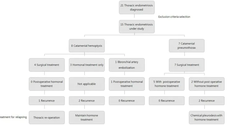
Fig. 2. Algorithm summarizing thoracic endometriosis management protocols employed at our institute. BAE, bronchial artery embolization.
may be suitable as a first line treatment. Because of the risks of thoracic surgery, operative management should be a last resort in cases where symptoms prevail. In these cases, minimally invasive, targeted resection, with routine adjuvant medical maintenance with a GnRH agonist, with or without progestins, should always be considered.
Postoperative recurrence rates are lower when adjuvant hor- monal therapy is employed, the standard approach being a GnRH agonist for 3 to 6 months [20]. In our study, recurrence was not detected in any of the 6 cases that received adjuvant hormonal therapy after surgery, including the patient that under- went bronchial artery embolization, which has important thera- peutic implications for management of thoracic endometriosis
Due to the paucity of thoracic endometriosis itself, there is a lack of good controlled evidence for the use of adjuvant GnRH agonists or oral contraceptives. Also with dienogest (Visanne), there is no description on its effectiveness for thoracic endome- triosis but taking its pharmacological mechanism into account, should be considered as a good option when patients are re- luctant to continue with GnRH agonists or oral contraceptives.
Further studies should be proceeded for the impact of thoracic endometriosis as with other extra-pelvic endometriosis.
In conclusion, the diagnosis and treatment of thoracic en- dometriosis requires a multidisciplinary approach and skillful differential diagnosis, based on a careful gynecological his- tory and inquiry into the cyclicity of pulmonary symptoms.
Although imaging findings are non-specific, laterality towards the right lung may be a feature. Since recurrence is more com- mon in those presenting with a pneumothorax, extra caution is warranted after surgery, with a strong recommendation for adjuvant medical therapy (Fig. 2).
Conflict of interest
No potential conflict of interest relevant to this article was reported.
References
1. Attaran M, Falcone T, Goldberg J. Endometriosis: still tough to diagnose and treat. Cleve Clin J Med 2002;69:647-53.
2. Szamatowicz M. Endometriosis: still an enigmatic disease.
What are the causes, how to diagnose it and how to treat
successfully? Gynecol Endocrinol 2008;24:535-6.
3. Augoulea A, Lambrinoudaki I, Christodoulakos G. Thoracic endometriosis syndrome. Respiration 2008;75:113-9.
4. Hilaris GE, Payne CK, Osias J, Cannon W, Nezhat CR. Syn- chronous rectovaginal, urinary bladder, and pulmonary endometriosis. JSLS 2005;9:78-82.
5. Visouli AN, Darwiche K, Mpakas A, Zarogoulidis P, Papa- giannis A, Tsakiridis K, et al. Catamenial pneumothorax: a rare entity? Report of 5 cases and review of the literature.
J Thorac Dis 2012;4 Suppl 1:17-31.
6. Simoglou C, Zarogoulidis P, Machairiotis N, Porpodis K, Simoglou L, Mitrakas A, et al. Abdominal wall endome- trioma mimicking an incarcerated hernia: a case report.
Int J Gen Med 2012;5:569-71.
7. Veeraswamy A, Lewis M, Mann A, Kotikela S, Hajhosseini B, Nezhat C. Extragenital endometriosis. Clin Obstet Gy- necol 2010;53:449-66.
8. Bagan P, Berna P, Assouad J, Hupertan V, Le Pimpec Barthes F, Riquet M. Value of cancer antigen 125 for di- agnosis of pleural endometriosis in females with recurrent pneumothorax. Eur Respir J 2008;31:140-2.
9. Haga T, Kataoka H, Ebana H, Otsuji M, Seyama K, Tatsumi K, et al. Thoracic endometriosis-related pneumothorax distinguished from primary spontaneous pneumothorax in females. Lung 2014;192:583-7.
10. Okeke TC, Ikeako LC, Ezenyeaku CC. Endometriosis. Ni- ger J Med 2011;20:191-9.
11. Huang H, Li C, Zarogoulidis P, Darwiche K, Machairiotis N, Yang L, et al. Endometriosis of the lung: report of a case and literature review. Eur J Med Res 2013;18:13.
12. Channabasavaiah AD, Joseph JV. Thoracic endometriosis:
revisiting the association between clinical presentation and thoracic pathology based on thoracoscopic findings in 110 patients. Medicine 2010;89:183-8.
13. Yoshioka H, Fukui T, Mori S, Usami N, Nagasaka T, Yokoi K. Catamenial pneumothorax in a pregnant patient. Jpn J Thorac Cardiovasc Surg 2005;53:280-2.
14. Orriols R, Munoz X, Alvarez A, Sampol G. Chest CT scanning:
utility in lung endometriosis. Respir Med 1998;92:876-7.
15. Suginami H. A reappraisal of the coelomic metaplasia the- ory by reviewing endometriosis occurring in unusual sites and instances. Am J Obstet Gynecol 1991;165:214-8.
16. Rousset-Jablonski C, Alifano M, Plu-Bureau G, Camilleri- Broet S, Rousset P, Regnard JF, et al. Catamenial pneumo- thorax and endometriosis-related pneumothorax: clinical
features and risk factors. Hum Reprod 2011;26:2322-9.
17. Tripp HF, Obney JA. Consideration of anatomic defects in the etiology of catamenial pneumothorax. J Thorac Car- diovasc Surg 1999;117:632-3.
18. Joseph J, Sahn SA. Thoracic endometriosis syndrome: new observations from an analysis of 110 cases. Am J Med 1996;100:164-70.
19. Shin SP, Park CY, Song JH, Kim HM, Min D, Lee SH, et al.
A case of catamenial hemoptysis treated by bronchial ar- tery embolization. Tuberc Respir Dis 2014;76:233-6.
20. Lee DY, Bae DS, Yoon BK, Choi D. Post-operative cyclic oral contraceptive use after gonadotrophin-releasing hormone agonist treatment effectively prevents endome- trioma recurrence. Hum Reprod 2010;25:3050-4.