https://doi.org/10.5468/ogs.2017.60.1.8 pISSN 2287-8572 · eISSN 2287-8580
Introduction
The corpus callosum connects two interhemispheres [1], and agenesis of corpus callosum (ACC) is the most common central nervous system defect [2]. ACC has been reported in 0.3% to 0.7% of the general population and in 2% to 3%
of those with developmentally retarded [3-5]. It may be either complete or partial [6]; the partial form of ACC is also called hypogenesis [7] of corpus callosum. If there are no other anomalies accompanying ACC, then it is defined as isolated ACC.
Since the 1990s, ACC has been diagnosed prenatally
Clinical outcomes and neurodevelopmental outcome of prenatally diagnosed agenesis of corpus callosum in single center of Korea
Sung Eun Kim 1 , Hye-In Jang 1 , Kylie Hae-jin Chang 1 , Ji-Hee Sung 1 , Jiwon Lee 2 , Jeehun Lee 2 , Suk-Joo Choi 1 , Soo-young Oh 1 , Cheong-Rae Roh 1 , Jong-Hwa Kim 1
Departments of
1Obstetrics and Gynecology,
2Pediatrics, Samsung Medical Center, Sungkyunkwan University School of Medicine, Seoul, Korea
Objective
With recent advances and frequent use of prenatal ultrasound, the antenatal diagnosis of agenesis of the corpus callosum (ACC) is not rare in obstetrics practices. However, information regarding the long-term neurological outcome remains uncertain. The aim of this study was to investigate clinical outcomes of prenatally diagnosed ACC and to analyze postnatal neurodevelopmental outcomes of ACC neonates born in our single center.
Methods
We retrospectively reviewed 56 cases of prenatally suspected ACC referred to our center.
Results
Fifty-six fetuses were diagnosed with ACC, and 12 of those were followed-up in our center until delivery. Of the remaining 44, 7 were delivered after being referred back to the original hospital, 23 were lost to follow-up, and 14 had unknown outcomes. Among all 56, 29 were considered to have isolated ACC and 27 were considered to have non-isolated ACC. Of the 10 live fetuses delivered in our center, four had isolated ACC, three had non-isolated ACC, and the rest had outcomes unrelated to ACC. Neurodevelopmental outcome was followed-up until approximately age 3 years. Of the four with isolated ACC, three (75%) had normal neurodevelopmental outcomes.
Conclusion
Similar to other studies, the results of our single-center study included positive neurodevelopmental outcomes for those with isolated ACC. However, despite our endeavor to counsel patients with prenatally diagnosed ACC, the delivery rate in our center was quite low. Therefore, larger, multicenter, retrospective studies including long-term neurological development outcomes are crucial and urgently needed to provide better counseling.
Keywords: Agenesis of corpus callosum; Isolated agenesis of the corpus callosum; Neurodevelopmental outcome
Articles published in Obstet Gynecol Sci are open-access, distributed under the terms of the Creative Commons Attribution Non-Commercial License (http://creativecommons.
org/licenses/by-nc/3.0/) which permits unrestricted non-commercial use, distribution, and reproduction in any medium, provided the original work is properly cited.
Copyright © 2017 Korean Society of Obstetrics and Gynecology Received: 2016.5.31. Revised: 2016.7.29. Accepted: 2016.8.6.
Corresponding author: Soo-young Oh
Department of Obstetrics and Gynecology, Samsung Medical Center, Sungkyunkwan University School of Medicine, 81 Irwon-ro, Gangnam- gu, Seoul 06351, Korea
Tel: +82-2-3410-3517 Fax: +82-2-3410-0630 E-mail: [email protected]
http://orcid.org/0000-0003-3002-0048
through ultrasound screening [8]. Prenatal diagnosis of ACC is considered important because it may be associated with central nervous system abnormalities [9,10] such as mental retardation, epilepsy, cerebral palsy, and others [11,12]. If
other anomalies are combined with non-isolated ACC, then outcomes, especially those regarding neurodevelopment, are usually poor [13]. Thus, termination of pregnancy is some- times recommended when non-isolated ACC is diagnosed
Fig. 1. Ultrasonographic findings in a normal fetus (left) and with agenesis of corpus callosum (right). (A) Transventricular view. (B) Tear-drop sign. (C) Normal sagittal view. Corpus callosum can be seen. (D) Bull’s head sign. Lateral displacement of anterior horns in the coronal plane. (E) Normal pericallosal artery. (F) Pericallosal artery is not visible on Doppler.
A B
C D
E F
[14]; however, this option varies by country .
Long-term studies, especially those regarding neurodevel- opmental outcomes, are not available worldwide. Although there is still much controversy [15], most patients with isolat- ed ACC have had good prognoses [16]. In those with isolated ACC, 72.2% showed normal development [16]; in several small sample studies, 100% showed normal development [17]. Normal or mildly delayed neurodevelopment resolves in 67% of isolated ACC, but in non-isolated ACC only 7% re- solves [18]. Therefore, distinguishing isolated ACC from non- isolated ACC is clinically important for prenatal counseling.
Few studies have targeted Korean patients regarding the long-term neurodevelopmental outcomes of ACC despite the common practice of targeted ultrasound. Therefore, based on our patients, we aimed to investigate clinical outcomes of prenatally diagnosed ACC and, importantly, to analyze the neurodevelopmental outcomes of those with prenatally diag- nosed ACC so that appropriate counseling can be provided.
Materials and methods
We retrospectively reviewed 70 cases of antenatally suspected ACC in fetuses referred to or diagnosed at our center be- tween 2008 and 2015. We excluded 14 patients (20%) with suspected ACC diagnosed at a referral hospital. Thus, only 56 fetuses (80%) with suspected ACC examined at our center were included in this study. Among these, 53 were referred to our center due to abnormal findings on ultrasonography per- formed at a referral hospital and three were followed-up at our center. The study was approved by the institutional review board.
We investigated the following data for each patient: gesta- tional age and ultrasonography findings at the time of referral or first diagnosis of ACC including ventriculomegaly and tear- drop sign; karyotype result if performed; brain ultrasound or magnetic resonance imaging (MRI) findings before or after birth; neonatal outcome; postnatal diagnosis; delivery mode;
delivery indication; gestation age at delivery; neonatal birth weight; and neonatal intensive care unit admission. Gestation age at diagnosis was recorded based on the gestation age at the time of transfer or time of diagnosis in our hospital. The diagnosis of ACC was made by using indirect signs of ACC on ultrasound, including the presence of a tear-drop sign and absence of cavum septum pellucidum (CSP), or by direct
nonvisualization of the corpus callosum on the sagittal plane (Fig. 1). Ventriculomegaly, an enlargement of the posterior horn more than 10 mm, was not regarded as an associated anomaly because it may be one of the characteristics of ACC.
Isolated ACC was defined when there was no other anom- aly on ultrasound or MRI based on prenatal or postnatal find- ings, respectively. The partial or complete form of ACC was not separately distinguished because they showed no signifi- cant difference with respect to functional subtyping of the callosum and neurological outcome [19].
We analyzed the neurodevelopmental outcomes based on medical records assessed and described by the pediatric neu- rology or rehabilitation doctors if delivery was performed in our center. Neurodevelopmental outcomes were followed-up until approximately age 3 years. To supplement neurodevel- opmental outcomes of isolated ACC, we also tried analyzing neurodevelopmental outcome of postnatally diagnosed ACC patients (n=9) who primarily visited the pediatric neurology or rehabilitation department of our center. We classified neuro- developmental outcomes into four categories; normal, mild, moderate, severe. ‘Mild’ included children with neurodevelop- ment delay within six months, ‘Moderate’ with 6 months to 12 months delay, and ‘Severe’ with neurodevelopment delay of more than 12 months. The assessment of neurologic devel- opment was performed through the clinical history taking of development milestones. Objective development evaluation includes hand evaluation, Alberta infant motor score, Activi- ties of Daily Living evaluation, Denver Developmental Screen- ing Test, etc. but which were not performed on every child.
Follow-up evaluation for all cases with prenatally diagnosed ACC (and postnatally diagnosed ACC) was performed by pe- diatricians and pediatric neurologists. The final neurodevelop- mental outcome was assessed by two pediatric neurologists (JWL and JHL) by review of medical record.
Since we could not obtain results by directly calling the patient, which was not permitted by IRB, we endeavored to ascertain follow-up or delivery results by calling the referral institution if delivery was not performed in our institution.
“Follow-up loss” meant that patients did not go back to the
original clinic or give birth there. Patients of hospitals that no
longer exist or of hospitals that could not provide information
were classified as “unknown.” To check whether ACC was
present, brain ultrasound was performed after birth. Some
patients underwent brain MRI when other brain anomalies
were suspected.
Results
This study included 56 patients with ACC prenatally diag- nosed by ultrasound examination. Through prenatal diagno- sis, we classified 56 patients as having either isolated or non- isolated ACC and summarized the clinical characteristics (Table 1). Among these 56 patients, 29 (51.8%) were considered to have isolated ACC and 27 (48.2%) were considered to have non-isolated ACC. These classifications were based on prena- tal sonographic findings alone and did not consider the post- natal diagnosis. Ventriculomegaly and a tear-drop sign were observed in approximately 70% to 80% cases of prenatally diagnosed ACC. The median atrial diameters were 16.3 mm and 12.6 mm for isolated ACC and non-isolated ACC, re- spectively. Prenatal chromosomal analyses were performed for 6/29 (20.7%) in the isolated ACC group and for 8/27 (29.6%) in the non-isolated ACC group. With the exception of un- known results, all fetuses in the isolated ACC group showed normal chromosomes and one of eight (12.5%) in the non- isolated ACC group had a chromosomal abnormality (trisomy 18). According to follow-up results, the rates of referral back
to the hospital were 17.2% for the isolated ACC group and 7.4% for the non-isolated ACC group.
We investigated the follow-up results of these fetuses with prenatally diagnosed ACC (Fig. 2). Twelve deliveries were performed in our center, including two terminations that oc- curred during the early study period; the other 44 patients were referred back to the original clinic or lost to follow- up. Among 44 patients who did not undergo delivery at our hospital, seven were referred back to the original clinic due to their long distance from our center and were confirmed as having delivered the pregnancy. Twenty-three patients did not go back to the original clinic or did not give birth there and were considered lost to follow-up. This group was suspected of undergoing termination. The remaining 14 patients of no longer existing hospitals or hospitals that could not provide information were classified as unknown. These follow-up data indicate that the overall percentage of follow-up loss for prenatally diagnosed ACC was 41%, but it increased up to 66% when the unknown category was included.
Clinical characteristics and outcomes for 12 fetuses with ACC delivered at our center are presented in Table 2. Among
Table 1. The characteristics of patients when prenatally diagnosed with ACC in our center (n=56)
Isolated ACC (n=29) Non-isolated ACC (n=27) P-value
Maternal age (yr) 30 (25–43) 32 (23–40) 0.036
Gestational age at diagnosis
a)(wk) 25 (20–37) 22 (17–36) 0.007
Ultrasound findings
Presence of ventriculomegaly 23 (79.3) 19 (70.4) 0.440
Size of ventriculomegaly (mm) 16.3 (10.4–22.8) 12.6 (10.5–25.4) 0.069
Presence of tear-drop sign 21 (72.4) 19 (70.4) 0.866
Chromosomal analysis 1.000
Normal 6 (100) 7 (87.5)
b)Abnormal 0 1 (12.5)
Prenatal brain MRI 6 (20.7) 4 (14.8) 0.566
Follow-up results 0.661
Refer back 5 (17.2) 2 (7.4)
Follow-up loss 12 (41.4) 11 (40.7)
Unknown 7 (24.1) 7 (25.9)
Delivery in our center 5 (17.2) 7 (25.9)
Data presented as median (minimum–maximum) or number (%).
ACC, agenesis of corpus callosum; MRI, magnetic resonance imaging.
a)
The gestational age at diagnosis was when the first ultrasonography gestational age was performed in our center and may not reflect the
exact time of diagnosis. Five out of 56 cases were transferred to our center after gestational age of 34 weeks and 0 days, in which 4 of those
cases delivered and are considered to have visited our center for the purpose of neonatal evaluation or neonatal intensive care unit care after
delivery;
b)One case is inv(9)(p12q13) which is normal variant.
10 live fetuses delivered, four had isolated ACC, three had non-isolated ACC, and the rest were found to have outcomes unrelated to ACC after birth. Excluding termination cases, three out of four suspected isolated ACC cases during the prenatal period were actually confirmed as isolated ACC postnatally, but the other one was finally diagnosed as non- isolated ACC combined with semilobar to lobar holoprosen- cephaly. On the contrary, one out of the six cases considered to be non-isolated ACC was finally proven to be isolated ACC because the degree of enlarged mega cisterna magna
was found to be normal on postnatal ultrasound. In the non- isolated ACC group, two patients had holoprosencephaly (patients 2 and 7) and one had cleft lip and right inguinal her- nia (patient 10). One of two patients had lobar holoprosen- cephaly, was followed-up for delayed development, and was lost to follow-up after 17 months (patient 2); another had semilobar to lobar holoprosencephaly and is currently being observed for symptomatic localization-related epilepsy (patient 7). Among our study population, there were two patients who were initially suspected with ACC but were subsequently
Fig. 2. Clinical characteristics of 56 patients with prenatally suspected agenesis of corpus cal- losum (ACC) based on postnatal findings. SMC, Samsung Medical Center; CoA, coarctation of aorta;
GMH, germinal matrix hemor- rhage.
*Isolated ACC was defined when there was no other anom- aly on ultrasound or MRI based on prenatal or postnatal find ings, respectively.
56 Suspect ACC by ultrasound
12 SMC follow-up 2 Termination
10 Delivery
7 ACC
4 Isolated ACC 3 Non-isolated ACC*
1 Cleft lip 2 Holoprosencephaly
1 Lobal type 1 Semilobar to lobar type
3 Not ACC
1 Hydrops-death 1 CoA, hypospadias 1 Cystic GMH, hydrocephalus
7 Refer back-delivery 23 Follow-up loss
14 Unknown
diagnosed as having complications of fe- tal hydrops and coarctation of the aorta.
ACC was originally diagnosed because the CSP was not visible on ultrasound.
Among the four diagnosed with isolated ACC, one was transferred to another hospital because of the mother’s personal preference (patient 3). The other three (patients 1, 4, and 6) were followed-up for at least 12 months and showed no de- velopmental delay, suggesting that at least 75% of patients with isolated ACC had normal neurodevelopment.
Table 3 shows the summary of clinical characteristics of 10 live births (with the exception of two terminations). Median gestation age at diagnosis was 27 weeks.
However, with the exclusion of four pa- tients admitted for delivery near term or at term, the median gestation age at prena- tal diagnosis was 22 weeks. Chromosomal study results were available for 8 of the 10 patients who delivered. Two did not undergo a chromosomal study (patients 1 and 3) because they planned for deliv- ery regardless of abnormal chromosome results of the fetuses. Of these eight who underwent chromosomal study, a normal karyotype was found in seven fetuses and one had chromosomal anomalies: arr Xp11.22(53,166,281–53,427,895)x1 (pa- tient 7). One patient who had a chromo- somal abnormality had non-isolated ACC and semilobar to lobar holoprosencephaly and was small for gestational age; this pa- tient later presented with epilepsy.
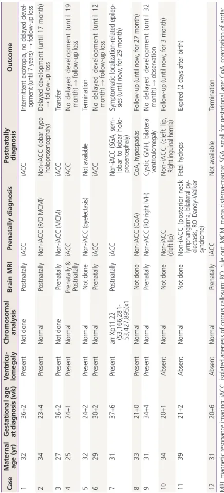
Supplementary analysis including postnatally diagnosed isolated ACC, in which follow-up was performed at least 9 months to maximum 4 years in the pediatric neurology or rehabilitation de- partment, demonstrated that 5 (55.6%) showed normal developmental outcome, 2 (22.2%) showed mild developmental delay, another 2 (22.2%) showed moder- Table 2. Summary of char acteristics and outcome in 12 cases with agenesis of corpus callosum delivered in our center Cas e Mater nal age (yr)
Gestational age at diagnosis (wk)
V entricu - lomegaly Chr
omosomal analysis
Brain MRI Pr enatally diagnosis
Postnatally diagnosis
Outcome 1 32 36+2 Pr esent Not done Postnatally iA CC iA CC In te rm itt en t ex ot ro pia, no d ela ye d d ev el - op m ent (u nti l 7 y ea rs ) → fol lo w -u p lo ss 2 34 23+4 Pr esent Normal Postnatally N on-i A C C (R /O M CM ) N on-i A C C ( lo ba r ty pe holo pr os enc ep ha ly) Delayed development (until 17 month) → follow-up loss 3 27 36+2 Pr esent Not done Pr enatally N on-i A C C (M CM ) iA CC Transfer 4 25 24+1 Pr esent Normal Pr enatally & Postnatally
iA CC iA CC N o d el ay ed d ev el op m en t ( un ti l 1 9 month) → follow-up loss 5 32 24+2 Pr esent Normal Not done N on-i A C C (p yele ct as is) No t a va ila ble Termination 6 29 30+2 Pr esent Normal Pr enatally iA CC iA CC N o d el ay ed d ev el op m en t ( un ti l 1 2 month) → follow-up loss 7 31 37+6 Pr esent arr Xp11.22 (53,166,281- 53,427,895)x1
Postnatally iA CC N on -iA CC (S G A , s em i- lo ba r to lo ba r holo - pr os enc ep ha ly)
Symptomatic localization‐r elated epilep - sies (until now , for 29 month) 8 33 21+0 Pr esent Normal Not done N on-i A C C (C oA ) Co A , h ypo spa di as Follow-up (until now , for 27 month) 9 31 34+4 Pr esent Normal Pr enatally N on -iA C C (R O ri gh t I VH ) C ys tic GMH , b ila te ra l ve nt ric ulo m eg al y N o d el ay ed d ev el op m en t ( un ti l 3 2 month) → observation 10 34 20+1 Absent Normal Not done N on-i A C C (c le ft lip ) N on-i A C C ( cl ef t l ip , Ri gh t i ngui na l h er ni a) Follow-up (until now , for 3 month) 11 39 21+2 Absent Normal Not done N on-i A C C ( po st er io r ne ck lym pha ng io m a, b ila te ra l p y- el ec ta sis , R O D an dy -W al ker sy ndro m e)
Feta l h yd ro ps Expir ed (2 days after birth) 12 31 20+6 Absent Normal Pr enatally iACC Not available Termination MRI, magnetic r esonance imaging; iACC, isolated agenesis of corpus callosum; RO, rule out; MCM, mega cister na-magna; SGA, small for gestational age; CoA, coar ctation of aorta; IVH, intraventricular hemorrhage; GMH, germinal matrix hemorrhage.
ate developmental delay, and none showed severe develop- mental delay.
Discussion
Our data demonstrated that approximately 75% of isolated ACC showed normal neurodevelopment. Although limited in number, our finding is in line with previous studies reporting that the rate of normal neurodevelopment for 16 patients with isolated ACC was 71.2% [20]. In our study, we also found that the follow-up loss rate for prenatally diagnosed ACC is substantial even for isolated ACC, despite our en- deavor to counsel these patients. In fact, each physician in our center generally provides information about normal neu- rodevelopmental outcomes being more than 80% for isolated ACC based on previous works [17,21]. Therefore, a conun- drum beyond proper counseling at the time of prenatal diag-
nosis of ACC and subsequent follow-up remains in our coun- try. However, the impact of proper counseling for prenatally diagnosed ACC was demonstrated by a study performed in another country. For instance, in France, the termination rate for ACC has decreased by almost 17%, from 13/35 (37.1%) in 2000–2003 to 9/44 (20.5%) in 2003–2006 [22]. This de- crease seems to be attributable to proper counseling based on information regarding the good prognosis for isolated ACC.
Therefore, although it is difficult to lower the termination rate for non-isolated ACC with associated anomalies, appropriate counseling regarding the higher percentage of normal neu- rodevelopment for isolated ACC is expected to reduce that termination rate.
When checking brain structures during the mid-trimester fetal anatomy survey, there is a tendency to obtain a mainly transverse view. However, a mid-sagittal view is more accurate for determining ACC. Moreover, indirect signs of ACC are either absent or not clearly visible at the time of mid-trimester screening ultrasonography, which is performed before 24 weeks of gestation in many cases [23]. The gestational age at diagnosis of ACC in other countries is 23 to 26 weeks [24,25], which appears rather late compared to that in our country, although this is probably due to the more frequent use of ultrasound in our country. ACC is frequently associated with ventriculomegaly; our data show that up to 70% to 80% of those with ACC have ventriculomegaly. Therefore, fetuses with ventriculomegaly shown by prenatal ultrasound require a more detailed examination for the existence of the corpus cal- losum. In fact, it is reported that there is a callosal abnormality in 13% of ventriculomegaly cases [18]. When indirect signs of ACC are suspected on mid-trimester ultrasound screen- ing, short-term follow-up and repeat ultrasound with a mid- sagittal view are recommended.
Sometimes it may be difficult to confirm ACC as being isolated on prenatal ultrasonography alone. Our data also showed that one out of four cases of prenatally diagnosed ACC was subsequently found to be non-isolated after birth.
In reality, it was reported that approximately 5% to 20% of cases are misdiagnosed as isolated ACC during the prenatal period [26]. On the contrary, 16% (1/6) of non-isolated ACC cases were finally proven to be isolated ACC, indicating that false-positive diagnoses are possible. To overcome this kind of false-negative or positive diagnosis, differential diagnoses such as septo-optic dysplasia and holoprosencephaly should be considered if there is no CSP on ultrasonography; in addi- Table 3. Characteristics of delivered case
a)in our center (n=10)
Variable Value
Maternal age (yr) 31.5 (25–39)
Gestational age at diagnosis
b)(wk) 27 (20–37) Gestational age at diagnosis
c)(wk) 22 (20–30) Ultrasound findings
Presence of ventriculomegaly 8
Presence of tear-drop sign 8
Chromosomal analysis 8
Normal 7 (87.5)
Gestational age at delivered (wk) 36.5 (31–39) Delivery mode
Cesarean section 5 (50)
Induction failure 1
Previous cesarean section 1
Fetal distress 2
Breech position 1
Vaginal delivery 5 (50)
Preterm delivery 5
Neonatal birth weight (g) 2,470 (960–3630)
NICU admission 4 (40)
Data presented as median (minimum–maximum), number, or num- ber (%).
NICU, neonatal intensive care unit.
a)