Incidence of Post-transplant Malignancy after Renal Transplantation:
Single Center Analysis
Department of Surgery, Hanyang University College of Medicine, Seoul, Korea
Seung Jo Choi, M.D., Dongho Choi, M.D. and Oh Jung Kwon, M.D.
Background: Immunosuppression after kidney transplantation is associated with increased risk of malignancy, which has become the second most common cause of death among kidney transplant recipients. In this review, we report the incidence of malig- nancies after kidney transplantation in a single center and evaluate the incidence, characteristics, relationship to im- munosuppressive drugs and discuss what clinicians must consider during a follow-up of patients after kidney transplantation.
Methods: Between May 1978 and September 2013, a total of 748 kidney transplant patients who were able to undergo a follow-up process through electronic medical records were enrolled in this retrospective cohort study to determine the potential incidence and types of malignancy that may occur after kidney transplantation and the associated impact on patients and graft survival.
Results: Among 748 patients, 63 cases of malignancy appeared in 54 patients (7.2%). Gastrointestinal cancer (12 cases, 19%) and post-transplant lymphoproliferative disorder (12 cases, 19%) were the two most common types of malignancy. The second most common type of malignancy was urinary tract malignancy in 10 patients. Two different types of malignancy were diagnosed in nine patients during our follow-up. The overall graft survival in malignancy patients was better, which may mean that malignancy did not affect the overall graft loss.
Conclusions: Clinicians should be aware of the incidence of malignancy in transplant patients and perform routine examinations for early detection of malignancy.
Key Words: Malignancy, Kidney transplantation, Incidence, Multiple primary cancer
중심 단어: 악성종양, 신이식, 발생률, 다발성 원발성 악성종양Received August 25, 2014 Revised November 13, 2014 Accepted November 13, 2014 Corresponding author: Oh Jung Kwon
Department of Surgery, Hanyang University College of Medicine, 222-1 Wangsimni-ro, Seongdong-gu, Seoul 133-792, Korea Tel: 82-2-2290-8485, Fax: 82-2-2295-4576
E-mail: [email protected]
INTRODUCTION
Kidney transplantation is clearly accepted as the best treatment for end stage renal disease patients who need re- nal replacement therapy. Several studies have proven that kidney transplantation is associated with lower risk of mor- tality and improves quality of life than patients on long-term dialysis(1-3). Although the development of many
immunosuppressive drugs has decreased the incidence of acute graft rejection after kidney transplantation, complica- tions including infection, malignancy, and cardiovascular events are increasing(4,5). And malignancy has become the second most common cause of death among kidney trans- plant recipients(1). Several studies showed that im- munosuppression after organ transplantation is associated with increased risk of posttransplant lymphoproliferative disorder (PTLD) and squamous cell carcinoma(6-8).
Beyond gender, age, genetic risk, conventional risk factors, pre-existing cancers, there are several mechanism theories that explain the contribution of immunosuppressive agents to tumor growth after transplantation. First, most of the im- munosuppressive protocols are designated to reduce lympho- cyte reactivity that results in decreased alloreactivity to the
transplant and less immune surveillance(9). In addition, ma- lignancy occurs by DNA damage or interfering with DNA repair mechanisms. Azathioprine (AZA) inhibits DNA re- pair and induces DNA mutations by codon misreads(10).
Cyclosporine (CsA) has been shown to accelerate carcino- genesis by interfering with DNA damage repair and by up-regulating expression of tumor growth factor and vas- cular endothelial growth factor(11). Furthermore, enhanced angiogenesis, tumor invasion, metastasis and Epstein-Barr virus-induced B-cell expansion have been shown in the use of calcineurin inhibitors(12).
In this review, we report the incidence of malignancies after kidney transplantation in a single center study and evaluate the incidence, characteristics, relationship to im- munosuppressive drugs and discuss what clinicians must consider on following up patients after kidney transplantation.
MATERIALS AND METHODS
Between May 1978 and September 2013, 748 kidney transplant patients who were able to follow-up through electronic medical records were enrolled in this retrospective cohort study to determine the incidence and types of malig- nancy occurring after kidney transplantation and their im- pact on patients and graft survival. All data were collected and analyzed from kidney transplant patients’ clinical notes and computerized records including pathology and radiology reports. The immunosuppressive therapy was based on CsA/tacrolimus, mycophenolate mofetil (MMF)/AZA and steroids. Before 1985, patients received AZA and steroids.
Between 1986 and 1998, patients received dual maintenance immunosuppression with CsA and AZA. After 1998, AZA was replaced by MMF as second maintenance drug, and ta- crolimus was used as other choice of CsA. Kidney transplant patients are routinely screened for detecting malignancies.
Blood tests including tumor markers were examined twice per year. Gastrointestinal (GI) endoscopic examinations and abdominal ultrasonography were performed annually.
Among these patients, 63 cases of malignancy from 54 pa- tients were diagnosed and treated. We compared these ma- lignancy patients with total population of renal transplant patients in a single center and studied this group with types
of malignancy, incidences, interval between transplantation and cancer diagnosis, prognosis, and graft survival. Data were analyzed with SPSS ver. 18.0 (IBM Co., Armonk, NY, USA) and Kaplan-Meier method was used to calculate graft survival curve.
RESULTS
1. Patient characteristics
Seven hundred forty-eight patients were enrolled in this study. The mean age of patients was 38.2±10.8 years (range, 15∼70) and the male/female ratio was 1.91:1.
Among these patients, 326 patients (43.6%) received kidney from living-related donor, 367 patients (49.1%) from liv- ing-unrelated donor, and 55 (7.4%) from deceased donor.
AZA was used in 51 patients (6.8%), CsA in 591 patients (79.0%), and tacrolimus in 106 patients (14.2%) as main maintenance immunosuppressive drug. Acute rejection was observed in 210 patients (28.1%).
Sixty-three cases of malignancy appeared in 54 patients (7.2%). Mean age of cancer patients group was 44.0±9.6 years (range, 28∼66) which was older than cancer-free pa- tients group (37.7±10.7 years) and the male/female ratio was 1.57:1. Thirteen malignancy patients (24.1%) received kidney from living-related donor and 38 patients (70.4%) from living-unrelated donor which was larger portion than cancer-free patients. Only three patients received kidney from deceased donor. Most of malignancy patients (88.9%) were using CsA as main immunosuppressive drug and six patients (11.1%) were using tacrolimus (Table 1).
2. Types and incidences of malignancy
GI cancer (12 cases, 19%) and PTLD (12 cases, 19%) were the two most common type of malignancy (Table 2).
Stomach cancer (seven cases, 11%) was the most common type in GI cancer. Both colon cancer and hepatocellular car- cinoma was seen in two patients and pancreas tumor in one patient. Twelve cases were PTLD which were affecting var- ious organs (two cases, stomach; two cases, intestine; five cases, neck; two cases, kidney; and one case, lung). Second most common type of malignancy was urinary tract malig- nancies in 10 patients (seven cases, native kidney; one case, transplanted kidney; and two cases, bladder). Female genital
Table 1. Characteristics of patients
Characteristic Total patients (n=748) Cancer-free patients (n=694) Cancer patients (n=54)
Age (yr)
Gender (male:female) Donor
Living-related Living-unrelated Cadaver Immunosuppression Azathioprine Cyclosporine Tacrolimus Acute rejection Viral infection CMV EBV
38.2±10.8 (15∼70) 491:257 (65.6:34.4)
326 (43.6) 367 (49.1) 55 (7.4)
51 (6.8) 591 (79.0) 106 (14.2) 210 (28.1)
37 (4.9) 4 (0.5)
37.7±10.7 (15∼70) 458:236 (66.0:34.0)
313 (45.1) 329 (47.4) 52 (7.5)
51 (7.3) 543 (78.2) 100 (14.5) 198 (28.5)
35 (5.0) 4 (0.6)
44.0±9.6 (28∼66) 33:21 (61.1:38.9)
13 (24.1) 38 (70.4) 3 (5.6)
0 48 (88.9) 6 (11.1) 12 (22.2)
2 (3.7) 0
Data are presented as mean±SD (range) or number (%).
Abbreviations: CMV, cytomegalovirus; EBV, Epstein-Barr virus.
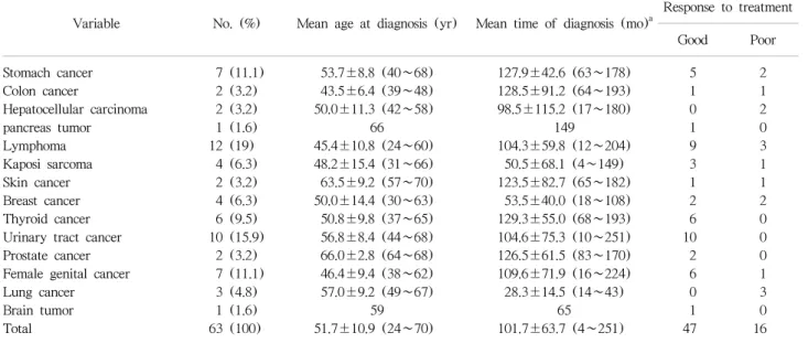
Table 2. Type of malignancy and time of diagnosis after renal transplantation
Variable No. (%) Mean age at diagnosis (yr) Mean time of diagnosis (mo)
aResponse to treatment
Good Poor
Stomach cancer Colon cancer
Hepatocellular carcinoma pancreas tumor
Lymphoma Kaposi sarcoma Skin cancer Breast cancer Thyroid cancer Urinary tract cancer Prostate cancer Female genital cancer Lung cancer Brain tumor Total
7 (11.1) 2 (3.2) 2 (3.2) 1 (1.6) 12 (19) 4 (6.3) 2 (3.2) 4 (6.3) 6 (9.5) 10 (15.9)
2 (3.2) 7 (11.1)
3 (4.8) 1 (1.6) 63 (100)
53.7±8.8 (40∼68) 43.5±6.4 (39∼48) 50.0±11.3 (42∼58)
66 45.4±10.8 (24∼60) 48.2±15.4 (31∼66) 63.5±9.2 (57∼70) 50.0±14.4 (30∼63) 50.8±9.8 (37∼65) 56.8±8.4 (44∼68) 66.0±2.8 (64∼68) 46.4±9.4 (38∼62) 57.0±9.2 (49∼67)
59 51.7±10.9 (24∼70)
127.9±42.6 (63∼178) 128.5±91.2 (64∼193) 98.5±115.2 (17∼180)
149
104.3±59.8 (12∼204) 50.5±68.1 (4∼149) 123.5±82.7 (65∼182) 53.5±40.0 (18∼108) 129.3±55.0 (68∼193) 104.6±75.3 (10∼251) 126.5±61.5 (83∼170) 109.6±71.9 (16∼224) 28.3±14.5 (14∼43)
65 101.7±63.7 (4∼251)
5 1 0 1 9 3 1 2 6 10 2 6 0 1 47
2 1 2 0 3 1 1 2 0 0 0 1 3 0 16 Data are presented as mean±SD (range).
a
The period from transplantation to the diagnosis of malignancy.
malignancies (uterus, cervix, and ovary) were seen in seven patients and prostate cancer in two patients. Kaposi sarcoma (four cases, 6.3%), skin cancer (two cases, 3.2%), breast cancer (four cases, 6.3%), thyroid cancer (six cases, 9.5%), lung cancer (three cases, 4.8%), and brain tumor (one case) were diagnosed during follow-up. The mean age at malig- nancy diagnosis was 51.7±10.9 (range, 24∼70) and the
mean time interval between malignancy diagnosis and trans- plantation was 101.7±63.7 months (range, 4∼251). Patient with diagnosis of PTLD at neck was the youngest patient at his age of 24- and 70-year-old male patient was the oldest patient who was diagnosed as basal cell carcinoma at nose.
Shortest time interval was Kaposi sarcoma (4 months) and longest one was urothelial carcinoma at renal pelvis of
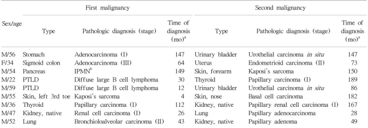
Table 3. Patients with multiple primary malignancies
Sex/age
First malignancy Second malignancy
Type Pathologic diagnosis (stage)
Time of diagnosis
(mo)
aType Pathologic diagnosis (stage)
Time of diagnosis
(mo)
aM/56 F/34 M/54 M/22 M/59 M/55 M/36 M/47 M/52
Stomach Sigmoid colon Pancreas PTLD PTLD
Skin, left 3rd toe Thyroid
Kidney, native Lung
Adenocarcinoma (I) Adenocarcinoma (III) IPMN
bDiffuse large B cell lymphoma Diffuse large B cell lymphoma Kaposi's sarcoma
Papillary carcinoma (I) Renal cell carcinoma (I) Bronchioloalveolar carcinoma (II)
147 64 149 30 12 4 112 26 43
Urinary bladder Uterus Skin, forearm Thyroid Urinary bladder Skin, nose Kidney, native Lung Kidney, native
Urothelial carcinoma in situ Endometrioid carcinoma (II) Kaposi's sarcoma
Papillary carcinoma (I) Urothelial carcinoma in situ Basal cell carcinoma
Papillary renal cell carcinoma (I) Papillary adenocarcinoma Papillary adenoma
147 73 150 189 86 182 167 28 49 Abbreviations: IPMN, intraductal papillary mucinous neoplasm; PTLD, posttransplant lymphoproliferative disorder.
a
The period from transplantation to the diagnosis of malignancy;
bIntraductal papilary mucinous neoplasm (radiologic diagnosis).
transplanted kidney (251 months). Two different types of malignancy were diagnosed in nine patients during our fol- low-up. All but one patient were diagnosed with malig- nancies in different periods. Most of them were male but various types of malignancy can occur in any period (Table 3).
3. Treatment modalities and prognosis
Among seven gastric cancer patients, four patients under- went subtotal gastrectomy, and three patients received endo- scopic mucosal resection (EMR). Two patients who under- went surgery recurred with peritoneal seeding, while other two patients with surgery and EMR treated patients were not recurred yet. Colon cancer patients underwent surgery and one patient recurred with liver and brain metastasis.
Both HCC patients had hepatitis B virus infection and they received radio frequency thermal ablation and transarterial chemoembolization repeatedly, but HCC recurred and ex- pired due to hepatic failure. Eight patients with lymphoma received chemotherapy and four patients received surgical resection. Among them, nine patients were in complete re- mission during follow-up, two patients were expired for other reasons and PTLD recurred in one patient. Three pa- tients with Kaposi sarcoma and two patients with other skin cancer were treated by surgical excision and their prognosis was good, but in one patient with Kaposi sarcoma who was treated with surgical excision, chemotherapy, and radio-
therapy recurred again. Three patients with breast cancer were treated by surgery and chemotherapy and one patient received chemotherapy alone due to multiple organ metastasis. All of thyroid cancer, urinary tract malignancies and prostate cancer patients showed good prognosis after surgical removal (nephrectomy, transurethral resection of bladder). All female genital malignancy patients were treated with surgical removal and combination chemotherapy in three patients, while lung metastasis was seen in one case of cervical cancer. Lung cancer patients show a poor prognosis despite treatment. Among 63 patients, 47 patients showed good response to treatment which means cancer was not re- curred during follow-up and 17 patients showed poor re- sponse to treatment which means cancer was recurred with peritoneal seeding or distant metastasis (Table 2).
4. Graft survival
Among 748 total patients, median follow-up period was 118.5 months, range from 0 to 419 months. Seventy cases died with a functioning graft due to other reasons (pneumonia, cardiovascular attack, myocardial infarction) and two cases with graft failure which was marked as 0 month follow-up. Overall graft survival in cancer patients was better than in cancer-free patients (
P
=0.035). In other words, malignancy did not affect overall graft loss (Fig. 1).Fig. 1. Cumulative graft survival of cancer patients and cancer free patients.
DISCUSSION
Although preferentially managing infectious and car- diovascular complications after kidney transplantation is crucial, posttransplant malignancy has become an essential cause of increasing mortality and morbidity nowadays.
According to several previous reports, most prevalent malig- nancies after kidney transplantation is Kaposi's sarcomas, non-Hodgkin lymphomas and nonmelanoma skin cancers with 10 to 20 times greater than in the general pop- ulation(13). Also viral-associated tumors and urogenital tu- mors seemed to take a large proportion of malignancy after kidney transplantation(14). Ro et al.(15) reported 4.3% ma- lignancy development in a 37-year follow-up in a single center study, and Kim et al.(16) showed 4.2% of malig- nancy incidence pattern after renal transplantation among 757 patients. In the previous Korean study, the incidence of malignancy was distributed 1.2% to 4.3%, and this was increased during the follow-up periods(16). In our study, malignancies after kidney transplantation were developed in 52 of 748 patients (7.2%) and GI malignancy (stomach can- cer) was the most common. Most of the other studies in Western countries and even in Eastern Asia, except in Japan, GI cancers occurred in relatively low rate compared to our study. Kaposi's sarcomas, non-Hodgkin lymphomas and nonmelanoma skin cancers seemed to be the most com- mon malignancies in those studies(17). There were four cas- es of Kaposi sarcoma in our study, and the mean interval
of diagnosis was 50.5±68.1 months in those cases. Among these patients, one patient was diagnosed as Kaposi sarcoma 4 months after renal transplantation which was the shortest time interval of diagnosis in our study. According to some reports, Kaposi sarcoma and lymphoma was diagnosed in the early stage of posttransplantation(18-20). Berber reported 50% of the patients displayed Kaposi sarcoma in the first posttransplant year(18). Dominant effect of the immuno- suppressive agents in the early stage of posttransplantation is associated with relatively early onset of specific malig- nancies such as Kaposi sarcoma and lymphoma(18).
We found nine patients diagnosed with two different types of malignancies after renal transplantation. The in- cidence rate of multiple primary cancer after kidney trans- plantation was 16% which was higher than recent study (8.1%) based on general population(21). Eight of nine pa- tients were male and all patients were diagnosed double pri- mary cancer before age 60. Only one patient was diagnosed with different malignancies at the same period who had stomach and urothelial cancer. Type of the malignancy seemed to be various and could be diagnosed at any period.
Further prospective research and concern will be required to figure out the risk, predisposing factors, and prognosis associated with different type of malignancy after trans- plantation.
Our study has some limitations. Due to a single center study, malignancy incidence after kidney transplantation was too small to compare with those in the general popu- lation. For more accurate comparison, calculating stand- ardized incidence rate would be helpful but there were limi- tations in our study. Tremblay et al. reported that in the era of using antithymocyte globulin and adding CsA or ta- crolimus, there is a statistically significant increase in cancer incidence compared to the era mainly using AZA and ste- roid(22). And several studies suggested that sirolimus might reduce the incidence of malignancy(23). Bang(24) showed relatively low malignancy rate using tacrolimus (3.4%) com- pared with CsA (7.6%), and Kasiske et al.(14) also sug- gested tacrolimus was associated with a lower incidence of skin cancer. In our study, 53 malignancy patients received kidney transplantation between 1986 and 1998 and 10 pa- tients received kidney transplantation after 1998. We couldn't figure out malignancy category specifically related
to immunosuppressive agents, because most of them (92.6%) were using CsA as immunosuppressive drug and only four patients (7.4%) were using tacrolimus. Another limitation is that, in our study, graft survival in cancer patient group was better than in cancer-free patient group (
P
=0.035). This value is statistically significant, but in clinical point of view, malignancy may not affect overall graft loss. To evaluation the graft survival result, adequate number of malignant pa- tient population and further study including multivariate analysis will be needed.CONCLUSION
Among these 63 cases of malignancy patients, 47 patients (74%) showed a good prognosis with early diagnosis and prompt management. However, other patients showed a poor prognosis with either recurrence of malignancy or per- itoneal seeding. Advanced stage at diagnosis might be the reason for poor prognosis. In conclusion, we suggest that clinicians and patients should be aware of incidence of ma- lignancy in transplant patients and perform routine exami- nations for early detection of malignancy for proper treatment.