• 검색 결과가 없습니다.

Multiple Primary Cardiac Malignant Peripheral Nerve Sheath Tumors in the Left Atrium: Case Report

N/A
N/A
Protected

Academic year: 2021

Share "Multiple Primary Cardiac Malignant Peripheral Nerve Sheath Tumors in the Left Atrium: Case Report"

Copied!
3
0
0

로드 중.... (전체 텍스트 보기)

전체 글

(1)

422 http://www.jchestsurg.org

JCS

Journal of Chest Surgery

Case Report

Multiple Primary Cardiac Malignant Peripheral Nerve Sheath Tumors in the Left Atrium: Case Report

Junfei Li, M.D.

1

, Qiansu Chen, M.D.

1

, Shaomei Yu, M.D.

2

, Siyuan Yang, M.D.

1

Departments of

1

Cardiac Surgery and

2

Ultrasound, Affiliated Hospital of Guizhou Medical University, Guiyang, China

ARTICLE INFO

Received

October 8, 2020

Revised

October 28, 2020

Accepted

October 28, 2020

Corresponding author

Siyuan Yang

Tel

86-17784950043

Fax

86-851-86773651

E-mail

[email protected]

ORCID

https://orcid.org/0000-0002-8087-7931

Malignant peripheral nerve sheath tumors are rare sarcomas of the heart. Herein, we re- port the case of a 24-year-old man who complained of dyspnea, cough, and upper left back pain. He was found to have multiple primary heart tumors obstructing the right su- perior pulmonary vein in the left atrium, which were diagnosed as malignant peripheral nerve sheath tumors. The patient underwent successful resection of the tumors and im- munohistochemistry was utilized for diagnosis.

Keywords: Neurogenic tumor, Neurofibrosarcoma, Heart neoplasms, Case report

Copyright©2021, The Korean Society for Thoracic and Cardiovascular Surgery

This is an Open Access article distributed under the terms of the Creative Commons Attribution Non-Commercial License (http://creativecommons.org/licenses/

by-nc/4.0) which permits unrestricted non-commercial use, distribution, and reproduction in any medium, provided the original work is properly cited.

Case report

A 24-year-old man presented to Affiliated Hospital of Guizhou Medical University with a 20-day history of dys- pnea, cough, and upper left back pain. He was previously healthy and had no relevant family history. Transthoracic echocardiography showed a large tumor inside the left atri- um; however, the origin and extent of the tumor could not be visualized (Fig. 1A). A subsequent thoracic-abdominal contrast-enhanced computed tomography (CT) scan re- vealed a large tumoral lesion (approximately 6.9 cm×4.0 cm×3.5 cm, 45 Hounsfield units) inside the left atrium, ex- tending toward and totally obstructing the right superior pulmonary vein (RSPV). Two smaller tumors were also de-

tected (Fig. 1B). Tumor markers were examined without positive findings.

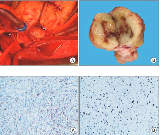
The patient was referred for surgical treatment. Cardio- pulmonary bypass was instituted and the patient was cooled to 32°C. The interatrial wall was opened and a giant hard mass was identified (Fig. 2A). The mass was located at the roof of the left atrium, with poor mobility and ex- tensive adhesion to the atrial septal wall. Exploring the pulmonary vein openings based on the CT images, we found that the mass intruded upon and completely blocked the RSPV. Palpation confirmed that the distal part of the RSPV was soft, and the RSPV was incised by approximate- ly 2 cm. The tumor in the RSPV was completely exposed.

We dissected the tumor from both the left atrium and

Fig. 1. (A) Transthoracic echocar- diogram showed the mass (labeled as M) in the left atrium. (B) Preoper- ative contrast-enhanced computed tomography angiography demon- strating a giant tumoral mass inside the left atrium, causing total ob- struction of the right superior pul- monary vein and 2 small lesions (arrow).

A B

x

M M

https://doi.org/10.5090/jcs.20.132

pISSN: 2765-1606 eISSN: 2765-1614

J Chest Surg. 2021;54(5):422-424

(2)

423

Junfei Li, et al. Multiple Malignant Tumors in the Left Atrium

http://www.jchestsurg.org

JCS

RSPV. The RSPV incision was closed with a 1-cm-wide au- tologous pericardial patch, and an 8-mm probe could be passed through the RSPV. The opening of the RSPV and the left atrium were reconstructed with a pericardial patch.

We detected 2 other small lesions lying on the endocardi- um 10 mm above the mitral annulus. The 2 small tumors were resected en bloc from the atrial wall. Transesophageal echocardiography after cardiopulmonary bypass showed that the RSPV was patent and the tumor had been totally removed. The biopsy of the biggest mass was glistening and myxoid, with focal hemorrhage and necrosis (Fig. 2B).

The patient had an uneventful postoperative course. The pathology diagnosis was malignant peripheral nerve sheath tumor (MPNST), a rare variety of soft tissue sarcoma. The immunohistochemistry results were as follows: creatine ki- nase (−), epithelial membrane antigen (−), vimentin (+) (Fig.

3A), CD34 (−), CD117 (−), DOG1 (−), desmin (−), smooth muscle actin (−), S100 (+) (Fig. 3B), Syn (−), CgA (−), CD56 (+), myogenin (−), MyoD1 (−), caldesmon (−), CD68 (focal +), and Ki-67 (40%). At 6 months after the surgical correction, the patient remained alive and showed no signs of recur- rence.

The study was approved by the Affiliated Hospital of Guizhou Medical University (IRB approval no., GY2020-

3154). This case is presented with written informed con- sent from the patient.

Discussion

MPNST, also known as neurofibrosarcoma, is a tumor that develops from nerve tissue. MPNST can occur any- where in the body, but most often occurs in the deep tissue of the arms, legs, or trunk [1]. The treatment of MPNST begins with surgery to remove as much of the tumor as possible. Radiation therapy and chemotherapy might be useful in some patients to decrease the chance of recur- rence [2]. Approximately 25%–50% of MPNSTs are associ- ated with a genetic condition known as neurofibromatosis type 1 [3]. Surgical excision is the gold standard, with me- dian survival ranging from 3 months to 1 year due to its high recurrence rate and metastasis. MPNST is considered an aggressive tumor because there is up to a 65% chance of recurrence after surgery, and an approximately 40%

chance of metastasis to distant parts of the body. Primary cardiac MPNST is very rare. In our case, the patient had multiple lesions, which is even more uncommon. Ogechuk- wu and colleagues reported a unique case of a primary cardiac sarcoma originating from the left atrium and ob-

Fig. 3. Immunohistochemical stain- ing showed positivity for vimentin (A, original magnification ×100) and intense expression of S100 (B, orig- inal magnification ×100).

A B

Fig. 2. (A) Intraoperative view of the tumoral mass (arrow) after open- ing of the interatrial wall from the right atrium. (B) The cut surface of the largest mass was glistening and myxoid, with focal hemorrhage and necrosis.

A B

(3)

424

https://doi.org/10.5090/jcs.20.132

http://www.jchestsurg.org

JCS

structing the right pulmonary vein [4]. Our case is the first report of multiple MPNST lesions, with 1 lesion obstruct- ing the RSPV.

The majority of MPNSTs show a fascicular spindle cell architecture histologically, as in our case. By immunohis- tochemistry, approximately 50% to 90% of MPNSTs are positive for S100 [5]. This same feature was seen in our case. Our case also showed intense expression of vimentin, positive expression of CD56, and focal positivity for CD68.

In conclusion, this is the first reported case of a patient with multiple MPNSTs causing total obstruction of the RSPV who underwent successful surgical correction. An immunohistochemical examination was required to con- firm diagnosis. Complete dissection was crucial. Chemo- therapy and radiotherapy might be useful for some patients with similar conditions.

Conflict of interest

No potential conflict of interest relevant to this article was reported.

Acknowledgments

We thank Dr. Zhu Gong for technical assistance and also sincerely mourn his passing. He was a good doctor as well

as a good friend.

ORCID

Junfei Li: https://orcid.org/0000-0001-7358-3372 Qiansu Chen: https://orcid.org/0000-0001-6542-3141 Shaomei Yu: https://orcid.org/0000-0002-3835-3747 Siyuan Yang: https://orcid.org/0000-0002-8087-7931

References

1. James AW, Shurell E, Singh A, Dry SM, Eilber FC. Malignant pe- ripheral nerve sheath tumor. Surg Oncol Clin N Am 2016;25:789- 802.

2. Rahman M, Cook DS, Ellis G, O’keefe PA. Malignant peripheral nerve sheath tumor of the heart. Asian Cardiovasc Thorac Ann 2006;14:425-7.

3. Kitamura M, Wada N, Nagata S, et al. Malignant peripheral nerve sheath tumor associated with neurofibromatosis type 1, with metasta- sis to the heart: a case report. Diagn Pathol 2010;5:2.

4. Ogechukwu CN, Christopher N, Christoph S, Etinosasere O, Jose B.

The insidious cardiac tumor: a primary left atrium intimal cardiac sarcoma in a young patient. Case Rep Cardiol 2019;2019:7245676.

5. Prifti E, Baboci A, Ikonomi M. A giant cardiac malignant peripheral

nerve sheath tumor presenting with total obstruction of the superior

vena cava. Ann Thorac Surg 2014;97:e7-9.

수치

Fig. 3. Immunohistochemical stain- stain-ing showed positivity for vimentin  (A, original magnification ×100) and  intense expression of S100 (B,  orig-inal  magnification ×100).

참조

관련 문서

Results : The expression of p21 was increased in boderline serous tumor and serous cystadenocarcinoma in contrast to benign serous tumors. The expression of

The purpose of this study was to analyze the impaction pattern of the impacted mandibular third molar and the relationship with the inferior alveolar nerve

Pilot Confirm runway 09 left for landing. ATC gives you further instructions to turn right heading 310. On the radar display, there was an area to avoid which requires a

Sizes of left and right pupils Intensities of left and right pupils Ellipse ratios of left and right

The following key words were used: artificial vision, blindness, cortical prosthesis, electrical stimulation electronic implants, macular degener- ation, optic nerve,

void makeTree(Object root, Object left, Object right): Creates a binary tree with root as the root element, left as the left subtree, right as the right

 include as foreign key attributes in S the primary keys of the relations that represent the participating entity types.  their combination will form the primary

To assess the association between SCD severity and plasma AβO levels or brain Aβ deposition, we applied multiple linear regression models in which the outcome variables