Hyoung-Sup Lim, Su-Gwan Kim, Ji-Su Oh
Department of Oral and Maxillofacial Surgery, School of Dentistry, Chosun University, Gwangju, Korea
Implant Survival Rates of Maxillary Sinus Augmentation:
a Literature Review of Graft Materials
Purpose: By reviewing literature on the subject, we compared the survival rate of implants placed in vari- ous graft materials used for maxillary sinus augmentation.
Materials and Methods: The search protocol used the Pubmed electronic database, with a time limit from 1998 to 2009. Keywords such as ‘sinus lift,’ ‘sinus augmentation,’ ‘sinus floor elevation,’ ‘sinus graft,’
‘bone graft,’ ‘implants,’ ‘oral implants,’and ‘dental implants’were used, alone and in combination, to search the database. We selected articles and divided them into three groups by type of graft materials:
Group 1. Autogenous bone group: autogenous bone alone; Group 2. Combined bone group: autogenous bone in combination with bone substitutes; and Group 3. Substitute group: bone substitutes alone or bone substitute combinations.
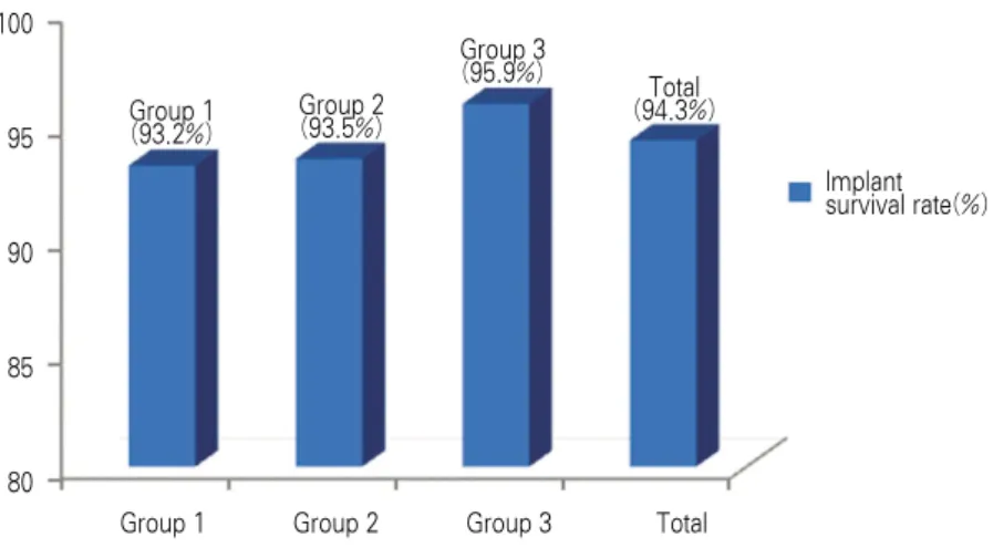
Results: We selected 37 articles concerning a total of 2,257 patients and 7,282 implants; 417 implants failed. The total implant survival rate (ISR, %) was 94.3%. In Group 1, 761 patients and 2,644 implants were studied; 179 implants failed and the ISR was 93.2%. In Group 2, 583 patients and 1,931 implants were studied; 126 implants failed and the ISR was 93.5%. In Group 3, 823 patients and 2,707 implants were studied; 112 implants failed and the ISR was 95.9%.
Conclusion: Implants inserted in grafts composed of bone substitutes alone or in grafts composed of autoge- nous bone in combination with bone substitutes may achieve survival rates better than those for implants using autogenous bone alone (P < 0.05).
Key words: Implant survival rate, Maxillary sinus augmentation
AbstractIntroduction
Bone quality in the posterior maxilla is poor, and pneumatization of the maxillary sinus and a insuffi- ciency in the vertical alveolar height after tooth loss may limit the placement of implants.
1-3)To resolve these problems, various methods, such as onlay graft, veneer graft, bone graft after performing LeFort I osteotomy simultaneously, and maxillary sinus augmentation, have been introduced.
4-6)The maxillary sinus augmentation has been reported to be comparable to the success rate of implants placed in the edentulous posterior maxilla with a sufficient
height. The maxillary sinus augmentation have been accepted as predictable and common method clinically.
7)Maxillary sinus augmentation using autogenous bone was introduced by Boyne and James in 1980,
6)and since then, various bone substitutes have been used. Among maxillary sinus graft materials, autoge- nous bone has been considered the gold-standard.
Autogenous bone has advantages of excellent bone
regeneration potential, biocompatibility, and absence
of immune response. However, its use can be
restricted by the morbidity of donor graft site, infec-
tion, resorption of grafted bone, and the difficulty in
obtaining a sufficient bone intraorally.
8,9)Thus variety of allobone, xenobone, and synthetic bone have recently been introduced. Many studies are ongoing to assess the outcomes of bone graft materials in maxillary sinus augmentation
In this study, the prognosis and success rate of implants using various graft materials in the maxil- lary sinus lift were examined through literature reviews.
Materials and Methods
Research papers listed in the Pubmed database from 1998 to 2009 using the terms ‘sinus lift,’
‘sinus augmentation,’ ‘sinus floor elevation,’ ‘sinus graft,’ ‘bone graft,’ ‘implants,’ ‘oral implants,’and
‘dental implants,’alone or in combination, were searched and collected.
Articles were selected according to the following inclusion criteria. 1) A lateral approach to the maxil- lary sinus was used, 2) limited to cases placing implants in humans, 3) limited to studies of bone grafts in more than 20 cases, 4) root-form implants was used, 5) mean follow-up time was more than 12 months after implant loading, and 6) the implant survival rate was clearly calculated data reported in the paper.
Reviews and technical reports were excluded. Also papers from same group of authors, with very similar databases of patients, materials, methods and out- comes, were excluded.
The collected papers were organized and divided to three groups based on the type of bone graft materi- als used.
�Group 1. Autogenous bone group; this group used only autogenous bone.
�Group 2. Autogenous bone + bone substitutes group; this group used autogenous bone in combi- nation with bone substitutes.
�Group 3. Bone substitutes group; this group used bone substitutes alone or bone substitute combina- tions.
Statistical analysis was performed using statistical software. SSPS (statistical package for the social sci- ence); Chi-square test was used to determine statis- tical significance among between groups. Difference
were considered significant at P value < 0.05.
Results
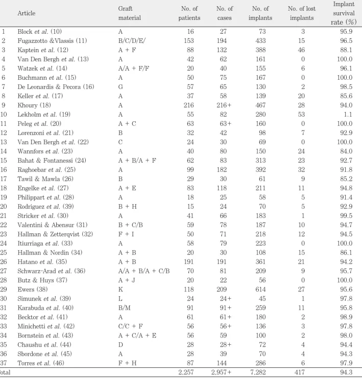
Through this search, 37 papers were selected and classified according to the year of publication (Table 1). In these reports, 7,282 implants were placed in a total of 2,257 patients; 417 implants failed, and the overall success rate of implants was 94.3%. The first, autogenous bone group included 15 papers; 2,644 implants were placed, 179 implants failed, and the success rate of implants was 93.2%. The second group, using autogenous bone in combination with bone substitutes, included 10 papers. The substitutes mixed with autogenous bone were deproteinized bovine bone (Bio-Oss
�; Geistlich Pharma, Wolhusen, Switzerland), hydroxyapatite (HA; Berkeley Advanced Biomaterials, Berkeley, CA, USA), dem- ineralized freeze-dried bone allograft (DFDBA;
Musculoskeletal Foundation, Holmdel, NJ, USA), Dentsply/Friadent/Ceramed, (Mannheim, Germany), tricalcium phosphate (TCP), and BioPlant HTR (Replacement Therapy Materials, Mechelen, Belgium). In the autogenous bone in combination with bone substitutes group, 1,931 implants were placed, 126 implants failed, and the success rate was 93.5%. The third bone substitutes group, using used bone substitutes alone or bone substitute combina- tions, included 16 papers. Bone substitutes included Bio-Oss
�, DFDBA, freeze-dried bone allograft (FDBA; Musculoskeletal Foundation) and Dentsply/Friadent/Ceramed, TCP, calcium sulfate, phyconic + fluro, marine algae, β-TCP (Cerasorb;
Curasan AG, Kleinostheim, Germany), Bio-Oss
�+ DFDBA, Bio-Oss
�+ PRP (Platelet-Rich Plasma;
SmartPrep, Harvest Technologies, Norwell, MA, USA), HA + DFDBA, HA + fibrin glue, and HA + PRP. In Group 3, 2,707 implants were placed, 112 implants failed, and the success rate was 95.9%
(Fig. 1, Table 2).
Discussion
Many different materials have been used to aug-
ment the maxillary sinus for the placement of
implants. The criteria for selection of the graft
Table 1.Articles selected according to criteria
Graft No. of No. of No. of No. of lost Implant
Article
material patients cases implants implants survival rate (%)
1 Block et al. (10) A 16 27 73 3 95.9
2 Fugazzotto &Vlassis (11) B/C/D/E/ 153 194 433 15 96.5
3 Kaptein et al. (12) A + F 88 132 388 46 88.1
4 Van Den Bergh et al. (13) A 42 62 161 0 100.0
5 Watzek et al. (14) A/A + F/F 20 40 155 6 96.1
6 Buchmann et al. (15) A 50 75 167 0 100.0
7 De Leonardis & Pecora (16) G 57 65 130 2 98.5
8 Keller et al. (17) A 37 58 139 20 85.6
9 Khoury (18) A 216 216+ 467 28 94.0
10 Lekholm et al. (19) A 55 82 280 53 1.1
11 Peleg et al. (20) A + C 63 63+ 160 0 100.0
12 Lorenzoni et al. (21) B 32 42 98 7 92.9
13 Van Den Bergh et al. (22) C 24 30 69 0 100.0
14 Wannfors et al. (23) A 40 80 150 24 84.0
15 Bahat & Fontanessi (24) A + B/A + F 62 83 313 23 92.7
16 Raghoebar et al. (25) A 99 182 392 32 91.8
17 Tawil & Mawla (26) B 29 30 61 9 85.2
18 Engelke et al. (27) A + E 83 118 211 11 94.8
19 Philippart et al. (28) A 18 25 58 5 91.4
20 Rodriguez et al. (39) B + H 15 24 70 5 92.9
21 Stricker et al. (30) A 41 66 183 1 99.5
22 Valentini & Abensur (31) B + C/B 59 78 187 10 94.7
23 Hallman & Zetterqvist (32) F + I 50 71 218 12 94.5
24 Itiurriaga et al. (33) A 58 79 223 0 100.0
25 Hallman & Nordin (34) A + B 20 30 108 15 86.1
26 Hatano et al. (35) A + B 191 191 361 21 94.2
27 Schwarz-Arad et al. (36) A/A + B/A + C/B 70 81 209 9 95.7
28 Butz & Huys (37) A + J 20 22 56 0 100.0
29 Ewers (38) K 118 209 614 27 95.6
30 Simunek et al. (39) L 24 24+ 45 1 97.8
31 Karabuda et al. (40) B/M 91 91+ 259 11 95.8
32 Becktor et al. (41) A 61 61+ 180 2 98.9
33 Minichetti et al. (42) C/C + F 56 56+ 136 3 97.8
34 Bornstein et al. (43) A + C/A + E 56 59 100 2 98.0
35 Chaushu et al. (44) D 28 28+ 72 4 94.4
36 Sbordone et al. (45) A 28 39 70 4 94.3
37 Torres et al. (46) F + H 87 144 286 6 97.9
Total 2,257 2,957+ 7,282 417 94.3
A: autogenous bone, B: Bio-Oss, C: demineralized freeze-dried bone allograft (DFDBA), D: freeze-dried bone allograft (FDBA), E:
tricalcium phosphate (TCP), F: hydroxyapatite (HA), G: calcium sulphate, H: platelet-rich plasma (PRP), I: fibrin glue, J: BioPlant HTR, K: marine algae, L: phycogenic material + fluorohydroxyapatite, M: Cerasorb
Table 2.Implant survival rates according to the graft materials
Group No. of paper No. of patients No. of implats No. of implant failures Implant survival rate (%)
Group 1 15 761 2644 179 93.2
Group 2 10 583 1931 126 93.5
Group 3 16 823 2707 112 95.9
Total 2257 7282 417 94.3
include
47): 1) the ability to produce bone in the sinus by cellular proliferation from viable transplanted osteoblasts or by osteoconduction of cells along the graft surface, 2) the ability to produce bone by osteoinduction, 3) the ability of the initially formed bone to remodel into mature lamellar bone, 4) the maintenance of the mature bone over time without loss, 5) the ability to stabilize the implants, 6) the low infection rate, 7) the easy availability, 8) the low antigenicity, and 9) high level of reliability.
In maxillary sinus augmentation, autogenous bone is considered the preferred material for bone regener- ation. Autogenous bone contains osteogenic cells that has an osteogenic potential and bone morphogenetic proteins (BMP) that induce osteoblasts with bone- forming potential and growth factors, and plays the role of the osteoconductive scaffold required for the process of new bone formation.
6)Additionally, autoge- nous bone is not immunogenic.
48)Allogeneic bone used as graft material is prepared from bone harvested from cadavers and treated by freeze-drying or by demineralization plus freeze dry- ing. The mineralized freeze-dried bone retain certain osteoconductive qualities.
49)Urist
50)first prepared decalcified freeze-dried allogeneic bone and reported their osteoconductive capacity. The demineralized
bone may result fibrous connective tissue or cartilage rather than bone in the sinus floor.
51)While Van den Bergh et al. reported that satisfactory results were obtained in cases with maxillary sinus lifts using demineralized freeze-dried bone (DFDB).
23)The xenogenic Bio-Oss
�is an inorganic material prepared by harvesting bovine bone, removing organ- ic substances by treating with ethylenediamine, and sterilizing. Its crystal structure, morphology, and physical characteristics are similar to human cancel- lous bone. Bio-Oss
�forms new bone through the osteoconduction process.
6,52,53)The xenogenic Bio-Oss
�can be regarded as an appropriate material in maxil- lary sinus augmentation for implant placement because slow resorption as physiologic remodeling.
48)The allogeneic or xenogeneic bone have the possible transmission of AIDS and infection via contaminated blood and tissues, and the risk of the infection with diseases from different species have been pointed out.
54,55)Thus, recently, alloplastic bone with excellent biocompatibility has been developed and is widely used as bone substitute materials. Advantages of synthetic bone include that it can be readily prepared and stored, clinical application is easy, and the risk of cross-infection is absent.
55-57)It does not have osteoinductive potential, but acts as a scaffold for
Group 1 Group 2 Group 3 Total
100
95
90
85
80
Group 1 (93.2%)
Group 2 (93.5%)
Group 3 (95.9%)
Total (94.3%)
Implant survival rate(%)
Fig. 1.Implant survival rates according to graft material.
�Group 1. Autogenous bone group; this group used only autogenous bone.
�Group 2. Autogenous bone + bone substitutes group; this group used autogenous bone in combination with bone substitutes.
�Group 3. Bone substitutes group; this group used bone substitutes alone or bone substitute combinations.
ingrowth of bone due to osteoconduction. Presently used synthetic bone graft materials include hydrox- yapatite, bioglass, bioceramic, polymer, calcium car- bonate, and tricalcium phosphate.
58-61)Medical risk factors seem to contribute to implant loss in patients who smoke, in post menopausal patients receiving estrogen replacement, and in patients with a history of antral sepsis.
17)moreover, in sinus augmentation procedures the implant’s sur- vival rate appears to be more influenced by the resid- ual bone height or by tobacco than by the type of bone graft.
46)And the highest failure rates are associ- ated with “severely resorbed”ridges, poverty of blood supply presumably is the cause.
24)No significant difference between the quality of newly formed bone tn the cases of the one-stage and the two-stage sinus lift has been found.
39)But, two- stage technique presented seems to be kept to be optibal because the crucial region is denuded only minimally duringimplant placement.
14,19, 23)Perforation of the sinus membrane does not com- promise the osseointegration process or the success of dental implants placed in the augmented maxillary sinus. However, a sealed sinus environment is important for optimal healing conditions after sinus lifting and implant placement surgery, and the size of membrane perforation is the most important factor in postponing of abandoning the augmentation and implant insertion.
40)Conclusion
We attempted to identify the published studies reporting survival rates for implants placed in aug- mented maxillary sinuses. The success rates of the bone substitutes group (Group 3; ISR: 95.9%) and autogenous bone + bone substitutes group (Group 2;
ISR: 93.5%) were comparable or superior to the autogenous bone group (Group 1; ISR: 93.2%) (P <
0.05). Based on the studies examined, the bone sub- stitutes alone had similar or better than autogenous bone in combination with bone substitutes and auto- genous bone alone in maxillary sinus augmentation.
References
1. Pietrokovski J : The bony residual ridge in man. J Prosthet Dent 34 : 456, 1975.
2. Misch CE : Treatment planning and implant dentistry.
Misch Implant Institute, 1985.
3. Misch CE, Judy KWM : Classification of partial edentu- lous arches for implant dentistry. Int J Oral Implantol 4 : 7, 1987.
4. Sailer HF : A new method of inserting endosseous implants in totally atrophied maxillae. J Craniomaxillofac Surg 17 : 299, 1989.
5. Tatum H : Maxillary and sinus implant reconstruction.
Dent Clin North Am 30 : 207, 1986.
6. Boyne PJ, James RA : Grafting of the maxillary sinus floor with autogenous marrow and bone. J Oral Surg 38 : 613, 1980.
7. Simsek B, Simsek S : Evaluation of success rates of imme- diate and delayed implants after tooth extraction. Chinese Med J 116 : 1216, 2003.
8. Marx RE : Autogenous grafting in oral and maxillofacial surgery : Philosophy and particulars of autogenous bone grafting. Oral Maxillofac Surg North Am 5 : 599, 1993.
9. Marx RE, Stevens MR : Morbidity from bone harvest in major jaw reconstruction : A randomized trial comparing the lateral anterior and posterior approaches to the ileum.
J Oral Maxillofac Surg 48 : 196, 1988.
10. Block MS, Kent JN, Kallukaran FU et al : Bone mainte- nance 5 to 10 years after sinus grafting. J Oral Maxillofac Surg 56 : 706, 1998.
11. Fugazzotto PA, Vlassis J : Long-term success of sinus augmentation using various surgical approaches and graft- ing materials. Int J Oral Maxillofac Implants 13 : 52, 1998.
12. Kaptein MLA, de Putter C, de Lange GL et al : Survival of cylindrical implants in composite grafted Maxillary sinuses. J Oral Maxillofac Surg 56 : 1376, 1998.
13. Van Den Bergh JPA, Ten Bruggenkate CM, Krekeler G et al : Sinus floor elevation and grafting with autogenous ili- ac crest bone. Clin Oral Implants Res 9 : 429, 1998.
14. Watzek G, Weber R, Bernhart T et al : Treatment of patients with extreme maxillary atrophy using sinus floor augmentation and implants: preliminary results. Int J Oral Maxillofac Surg 27 : 428, 1998.
15. Buchmann R, Khoury F, Faust C et al : Peri-implant con- ditions in periodontally compromised patients following maxillary sinus augmentation: a long-term post-therapy trial. Clin Oral Implants Res 10 : 103, 1999.
16. Leonardis DD, Pecora GE : Augmentation of the maxillary sinus with calcium sulfate : one-year clinical report from a perspective longitudinal study. Int J Oral Maxillofac Implants 14 : 869, 1999.
17. Keller EE, Tolman DA, Eckert SE : Maxillary antral-nasal inlay autogenous bone graft reconstruction of compromised maxilla : a 12-year retrospective study. Int J Oral Maxillofac Implants 14 : 707, 1999.
18. Khoury F : Augmentation of the sinus floor with mandibu- lar bone block and simultaneous implantation : a 6-year clinical investigation. Int J Oral Maxillofac Implants 14 : 557, 1999.
19. Lekholm U, Wannfors K, Isaksson S et al : Oral implants in combination with bone grafts. A 3-year retrospective multicenter study using the Bra�nemark implant system.
Int J Oral Maxillofac Surg 28 : 181, 1999.
20. Peleg M, Mazor Z, Garg AK : Augmentation grafting of the maxillary sinus and simultaneous implant placement in patients with 3 to 5 mm of residual alveolar bone height.
Int J Oral Maxillofac Implants 14 : 549, 1999.
21. Lorenzoni M, Pertl C, Wegscheider W et al : Retrospective analysis of Frialit-2 implants in the augmented sinus. Int J Periodontics Restorative Dent 20 : 255, 2000.
22. Van Den Bergh JPA, ten Bruggenkate CM, Krekeler G et al : Maxillary sinusfloor elevation and grafting with human demineralized freeze dried bone. Clin Oral Implants Res 11 : 487, 2000.
23. Wannfors K, Johansson B, Hallman M et al : A prospec- tive randomized study of 1- and 2-stage sinus inlay bone grafts : 1-year follow-up. Int J Oral Maxillofac Implants 15 : 625, 2000.
24. Bahat O, Fontanessi RV : Efficacy of implant placement after bone grafting for three-dimensional reconstruction of the posterior jaw. Int J Periodontics Restorative Dent 21 : 221, 2001.
25. Raghoebar GM, Timmenga NM, Reintsema H et al : Maxillary bone grafting for insertion of endosseous implants : results after 12-24 months. Clin Oral Implants Res 12 : 279, 2001.
26. Tawil G, Mawla M : Sinus floor elevation using a bovine bone mineral (Bio-Oss) with or without the concomitant use of a bilayered collagen barrier (Bio-Gide) : a clinical report of immediate and delayed implant placement. Int J Oral Maxillofac Implants 16 : 713, 2001.
27. Engelke W, Schwarzwa¨ller W, Behnsen A et al : Subantroscopic laterobasal sinus floor augmentation (SAL- SA) : an up-to-5-year clinical study. Int J Oral Maxillofac Implants 18 : 135, 2003.
28. Philippart P, Brasseur M, Hoyaux D et al : Human recombinant tissue factor, platelet-rich plasma, and tetra- cycline induce a high-quality human bone graft : a 5-year survey. Int J Oral Maxillofac Implants 18 : 411, 2003.
29. Rodriguez A, Anastassov GE, Lee H et al : Maxillary sinus augmentation with deproteinated bovune bone and platelet rich plasma with simultaneous insertion of endosseous implants. J Oral Maxillofacial Surg 61 : 157, 2003.
30. Stricker A, Voss PJ, Gutwald R et al : Maxillary sinus floor augmentation with autogenous bone grafts to enable placement of SLA-surfaced implants: preliminary results after 15-0 months. Clin Oral Implants Research 14 : 207, 2003.
31. Valentini P, Abensur D : Maxillary sinus grafting with anorganic bovine bone : a clinical report of long-term results. Int J Oral Maxillofac Implants 18 : 556, 2003.
32. Hallman M, Zetterqvist L : A 5-year prospective follow-up study of implant-supported fixed prostheses in patients subjected to maxillary sinus floor augmentation with an 80 : 20 mixture of bovine hydroxyapatite and autogenous bone. Clin Implant Dent Relat Res 6 : 82, 2004.
33. Itiurriaga MTM, Ruiz CC : Maxillary sinus reconstruction with calvarium bone grafts and endosseous implants. J Oral Maxillofac Surg 62 : 344, 2004.
34. Hallman M, Nordin T : Sinus floor augmentation with bovine hydroxyapatite mixed with fibrin glue and later placement of nonsubmerged implants : a retrospective study in 50 patients. Int J Oral Maxillofac Implants 19 : 222, 2004
35. Hatano N, Shimizu Y, Ooya K : A clinical long-term radi- ographic evaluation of graft height changes after maxillary sinus floor augmentatin with a 2 : 1 autogenous
bone/xenograft mixture and simultaneous placement of dental implants. Clin Oral Implants Res 15 : 339, 2004.
36. Schwarz-Arad D, Herzberg R, Dolev E : The prevalence of surgical complications of the sinus graft procedure and their impact on implant survival. J Periodontol 75 : 511, 2004.
37. Butz SJ, Huys LW : Long-term success of sinus augmen- tation using a synthetic alloplast : a 20 patients, 7 years clinical report. Implant Dent 14 : 36, 2005.
38. Ewers R : Maxilla sinus grafting with marine algae derived bone forming material : a clinical report of long- term results. J Oral Maxillofac Surg 63 : 1712, 2005.
39. Simunek A, Cierny M, Kopecka D et al : The sinus lift with phycogenic bone substitute. A histomorphometric study. Clin Oral Implants Res 16 : 342, 2005
40. Karabuda C, Arisan V, Hakan O : Effects of sinus mem- brane perforations on the success of dental implants placed in the augmented sinus. J Periodontol 77 : 1991, 2006.
41. Becktor JP, Hallstrom H, Isaksson S et al : The use of particulate bone grafts from the mandible for maxillary sinus floor augmentation before placement of surface-mod- ified implants : results from bone graftgin to delivery of the final fixed prosthesis. J Oral Maxillofac Sur 66 : 780, 2008.
42. Minichetti JC, D'Amore JC, Hong AY : Three-year analy- sis of tapered screw vent implants placed into maxillary sinuses grafted with mineralized bone allograft. J Oral Implantol 34 : 135, 2008.
43. Bornstein MM, Chappuis V, von Arx T et al : Performance of dental implants after staged sinus floor elevation proce- dures : 5-year results of a prospective study in partially edentulous patients. Clin Oral Implants Res 19 : 1034, 2008.
44. Chaushu G, Mardinger O, Calderon S et al : The use of cancellous block allograft for sinus floor augmentation with simultaneous implant placement in the posterior atrophic maxilla. J Periodontol 80 : 422, 2009.
45. Sbordone L, Toti P, Menchini-Fabris G et al : Implants success in sinus-lifted maxillae and native bone : a 3-year clinical and computerized tomographic follow-up. Int J Oral Maxillofac Implants 24 : 316, 2009.
46. Torres J, Tamimi F, Martinez PP et al : Effect of platelet- rich plasma on sinus lifting: a randomized-controlled clini- cal trial. J Clin Periodontol 36 : 677, 2009.
47. John NK, Michael SB : Augmentation of maxillary sinus floor with autogenous bone for placement of endosseous implants : A preliminary report. J Oral Maxillofac Surg 51 : 1203, 1993.
48. Yildirim M, Spiekermann H, Biesterfeld S et al : Maxillary sinus augmentation using xenogenic bone substitute mate- rial Bio-Oss in combination with venous blood. Clin Oral Impl Res 11 : 217, 2000.
49. Feinberg SE, Fonseca FJ : Biologic aspects of transplanta- tion of grafts, in Fonseca RJ, Davis WH(eds).
Reconstructive Preprosthetic Oral and Maxillofacial Surgery. Philadelphia, PA, Saunders 1986.
50. Urist MR : bone formation by autoinduction. Science 150 : 893, 1965.
51. Loukota RA, Isaksson SG, Linner ELJ et al : A technique for inserting endosseous implants in the atrophic maxilla in a single stage procedure. Br J Oral Maxillofac Surg 30 : 46, 1992.
52. Smiler DG, Johnson PW, Lozada JL et al : Sinus lift grafts and endosseous implants : Treatment of the atroph- ic posterior maxilla. Dent Clin North Am 36 : 151, 1992.
53. McAllister BS, Cogan AG, Hollinger JO : Eighteen-mouth radiographic and histologic evaluation of sinus grafting with anorganic bovine bone in the chimpanzee. Int J Oral Maxillofac Implants 14 : 361, 1999.
54. Buck BE, Melinin TI, Brown MD : Bone transplantation and human immuno-deficiency virus. An estimate of risk of acquired immunodeficiency syndrome (AIDS). Clin Orthop 240 : 129, 1989.
55. Bell WH : Resorption characteristics of bone and bone substitutes. Oral Surg 17 : 650, 1964.
56. Shetty V, Han TJ : Alloplastic materials in reconstructive periodontal surgery. Dent Clin North Am 35 : 521, 1991.
57. Ouhayoun JP, Shabana AHM, Issahakian S et al : Histological evaluation of natural coral skeletal as a graft- ing material in miniatureswine mandible. J Mat Sci :
materials in medicine 3 : 222, 1992.
58. Yukna RA : Synthetic bone grafts in periodontics.
Periodontology 2001 : 92, 1993.
59. Fabbro M, Testori T, Francetti L et al : Systematic Review of Survival Rates for Implants Placed in the Grafted Maxillary sinus. Int J Periodontics Restorative Dent 24 : 565, 2004.
60. Tong D, Rioux K, Drangsholt M et al : A review of sur- vival rates for implants placed in grafted maxillary sinuses using meta-analysis, Int J Oral Maxillofac Implants 13 : 175, 1998.
61. Tidwell JK : Composite graft of the maxillary sinus fot placement of endosteal implants. Int J Oral Maxilliofac Sur 21 : 204, 1992.
저자 연락처
우편번호 501-759
광주광역시 동구 서석동 375번지
조선대학교 치과대학 구강악안면외과학교실 김 수 관
원고 접수일 2010년 03월 22일 게재 확정일 2010년 06월 21일
Reprint Requests Su-Gwan Kim
Department of Oral and Maxillofacial Surgery, School of Dentistry, Chosun University
375, Seosuk-dong, Dong-gu, Gwangju, 501-759, Korea Tel: +82-62-220-3815 Fax: +82-62-228-7316 E-mail: [email protected]
Paper received 22 March 2010 Paper accepted 21 June 2010