Copyright ⓒ 2011, The Korean Academy of Oral Biology
117
Journal of Oral Biology
Blood Vessel Regeneration using Human Umbilical Cord-derived Endothelial Progenitor Cells in Cyclophosphamide-treated
Immune-deficient Mice
Soon-Keun Kwon
1,2#, Yu-Jin Ko
1,2#, Tae-Jun Cho
1,2, Eu-Gene Park
1,2, Byung-Chul Kang
2,3, Gene Lee
1,2, and Jaejin Cho
1,2*1
Lab of Dental Regenerative Biotechnology Major, School of Dentistry, Seoul National University, Korea
2
Dental Research Institute, Seoul National University
3
Graduate School of Immunology, College of Medicine, Seoul National University, Korea
4
Department of Experimental Animal Research, Biomedical Research Institute, Seoul National University Hospital (received May 12, 2011 ; revised June 10, 2011 ; accepted July 22, 2011)
Endothelial cells are a vital constituent of most mammalian organs and are required to maintain the integrity of these tissues. These cells also play a major role in angiogenesis, inflammatory reactions, and in the regulation of thrombosis.
Angiogenesis facilitates pulp formation and produces the ves- sels which are essential for the maintenance of tooth homeo- stasis. These vessels can also be used in bone and tissue regeneration, and in surgical procedures to place implants or to remove cancerous tissue. Furthermore, endothelial cell re- generation is the most critical component of the tooth gener- ation process. The aim of the present study was to stimulate endothelial regeneration at a site of acute cyclophosphamide (CP)-induced endothelial injury by treatment with human umbilical cord-derived endothelial/mesenchymal stem cells (hEPCs). We randomly assigned 16 to 20-week-old female NOD/SCID mice into three separate groups, a hEPC (1 × 10
5cells) transplanted, 300 mg/kg CP treated and saline (control) group. The mice were sacrificed on days 5 and 10 and blood was collected via the abdominal aorta for analysis.
The alanine transaminase (ALT), aspartate aminotransferase (AST), serum alkaline phosphatase (s-ALP), and albumin (ALB) levels were then evaluated. Tissue sections from the livers and kidneys were stained with hematoxylin and eosin (HE) for microscopic analysis and were subjected to immuno- histochemistry to evaluate any changes in the endothelial layer. CP treatment caused a weight reduction after one day.
The kidney/body weight ratio increased in the hEPC treated animals compared with the CP only group at 10 days. More- over, hEPC treatment resulted in reduced s-ALP, AST, ALT levels compared with the CP only group at 10 days. The CP only animals further showed endothelial injuries at five days which were recovered by hEPC treatment at 10 days. The number of CD31-positive cells was increased by hEPC treat- ment at both 5 and 10 days. In conclusion, the CP-induced disruption of endothelial cells is recovered by hEPC treat- ment, indicating that hEPC transplantation has potential benefits in the treatment of endothelial damage.
Key words: human umbilical cord derived endothelial pro- genitor cell, cyclophosphamide, stem cell transplantation, endothelial injury, regeneration.
서 론
혈관을 구성하는 혈관 내피세포는 우리몸의 대부분의 조 직을 구성하고 유지하는데 매우 중요하며 혈관생성, 염증 반응, 혈전증 조절에도 중요한 역할을 한다(Levenberg, 2005).
혈관 내피세포는 혈관을 생성시키고 이러한 혈관계 질환을 치료하는 잠재력으로 인해 연구의 주제로 각광 받고 있다.
Vasculogenesis는 기능적인 혈관계를 형성하는 것을 뜻하 며 이는 endothelial progenitor cell (angioblast)이 중배엽 에서 분화되고, 이들이 융합하여 cord를 형성하고, 연속적 인 network를 구성하는 tube가 형성되는 과정을 의미한다 (Vokes et al., 2004). Angiogenesis는 이미 존재하는 혈관
#
Equally contributed
*Corresponding author: Jaejin Cho, Dental Regenerative Biotech- nology Major, School of Dentistry, Seoul National University, 28 Yongun-Dong, Chongno-gu 110-749, Korea.
Tel: +82-2-740-8666, Fax: +82-2-3676-8730
E-mail: [email protected]
으로부터 미세혈관 구조가 형성 되는 것으로 혈관의 확장 (expansion), 가지침(branching), 재구조화(reorganization)를 의미한다(Wilting and Christ, 1996). 이것은 성인에서 새 로운 혈관 재생에 큰 역할을 하며 허혈성 조직에서 분비 하는 VEGF, FGF 등의 local angiogenic growth factor에 의해 개시된다(Schatteman and Awad, 2004).
성인에서 angiogenesis만 일어나는 것이 아니라 vas- culogenesis도 일어나는데 허혈이 발생된 부위로 순환하던 전구세포가 모여 혈관을 생성한다(Schatteman, 2004). 골 수 등에 존재하던 세포들은 허혈성 조직이나 암조직에서 발현하는 cytokine에 의해 mobilization 되어 peripheral blood로 방출된다(Takahashi et al., 1999).
줄기세포는 여러 source로부터 얻을 수 있는데 최근 많 이 사용되고 있는 것은 골수, 재대혈, 태반 등이다(Levenberg, 2005). 골수에서 얻을 수 있는 stem cell은 일정 시간 배 양하면 MAPA (Multipotent adult progenitor cell)을 얻을 수 있지만, 침습적으로 채취를 해야 한다는 단점이 있다.
재대혈에서 분리하여 얻을 수 있는 태아의 줄기세포는 성 인에서 얻어지는 줄기세포에 비해 더 큰 분화능을 가지고 있으며 면역거부반응의 장벽이 낮은 장점이 있지만 얻을 수 있는 양이 매우 적다(Rafii and Lyden, 2003). 본 실험 에서는 사람 탯줄 유래 줄기세포를 사용하였는데, 이것은 풍부하게 얻을 수 있으며, 채취도 용이하고 donor에 해를 입히지 않으며 성인에서 얻어지는 줄기세포보다 좀더 조직 적합성이 뛰어나고, 빠르고 더 오래 재생하는 특성을 가지 고 있기 때문에 연구에 널리 이용된다(Rafii and Lyden, 2003).
심혈관계 질환을 가진 환자의 심장에 혈관 내피 줄기 세 포를 주입해 줄기 세포를 이용한 치료의 가능성을 확인하 였다(Stamm et al., 2003). 혈관내피세포를 기반으로 한 재 생치료는 혈관 질환의 치료에 응용 되어 인공혈관을 만들 고 손상된 혈관을 재생시키는데 임상적으로 유용하게 쓰 일 수 있다(Kaushal et al., 2001).
이러한 혈관 재생은 치과 영역에서도 응용 될 수 있다.
사람 치아의 치수 안에는 혈관이 발달되어 있고 혈관내피 전구세포(endothelial progenitor cell, HEPC)도 존재한다 (Gronthos et al., 2002). 치수내의 혈관내피 세포는 치수 안으로 lymphocyte와 leukocyte가 이용해 들어오는데에 큰 역할을 하며 이것으로 치수의 염증 반응을 조절할 수 있 다(Sawa et al., 1998). 혈관 형성(vasculogenesis)은 치수 가 형성 되는 것을 돕고, 치아 항상성 유지에 없어서는 안 되는 혈관을 생성한다(Trubiani et al., 2003). 또한 임플란 트 식립, 치주 수술, 암 치료시 골이나 조직의 재생에도 적용 가능하다(Meijer et al., 2007). 골절시 치유, 외상으 로 인한 골결손의 회복, 감염, 악안면 기형 등에 응용 될 수 있다(Meijer et al., 2007). 혈액이 공급되지 않으면 골 이나 조직의 재생이 일어날 수 없다. 임플란트 수술시 골 이식이나 구강악안면의 재건 수술시 혈관의 역할은 매우
중요하므로 혈관 재생이 수술의 성공을 좌우 하는 중요한 요소가 된다. 최근 치아 재생 연구가 활발히 진행되고 있 는데, 그 한계점은 크기가 작고 3차원적 구조도 성인 치아 와는 다른 치아형태가 형성 된다는 점이다(Ohazama et al., 2004). 영양분과 대사 산물의 확산에 혈관 형성이 큰 역할 을 할 뿐만 아니라 치근단에 단 한 개의 혈관입구를 가지 는 치아의 특징상 이러한 한계를 가지는 원인으로 혈관 형 성이 치아 재생의 가장 중요한 요소일 가능성을 고려해 볼 수 있다(Hacking and Khademhosseini, 2009; Chai and Slavkin, 2003). 최근 exogenous 하게 vacular progenitor를 공급하여 조직의 revascularization을 일으키는 것에 대한 관심이 증가 되고, 그에 관한 연구가 많이 진행되고 있다 (Levenberg, 2005). 치아에서 신생 혈관 형성 연구는 어려 움이 있기 때문에 cyclophosphamide로 전신적 혈관 손상 을 유도한 후 신장과 간에서 손상을 확인하는 실험을 계 획하였다. 이에 본 연구는 NOD/SCID 면역결핍 마우스 (immune-deficient mice)에 CP를 이용한 혈관 손상 모델을 확립하고, 신생아 탯줄 유래 혈관 내피 줄기 세포를 이 모 델 동물에 이종 이식하여 손상된 혈관에 이식된 줄기세포 가 engraftment, integration, 증식, 분화 및 혈관의 재생에 기여할 수 있는지 여부를 관찰한다. 이를 토대로 하여 앞서 언급한 치의학 분야에서 치아 혈관 형성 연구 등 다양한 응용에 대한 발판을 마련할 수 있을 것이다.
재료 및 방법
Laboratory animals
16~23 g의 암컷 NOD/SCID (Orient Bio, Namyanju, Korea) 면역결핍 마우스가 사용되었다. 서울대학교병원 전 임상실험부의 Specific Pathogen Free (SPF) 동물 사육실에 서 조절된 22oC와 12 h / 12 h light-dark condition 하에서 사육되었고 동물은 자유롭게 물을 마실 수 있도록 하고 표 준화된 사료를 공급하였다. 동물을 희생하기 전 12시간 동 안 절식을 시켰다. 실험에 사용된 모든 동물은 Institutional Animal Care and Use Committee (IACUC)의 승인하에 실 험에 사용되었다. (IACUC No. 09-0024)
Human umbilical cord derived endothelial progenitor cells 서울대학교 의과병원에서 정상 분만시의 신생아의 탯줄 을 얻었고, 연구에 대한 서울대학교 치의학대학원 치의학 연구윤리(Institutional Review Board, IRB)의 승인을 받았 다(IRB No. S-D20090001). HBSS (Hank's balanced salt solution, WelGENE Inc, Daegu, Korea) 에서 보관하였고, 2시간 이내에 세포 분리를 하였다. 탯줄을 phosphate-buffered saline (PBS)로 두 번 세척하여 혈액을 완전히 제거하였다.
전체 탯줄의 길이를 4등분 하여 정맥을 절개하여 내부가 완전히 드러나게 한 후 0.05% collagenase type I (Sigma,
St. Louis, MO, USA)을 처리하여 분리하였다. 세포는 35 mm dish에 seeding 하여 본 연구실에서 확립한 conditioned medium에서 배양하였다. 3일 후 PBS로 세척 하여 부착되 지 않은 세포들은 제거하였고 계대배양 하였다. 사용한 cell 은 passage 5 였다.
Cyclophosphamide treatment
면역결핍 쥐에 이식한 세포의 기능성 분석을 위해 전식 적으로 혈관을 손상 시켜 동물 모델을 확립하였다. 면역 결핍쥐에 혈관을 손상시키는 cyclophosphamide (CP:Sigma, St. Louis, MO, USA) 300 mg/kg을 intraperitoneal (i.p.) 로 투여하였다. 생명에 지장이 없는 농도이며 혈관 내피세 포의 self- healing과 short-term experiment임을 감안해 다 른 연구에서 사용한 여러 다양한 농도 중 가장 높은 농도 를 정하였다. CP를 투여하여 혈관내피세포의 손상 모델을 유도하였다.
Experimental design
16~20주의 암컷 NOD/SCID 마우스를 임의적인 세 그룹 으로 분류하였다. HEPC group은 CP 300 mg/kg를 복강에 투여하고 다음날 사람 탯줄 유래 혈관내피전구세포 (endo- thelial progenitor cell, HEPC)을 꼬리 정맥을 통해 1 × 10⁵/ mL씩 주사하였고, CP만을 투여한 군을 양성 대조군으로 설정하였으며, saline만을 투여한 군을 음성 대조군으로 설 정하였다. 시험 시작 후 5일 및 10일에 면역결핍 마우스 를 희생하였다.
Body weight
CP가 혈관 내피세포를 파괴하여 체중에 영향을 미쳤는지 여부를 확인하기 위하여 마우스의 체중을 측정하였다. CP 를 투여하여 혈관을 파괴 시키고 cell을 주입한 군과 saline 을 주입한 군, CP를 투여하지 않은 군의 체중을 비교하였다.
Blood analysis
CP의 투여가 간 기능 손상을 유발하는지와 HEPC의 이 식이 어떠한 영향을 미쳤는지 알아보기 위해 간 기능 검 사를 시행하였다. 간 기능의 손상을 나타내는 alanine trans- aminase (ALT), aspartate aminotransferase (AST), serum alkaline phosphatase (s-ALP)의 상승, 간 기능 저하 및 영 양 상태 불능을 나타내는 albumin (ALB)의 감소 등이 나 타나는지 확인하였다.
Microscopic analysis and immunohistochemistry
간 조직 내부에 glycogen을 최소화 하기 위해 희생 하루 전 12시간 절식을 시행하였고 isoflurane 흡입 마취하에서 복대동맥혈을 채취하고 심장, 간, 신장을 적출하여 무게를 측정하였다. 간은 가장 큰 lobe의 끝 부위를 잘랐고, 신장 은 한 개는 longitudinal로 다른 한 개는 cross-sectional 로
절단하였다. 잘린면이 바닥으로 오게 하여 optimum cutting temperature (O.C.T.) compound에 포매하였다. 면역조직화 학법을 사용하기 위해 조직을 5 µm의 두께로 절단하여 조 직 절편를 제작하였다. 그리고 증류수에서 3분간 흔들어 세 척하고 3% 에탄올에 10분간 두었다. 그 후에 PBS로 5분 씩 3번 세척 하고, 10% normal goat serum에 1시간 동안 담가 놓아 blocking을 시행하였다. Normal goat serum을 제거한 후에 30 µl의 일차 항체를 처리하였다. 일차 항체는 mouse anti-human CD31 (Dako, Glostrup, Denmark)을 1% Bovine serum albumin (BSA) in PBS로 1: 100의 농 도가 되도록 희석하여 만들었다. 상온에서 1시간 동안 배 양한 후에 PBS로 5분간 shaking 하면서 3회 세척하였다.
물기를 제거한 후 30 µl의 이차 항체를 1시간 동안 처리하 였다. 이차 항체는 1: 300으로 희석한 peroxidase-conjugated goat anti-mouse IgG (Jackson, PA, USA)를 사용하였다. 다 시 PBS로 5분간 shaking 하면서 3회 세척하였다.
30 ul diaminobenzidine (DAB) 용액을 처리하고 발색이 되기까지 기다린 후 흐르는 물에 세척하였다. Hematoxylin 을 10초 가량 처리하여서 핵을 염색하였다. 70% 에탄올 로부터 100% 에탄올까지 순차적으로 옮겨 가며 탈수시킨 후에 mounting medium으로 처리하고 커버글라스를 덮어 주었다. 염색이 끝난 후 광학 현미경 하에서 관찰하였다.
결 과
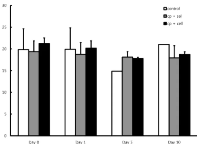
CP를 투여한 모든 군에서는 다음날 체중의 감소가 나타 났으나, 투여하지 않은 대조군에서만 유일하게 미약하게나 마 체중의 증가를 보였다. 이를 통해 CP의 투여가 마우스 의 체중 감소에 영향을 미쳤다고 추측할 수 있다.
체중 대비 간과 신장의 무게 비율(%)을 측정한 결과, 시
Fig. 1. The weight of immune-deficient mice. (CP + sal: after
injecting CP 300 mg/kg in abdominal cavity of mice the next day
injection of saline, CP + cell: after injecting CP 300 mg/kg the next
day injection of endothelial progenitor cell (HEPC:1 × 10
5/mL),
control: only saline injection).
험 시작 10일의 간의 무게 비가 5일의 간의 무게 비에 비 해 다소 증가하였다. 신장의 무게비는 큰 차이를 보이지 않았다(Fig. 2).
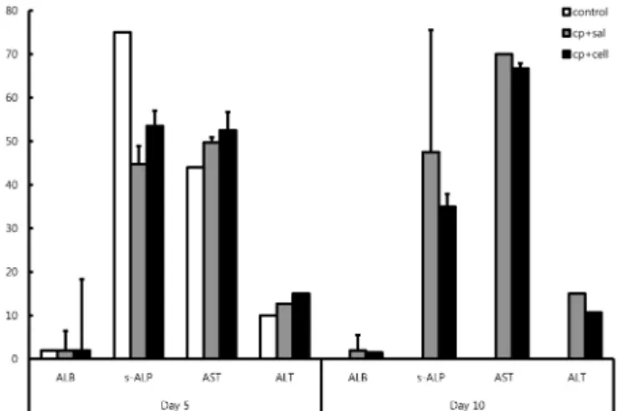
간 기능 저하 및 영양 상태 불능시에 albumin (ALB)의 감소가 나타나고, 간 기능의 손상시 alanine transaminase (ALT), aspartate aminotransferase (AST), serum alkaline phosphatase (s-ALP)의 상승이 나타난다. 시험 시작 10일 의 HEPC 이식군에서는 CP만 투여한 양성 대조군에 비해 s-ALP, AST, ALT의 수치가 감소하였다(Fig. 3).
시험 시작 5일 후 CP를 투여한 군에서는 혈관 내피세포 가 손상된 혈관들이 비교적 많이 관찰되었다. 시험 시작 5 일 된 HEPC 이식군에서는 손상된 혈관과 비교적 intact 한 혈관이 비슷한 비율로 관찰 되었다(Fig. 4). 시험 시작
10일 후 CP를 투여한 군과 HEPC를 이식한 군에서는 비 교적 intact한 혈관 내피세포가 관찰되는 혈관들을 볼 수 있었다(Fig. 5).
시험 시작 5일과 10일 된 HEPC 이식군에서도 anti-human
Fig. 2. The weight rate of river and kidney (left and right) at 5 and
10 days after experimental treatment. (CP + sal: after injecting CP 300 mg/kg in abdominal cavity of mice the next day injection of saline, CP + cell: after injecting CP 300 mg/kg the next day injec- tion of endothelial progenitor cell (HEPC:1 × 10
5/mL), control:
only saline injection).
Fig. 3. ALB, s-ALP, AST and ALT analysis at 5 and 10 days after experimental treatment. AT 10 days ALB, s-ALP, AST and ALT of HEPC treated group were decreased than only CP group. (CP + sal: after injecting CP 300 mg/kg in abdominal cavity of mice the next day injection of saline, CP + cell: after injecting CP 300 mg/
kg the next day injection of endothelial progenitor cell (HEPC:
1 × 10
5/mL), control: only saline injection).
Fig. 4. Many damaged vascular endothelial cells were observed in blood vessels (A). But the rate of damaged blood vessels and intact blood vessels in HEPC treated group was similar (B). (A: only CP treated group at 5 days, B: HEPC treated group at 5 days, HE stain- ing × 400).
Fig. 5. Many intact vascular endothelial cells were observed in all groups. (A: only CP treated group at 10 days, B: HEPC treated group at 10 days, HE staining × 400).
Fig. 7. Anti-human CD31 in only CP group was not expressed by immunohistochemistry staining (A). But anti-human CD 31 in HEPC treated group was expressed by immunohistochemistry staining. (A: only CP treated group at 10 days, B: HEPC treated group at 10 days, × 400).
Fig. 6. Anti-human CD31 in only CP group was not expressed by
immunohistochemistry staining (A). But anti-human CD 31 in
HEPC treated group was expressed by immunohistochemistry
staining. (A: only CP treated group at 5 days, B: HEPC treated
group at 5 days, × 400).
CD 31의 발현을 관찰 할 수 있었다(Fig. 7). HEPC를 투여 하지 않는 군에서는 5일과 10일 모두에서 human CD 31이 발현되지 않아 혈관내피세포가 염색 되지 않았다(Figs. 6, 7).
고 찰
혈관내피세포의 자연적 회복을 고려하여 시험 기간을 10 일로 설정하였다(Xie, 1985; Anton, 1997). 마우스를 희생 하기 전 절식의 영향으로 모든 군에서 체중이 감소하여 CP 에 의한 순수한 체중 변화를 측정하는데 어려움이 있었다.
체중 감소량의 차이를 비교함으로서 세포 이식군과 비이식 군의 차이를 살펴보고자 하였다. 쥐의 caudal vein에 cell을 주입하는 과정에서 한 마리가 사망했다. 사망원인은 너무 빠른 주입 속도로 생각된다. 또한 혈액채취 시 용혈이 되 어 AST, ALT 수치가 상승되어 결과 해석에 어려움을 초 래했다. 그러나 용혈되어 높은 값으로 나타난 수치를 포함 시킨 결과도 수치의 증가와 감소 양상은 동일 하였다.
마우스의 체중 변화를 분석한 결과 CP 투여군에서만 다 음날 체중 감소가 나타났고, 투여하지 않은 군에서는 체중 의 증가가 나타난 것으로 미루어 볼 때, CP가 마우스의 체중 감소에 영향을 미쳤다고 생각할 수 있다. 또한 Day 5에 비해 Day 10의 간의 무게 비가 사람혈관내피전구세 포를 이식한 군에서 다소 증가한 것을 확인하였다.
혈청 분석 결과 Day 10의 hHEPC 이식 군에서 CP 투 여군에 비해 s-ALP, AST, ALT의 수치가 감소한 양상을 확인하였다. 이를 통해 혈관 내피줄기세포에 의해 간손상 이 회복되었을 가능성을 생각해 볼 수 있다.
H&E 염색을 통해 관찰한 조직의 양상에서 Day 5의 CP 투여군에서 비교적 혈관 내피세포가 손상된 혈관들을 많이 관찰할 수 있었다. 하지만 Day 10의 CP 투여군과 hHEPC 이식군에서 모두 비교적 intact한 혈관 내피세포가 많이 관 찰되어, 두 군의 차이가 뚜렷하게 나타나지 않았다. 이를 통해 혈관 내피세포의 자발적 회복 가능성을 고려해 볼 수 있다. 면역 형광 염색 결과 day 5와 day 10의 hHEPC 이 식군에서 human CD 31이 발현된 혈관 내피세포를 관찰 할 수 있었는데, 이식한 세포가 마우스의 혈관 내피계에 어느정도 대체 된 것으로 추정된다.
실험 동물의 수를 늘려서 표준편차를 줄이고, 직접적인 인간 특이 유전자 (slu-sx)등의 정량 분석을 하여 human cell의 이식을 확인하는 추가적인 실험도 유용할 것으로 생 각되며, human centromere detection 을 위한 in situ hy- bridization 을 통한 이식세포 분포 관찰 연구도 할 수 있 을 것이다. 이 모델을 특이 조건으로 배양된 사람 줄기 세 포가 치아 재생시 혈관 재형성 연구에 응용될 수 있는 발 판을 마련했다.
혈관 손상 시 자발적으로 HEPC가 순환계로 mobilization 된다. 이러한 현상으로 인해 혈관의 자발적 회복이 일어난
다. 그러나 심혈관계 질환을 가지고 있거나 가역적인 손상 을 받은 환자의 경우, 이러한 현상만으로 손상이 자발적으 로 회복되지는 않는다. 그 이유는 두 가지로 설명할 수 있 다. 이렇게 mobilization 되는 HEPC, CEPs가 손상된 조 직에 가서 incorporation 되기에 충분하게 성숙되지 않았 기 때문이라는 설명과, 허혈된 조직의 혈액 공급 저하로 인해 HEPC, CEP가 손상된 vasculature를 인식하지 못하 기 때문이다(Rafii and Lyden, 2003).
종양, 혈전증 등의 병적인 상태에서 HEPC, CEP, hema- topoietic cell을 정맥 주사하는 것은 부작용을 일으킬 수 있다(Celletti et al., 2001). 쥐를 이용한 실험에서 HEPC 와 monocytes를 mobilization 한 후 atheroma formation 이 증가된 연구 결과도 있다(Moulton et al., 1999). 다른 연구에서는 종양형성, 동맥경화, retinopathy등이 발생함을 보고한 바 있다(Lyden et al., 2001). 이러한 부작용을 줄 일 수 있는 방법 중의 하나는 손상된 조직에 직접 주입하 는 것이다.
앞서 언급하였듯이 이러한 줄기세포를 이용한 치료는 유 용하게 사용될 수 있는데, 너무 적은 양의 HEPC population 으로 인해 임상 적용의 한계가 있다. 이것을 극복하기 위 해 제대혈, 골수, embryonic stem cell등 다른 곳으로부터 더 많은 HEPC를 얻고, 이것을 drugs나 gene transfer를 조합하여 사용하면 HEPC의 효율이 증가할 것이다(Taniguchi et al, 2006; Kalka et al, 2000).
감사의 글
This study was supported by grants from the Korea Science & Engineering Foundation of the Korean Ministry of Science & Technology (M10646010002-06N4601-00210 and M10641520002-06N4152-00210).
참 고 문 헌