The Protective Effect of Zizania latifolia Extract against t-BHP-induced Oxidative Stress in HepG2 Cells
Se-Ho Park
1,2, Jae-Yeul Lee
1,2, Seun-Ah Yang
3, Daesuk Bang
4and Kwang-Hwan Jhee
1*
1Department of Applied Chemistry, Kumoh National Institute of Technology, Gumi 39177, Korea
2Institute of Natural Science, Keimyung University, Daegu 42601, Korea
3Department of Food Science and Technology, Keimyung University, Daegu 42601, Korea
4Department of Chemical Engineering, Kumoh National Institute of Technology, Gumi 39177, Korea Received December 1, 2020 /Revised December 14, 2020 /Accepted December 14, 2020
Zizania latifolia has long been used as a tea for both edible and medicinal purposes. However, research
into the use of Z. latifolia as a high value-added edible material is lacking. In a previous study, we confirmed that tricin is the major component in Z. latifolia. In this study, we investigated the pro- tective effect of a Z. latifolia extract (ZLE). Toxicity tests of ZLE or tricin on HepG2 cells revealed no toxicity due to ZLE or tricin at all concentrations used. The reduction in cell viability by tert-butyl hydroperoxide (t-BHP) was suppressed by treatment with ZLE or tricin. In addition, ZLE or tricin ef- fectively inhibited the production of reactive oxygen species (generation of hydrogen peroxide, alkoxy free radicals, and peroxyl free radicals by t-BHP) and oxidative damage. ZLE or tricin treatments also increased the protein expression of superoxide dismutase 1 (SOD1), catalase, heme oxygenase-1 (HO- 1), and nuclear factor erythroid-related factor 2 (Nrf2), which are known as antioxidant enzymes, sug- gesting that the protective effect of ZLE is related to activation of tricin. Taken together, the results indicate that Z. latifolia can be developed as a functional food material for improving liver function.
Key words : Oxidative stress, protective effect, reactive oxygen species, Zizania latifolia
*Corresponding author
*Tel : +82-54-478-7837, Fax : +82-54-478-7710
*E-mail : [email protected]
This is an Open-Access article distributed under the terms of the Creative Commons Attribution Non-Commercial License (http://creativecommons.org/licenses/by-nc/3.0) which permits unrestricted non-commercial use, distribution, and reproduction in any medium, provided the original work is properly cited.
서 론
현대 사회에 들어서 건강에 대한 관심이 높아지고 있으며 특히 불규칙한 식습관과 생활패턴의 영향으로 간의 건강 관리 에 대한 관심이 증가하고 있다. 간은 신체 내 물질의 정화 작용 을 하는 중요한 필수조직 중 하나이며 다른 조직들을 조화롭 게 관리하는 역할을 한다. 간의 기능으로는 에너지 저장, 영양 소의 대사기능, 해독기능 등이 알려져 있다. 간 손상의 원인으 로는 음주와 흡연, 바이러스에 의한 감염, 독물, 약제 중독 등 이 있으며 산화적 스트레스 기반 반응성 유해 산소종(reactive oxygen species, ROS)의 생성으로 간섬유화[21, 26], 신장염 [29, 33], 피부질환[4, 5], 당뇨병[6, 30] 등의 여러가지 질환의 원인이 되고 있다. Hydroxyl radical, superoxide, hydrogen peroxide 등의 반응성이 우수한 ROS는 호기 대사를 통해 생체 내에서 생성되며, 지질, 단백질 및 DNA 등의 세포 거대분자에 손상을 유발시켜 노화, 염증, 발암, 동맥경화 등의 다양한 질병 을 일으킨다[31]. 특히 산화적 스트레스에 의한 국수적인 손상
은 지질과산화를 유도하여 신체 손상을 일으킨다. 이러한 지 질과산화를 일으키는 대표적인 물질로 tert-butyl hydroper- oxide (t-BHP)가 있다. t-BHP는 간세포의 cytochrome P-450 효소에 의해 세포 구성물들의 산화를 일으킬 수 있는 alkoxy free radical이나 peroxyl free radical로 분해되어 간세포의 DNA의 손상을 가져와 세포의 사멸을 유도하게 된다. t-BHP 로 산화적 손상을 유도한 HepG2 세포 모델은 산화적 손상 예방 소재의 탐색에 사용하는 대표적인 모델 중 하나이다[13, 18, 34].
고장초(Zizania latifolia)는 벼과(Gramineae)에 속하는 여러 해살이 풀로 줄풀 또는 야생미(wild rice)라고도 불리고 있으 며, 한국, 베트남, 일본, 중국 등 동아시아에 분포하는 자생식 물이다. 고장초의 줄기와 종자는 식용 및 약용으로 사용되고 있다. 고장초 종자의 항당뇨[6], 당 대사 개선, 항비만[8], 항산 화능[35], 혈중 지질 개선 이 보고되었고, 고장초 줄기의 신경 세포 보호 효과[24], 항알러지[14, 15, 17], 항주름[2, 20, 23] 기 능 등이 알려져 있다. 또한, 고장초 줄기에서 분리된 tricin 유 도체들의 항염 및 항알러지 활성[15, 17] 등이 최근 보고되었으 나, 고장초의 HepG2 세포에 대한 보호에 대한 보고는 없다.
따라서, 본 연구에서는 오랫동안 민간에서 사용되어 안전성에
문제가 없으며 최근 과학적으로 유효성이 알려지면서 관심이
높아지고 있는 Z. latifolia의 HepG2 세포에 대한 보호 관련
연구를 수행하였다.
재료 및 방법
실험재료
t-BHP, [3-(4,5-dimethylthiazol-2-yl)-5(3-carboxymethony-
phenol)-2-(4-sulfophenyl)-2H-tetrazolium] (MTT), polyacryl- amide (37.5:1), sodium dodecylsulfate (SDS), tetramethyle- thylenediamine (TEMED), phosphate buffer saline (PBS), Tween-20은 Sigma-Aldrich Chemical Co. (St. Louis, MO, USA)에서 구입하였으며, dimethyl sulfoxide (DMSO)는 ㈜대 정화금(Siheung, Korea)에서 구입하였다. Tricin은 CromaDex (Los Angeles, CA, USA)에서 구입하였으며, 2',7'-dichlor- odihydrofluorescein diacetate (DCF-DA), 4′,6-diamidino-2- phenylindole (DAPI), bicinchoninic acid (BCA)는 Thermo Fisher Scientific (Whaltham, MA, USA)에서 구입하였다.
고장초 추출물 제조 및 수율
본 실험에 사용된 고장초 에탄올(Z. latifolia ethanol extract, ZLE)은 ㈜비티씨(Ansan, Korea)로부터 제공받았다. 건조된 고장초를 세절하여 1 kg을 칭량하여 70% 에탄올 30 l를 혼합 하여 80℃에서 6시간 동안 교반하고 1회 추출하였다. 추출물 은 부직포로 여과한 후, 고속 추출 농축기를 이용하여 농축시 킨 뒤 evaporator (EYELA N-3010, Tokyo Rikakikai, Japan)로 건조하여 분말 형태로 회수하였다. 회수한 추출물은 -20℃에 서 보관하여 사용하였다. 실험은 추출물의 DMSO의 최종 농 도가 0.1%가 되도록 사용하였다. 추출물의 수율은 추출 전 고 장초 원물 무게에 대한 동결 건조 후 추출물 무게를 측정하여 나타내었다.
세포 배양
본 연구에 사용된 HepG2 세포는 한국세포주은행(Korean Cell Line Bank, Seoul, Korea)에서 분양 받았으며, 1% pen- icillin-streptomycin (WELGENE Inc., Gyeongsan, Korea) 및 10% fetal bovine serum (FBS, WELGENE Inc.)이 포함된 Dulbecco's Modified Eagle's Medium (DMEM, WELGENE Inc.)를 사용하여 37℃, 5% CO
2조건하에서 배양하였다.
세포 생존율 측정
세포 생존율 측정을 위하여 24-well plate에 HepG2 세포를 1×10
5cells/well로 분주하고 24시간 동안 배양한 후 ZLE (0, 50, 100, 250, 500 μg/ml) 또는 tricin (50, 100, 250 ng/ml)을 농도별로 첨가하여 24시간 처리하였다. 최종 농도가 5 μg/ml 이 되도록 MTT 용액을 넣고 37℃, 5% CO
2배양기에서 4시간 반응시킨 후, microplate reader (Molecular Devices, CA, USA)를 이용하여 550 nm에서의 흡광도를 측정하여 대조군에 대한 세포 생존율을 백분율로 표시하였다.
HepG2 세포 보호능 측정
ZLE의 HepG2 세포의 보호능을 측정하기 위해 MTT assay 를 수행하였다[28]. 먼저, HepG2 (1×10
5cells/well) 세포를 24-well cell culture plate에 분주하고 24시간 동안 배양하였 다. 이후 ZLE (0, 50, 100, 250, 500 μg/ml) 또는 tricin (50, 100, 250 ng/ml)을 농도별로 처리하고 24시간 배양하였다. 24 시간 배양 이후 HepG2 cell에 t-BHP (1 mM)를 처리하고 1시 간 반응시켰다. 1시간 반응 이후 최종 농도가 5 μg/ml이 되도 록 MTT 용액을 넣고 37℃, 5% CO
2배양기에서 4시간 반응시 킨 후, microplate reader를 이용하여 550 nm에서의 흡광도를 측정하여 대조군에 대한 세포 생존율을 백분율로 표시하였다.
HepG2 세포의 산화적 손상 보호 효과 관찰
ZLE의 HepG2 세포 내부의 산화적 손상 보호 효과를 관찰 하기 위해 DCF-DA assay를 수행하였다[27]. 먼저, HepG2 (1×10
5cells/well) 세포를 24-well cell culture plate에 분주하 고 24시간 동안 배양하였다. 이후 ZLE (0, 50, 100, 250, 500 μg/ ml) 또는 tricin (250 ng/ml)을 농도별로 처리하고 24시간 배양하였다. 24시간 배양 이후 HepG2 세포에 t-BHP (1 mM) 를 처리하고 1시간 반응시켰다. 1시간 반응 이후 PBS로 3회 세척하고 4% formaldehyde (with PBS)로 각 세포를 고정하였 다. 이후 PBS로 3회 세척하고 DCF-DA (20 μM)과 DAPI (0.5 μg/ ml)를 각 well에 첨가하여 20분간 반응시켰다. 이 후 PBS로 세척하고 형광현미경(fluorescence microscope, DMIRE2, Leica, Wetzlar, Gemany)으로 각 세포를 관찰하였다.
HepG2 세포의 산화적 손상 관련 단백질 발현 확인 ZLE의 HepG2 세포 내부의 산화적 스트레스 예방 관련 단 백질 발현 확인을 위해 Western blot analysis를 수행하였다.
먼저, HepG2 (5x10
5cells/well) 세포를 6-well cell culture plate에 분주하고 24시간 동안 배양하였다. 이후 ZLE (0, 50, 100, 250, 500 μg/ml) 또는 tricin (250 ng/ml)를 농도별로 처리 하고 24시간 배양하였다. 24시간 배양 이후 HepG2 세포에
t-BHP (1 mM)를 처리하고 1시간 반응시키고, PBS로 3회 세척하고 RIPA lysis buffer (1 ml)를 넣어 세포를 녹이고 13,000 rpm, 4℃, 10분 동안 원심분리 하였다. 원심분리 이후 상등액 을 취하고 BCA assay (Thermo Fisher Scientific)를 이용하여 단백질을 정량하였다. 추출한 단백질(20 μg)을 10% polyacryl- amide gel에 넣고 sodium dodecyl sulfate-polyacrylaminde gel electrophoresis (SDS-PAGE)를 수행하였다(120 V, 1시간).
이후 수화된 PVDF membrane (Whatman GmbH, Dassel,
Germany)에 transfer하고(100 V, 1시간), 5% skim milk (with
Tris-buffered saline, TBS)에 blocking하였다(RT, 1시간). 각
membrane은 superoxide dismutase-1 (SOD1, #2770), cata-
lase (#12980), hemeoxygenase-1 (HO-1, #70081), nuclear fac-
tor erythroid-related factor 2 (Nrf2, #12721) 항체(Cell Signal-
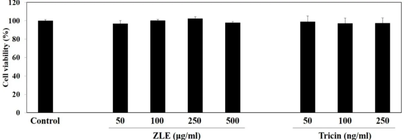
Fig. 1. Cell viability of HepG2 cells according to ZLE concentration. HepG2 cells were seeded into a 24-well cell culture plate (1×10⁵ cells/well) and incubated for 24 hr. The cells were treated with various concentration of ZLE (50, 100, 250, 500 μg/ml) or tricin (50, 100, 250 ng/ml). After incubation for 24 hr, MTT reagent in PBS (5 mg/ml, 20 μl) was added to each well.
After 4 hr incubation, each well was measured at 550 nm using a microplate spectrophotometer. The value represents mean
± SD of three different experiments (*p<0.05, **p<0.01, ***p<0.001 compared to the control).
ing Technology, Beverly, MA, USA)에 24시간 동안 4℃에서 반응시켰다. Membrane을 TBST buffer로 3회 세척하고 secon- dary antibody (#7074, Cell Signaling Technology)로 1시간, 상온에서 반응시키고 TBS with 0.1% Tween-20 (TBST)으로 3회 세척하였다. 이 후 enhanced chemiluminescence (ECL) solution을 10분간 반응시키고 band를 확인하였다(UVP Vis- ion Works
®LS Image Acquisition & Analysis Software, Upland, CA, USA). 각 band는 β-actin의 발현량을 기준하여 변화되는 단백질 발현 여부를 정량, 확인하였다.
통계학적 분석
본 실험에서 얻어진 결과에 모든 값은 3회 이상의 반복실험 의 측정값의 평균±표준오차로 나타내었으며, 실험 결과 값에 대한 통계적 분석은 Student’s t-test를 이용하여 분석하였으 며, p값이 0.05 이하이면 유의성이 있다고 판단하였다.
결 과
ZLE 또는 tricin의 HepG2 세포에 대한 세포 생존율 및 세포 보호 효과
ZLE 또는 tricin의 HepG2 세포에 대한 세포 생존율을 MTT assay로 측정한 결과, 본 연구에서 사용된 범위의 ZLE 또는 tricin의 처리 농도에서 세포 생존율의 유의적 변화가 나타나 지 않았다(Fig. 1).
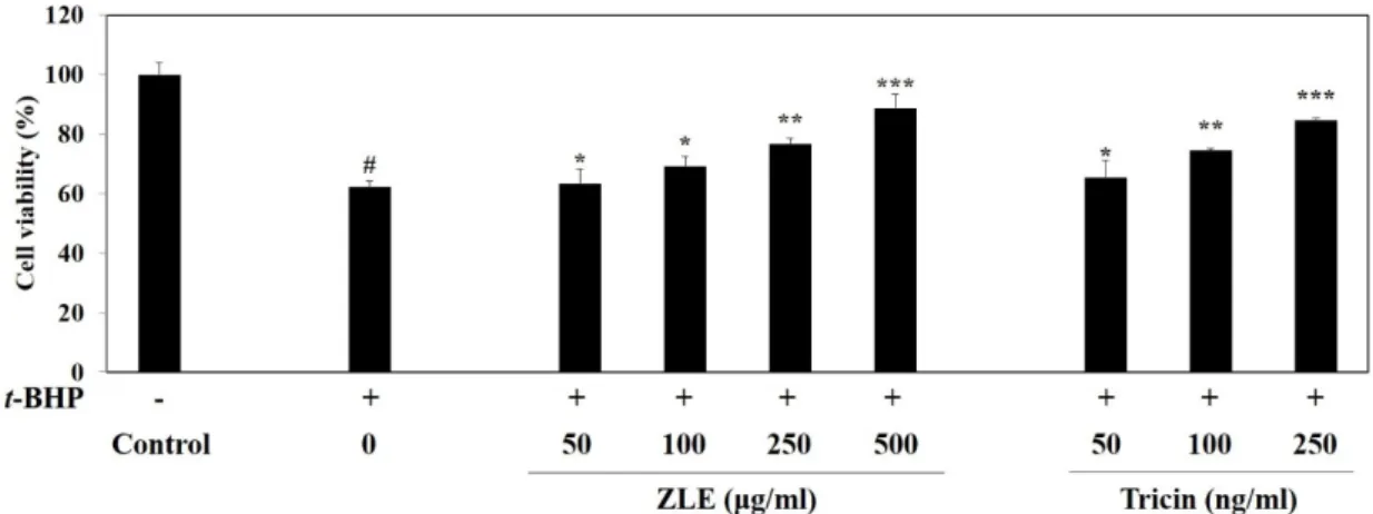
ZLE 또는 tricin의 HepG2 세포에 대한 세포 보호 효과를 MTT assay로 측정하였다. 그 결과, t-BHP가 처리된 HepG2 세포는 49%의 세포 생존율을 보임으로써 산화적 스트레스에 의한 HepG2 세포의 손상이 유도되었음을 확인하였다. t-BHP 로 산화적 스트레스에 의해 손상이 유도된 HepG2 세포는 ZLE 의 처리 농도가 증가함에 따라 세포 생존율이 각각 79.25, 87.54,
96.64%로 증가하는 것을 확인하여 HepG2 세포 손상에 대한 보호 효과가 우수함을 확인하였으며 tricin의 처리에 의해 세 포 생존율이 86.48%까지 증가하는 것을 확인하였다(Fig. 2).
ZLE 또는 tricin의 HepG2 세포의 활성산소 억제 ZLE 또는 tricin의 HepG2 세포의 활성산소 억제를 확인하 기 위해 DCF-DA assay를 수행하였다. t-BHP가 처리된 HepG2 세포에서는 정상세포에 비해 ROS가 다량 발생하는 것을 관찰 할 수 있었으며 ZLE의 처리 농도에 따라 세포내 ROS의 발생 량이 감소하고 고농도의 tricin의 처리로 효과적으로 ROS가 억제되는 것을 관찰할 수 있었다(Fig. 3). 이 결과는 ZLE의 주 요 성분인 항산화 효능을 갖는 tricin에 의해 HepG2 세포를 보호함을 시사한다.
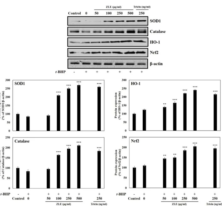
ZLE 또는 tricin의 HepG2 세포에 대한 단백질의 발현 확인
ZLE의 HepG2 세포에 대한 항산화 관련 단백질의 발현
은 Western blot analysis을 이용하여 확인하였다. SOD1,
catalase, HO-1, Nrf2는 세포 내 산화적 스트레스를 제거하
는 단백질로 알려져 있다[9, 32]. 본 연구에서는 ZLE의 산화
적 스트레스 완화 효과를 확인하기 위해 위의 4종의 단백질
의 발현 여부를 측정하였다. t-BHP가 처리된 HepG2 세포에
서 ZLE의 처리 농도(250 μg/ml)에 의해 각각의 단백질의
발현이 2.5, 2.0, 2.2, 1.9배 증가하였다(Fig. 4). 또한, t-BHP
가 처리된 HepG2 세포에 tricin (250 ng/ml)이 처리되면
SOD, catalase, HO-1, Nrf2의 발현이 각각 2.6, 1.8, 2.2, 2.0
배 증가하는 것을 확인하였다. 이 결과는 t-BHP에 의해 산
화적 손상이 유도된 HepG2 세포 내부의 항산화 단백질을
ZLE의 tricin이 과발현시켜 HepG2 세포 보호 효과를 나타
낸다고 판단된다.
Fig. 2. Protective effect of ZLE on t-BHP-induced oxidative stress in HepG2 cells. HepG2 cells were seeded into a 24-well cell culture plate (1×10⁵ cells/well) and incubated for 24 hr. The cells were treated with various concentration of ZLE (50, 100, 250, 500 μg/ml) or tricin (50, 100, 250 ng/ml). After incubation for 24 hr, the cells were treated with 1 mM t-BHP for 1 hr and then MTT reagent in PBS (5 mg/ml, 20 μl) was added to each well. After 4 hr incubation, each well was measured at 550 nm using a microplate spectrophotometer. The value represents mean ± SD of three different experiments (*p<0.05,
**p<0.01, ***p<0.001 compared to t-BHP-treated cells, #p<0.05 compared to the control).
Fig. 3. Inhibition of ROS generation at various concentration of ZLE against t-BHP-induced oxidative stress in HepG2 cells. HepG2 cells were seeded at a density of 1×10⁵ cells/well in 24-well cell culture plate and incubated for 24 hr. The cells were treated with various concentration of ZLE (50, 100, 250, 500 μg/ml) or tricin (250 ng/ml). After incubation for 24 hr, the cells were treated with 1 mM t-BHP for 1 hr and then the cells were washed twice with PBS. The cells were fixed with 4% formaldehyde with PBS. Cells loaded with DCF-DA (20 μM) and DAPI (0.5 μg/ml) for 20 mins, and then the cells were washed twice with PBS. The DCFDA/DAPI-stained cells were pictured by a fluorescence microscope (Scale bar=100 μm).
Fig. 4. Effect of ZLE or tricin on SOD1, catalase, HO-1, and Nrf2 protein expressions in t-BHP-induced oxidative stress in HepG2 cells. HepG2 cells were seeded at a density of 5×10⁵ cells/well in 6-well cell culture plate and incubated for 24 hr. The cells were treated with various concentration of ZLE (50, 100, 250, 500 μg/ml) or tricin (250 ng/ml). After incubation for 24 hr, the cells were treated with 1 mM t-BHP for 1 hr and then the cells were washed twice with PBS. Each protein (20 μg/lane) were electrophoresis on 10% SDS-PAGE and transferred onto PVDF membrane. The immunoblot was incubated overnight with blocking solution (5% skim milk) at 4℃, followed by incubation with primary antibodies. Blots were washed twice with TBST and incubated with secondary antibody for 1 hr at room temperature. Blots were again washed twice with TBST and then developed by ECL solution. β-actin was used as a housekeeping protein. The value represents mean
± SD of three different experiments (*p<0.05, **p<0.01, ***p<0.001 001 compared to t-BHP- treated cells, #p<0.05 compared to the control).
고 찰
본 연구는 ZLE의 HepG2 세포 보호 효과가 산화적 스트레 스를 효과적으로 억제한 것임을 확인한 첫 연구이다. 이전 연 구에서는 ZLE의 용매 추출 조건별 항산화 효능을 알아보았다.
결과 추출 용매(에탄올)의 비율이 증가함에 따라 DPPH,
ABTS 라디칼 소거능의 향상을 확인하였다. 특히 에탄올 70%
의 추출에서 가장 우수한 항산화 효능을 확인하여 본 연구에 서는 70% 에탄올로 80℃, 6시간 동안 추출한 ZLE을 사용하였 다[22].
본 연구는 HepG2 세포에 대한 ZLE와 tricin의 HepG2 세포
의 보호 효과를 검증하기 위해 t-BHP에 의한 산화적 손상으로
감소한 HepG2 세포의 생존율이 ZLE와 tricin의 처리에 의해 증가하는 것을 확인하였다. 타 보고에 의하면 황칠나무 잎 추 출물[11]의 100 μg/ml의 농도에서 60% 미만의 세포 생존율을 보였으며, 만병초 추출물[25]에서는 100 μg/ml의 농도에서 57.9%의 세포 생존율을 나타내었다. 이는 동일 농도로 처리한 경우(100 μg/ml) , ZLE의 HepG2 세포 보호 효과가 타 추출물 에 비해 우수함을 보였다.
또한, HepG2 세포 내에서 ZLE와 tricin의 산화적 손상 억제 를 정성적으로 관찰하였으며, t-BHP의 처리에 의해 HepG2 세포 내에 과생성된 ROS가 ZLE와 tricin의 처리에 의해 효과 적으로 억제되는 것을 관찰하였다. Lee 등의 연구 보고[25]에 따르면 만병초 추출물의 처리 농도가 100 μg/ml에서 ROS의 생성을 억제하는 것을 확인할 수 있었으며, 이고들빼기 추출 물은 40 μg/ml 농도에서 ROS의 억제 효과를 나타냄을 보고하 였다[10]. 따라서 본 실험 결과 ZLE도 활성산소의 억제를 통해 HepG2 세포 보호효과를 나타내는 것으로 사료된다.
HepG2 세포 내 과생성된 ROS를 ZLE와 tricin이 효과적으 로 억제함을 관찰한 결과를 토대로 ZLE와 tricin이 HepG2 세 포 내 항산화 관련 단백질의 발현을 조절하는지의 유무를 확 인하였다. 그 결과 SOD1, catalase, HO-1, Nrf2의 단백질 발현 이 과생성됨을 정성적, 정량적으로 확인하였다. 항산화 관련 단백질의 발현을 증가시켜 HepG2 세포 보호 효과를 나타내는 사례로 수련 추출물[1, 3], 캄보디아 생약 추출물[12], 삼 추출 물[16], 세이지 추출물[19] 등이 있으며, ZLE 또한 위 사례의 추출물과 비교하면 항산화 관련 단백질의 발현량이 우수한 것으로 확인되어 ZLE의 HepG2 세포에 대한 보호 효과가 우 수함을 입증하였다.
따라서 본 연구에서 산화적 손상이 유도된 HepG2 세포의 ZLE의 보호 효과가 100 μg/ml의 농도에서 나타나고, 세포내 발생된 ROS의 효과적인 억제 및 ROS의 제거를 담당하는 단백 질의 증가를 종합하면, ZLE의 HepG2 세포 보호 효과는 세포 내 ROS의 제거 단백질 과발현을 ZLE가 유도하여 세포내 ROS 발생량을 억제하는 메커니즘으로 세포 보호 효과를 나타내는 것으로 판단된다. 아직까지 HepG2 세포에 대한 ZLE의 보호 효과 중 산화적 스트레스의 제거에 의한 HepG2 세포의 보호 효과만을 규명한 연구로 본 연구를 기초로 향후 ZLE이 간을 보호하기 위한 기능성 식품 소재로 활용될 수 있을 것으로 기대된다.
감사의 글
본 연구는 금오공과대학교 학술연구비로 지원되었음.
The Conflict of Interest Statement
The authors declare that they have no conflicts of interest with the contents of this article.
References
1. Alam, M. B., Ju, M. K. and Lee, S. H. 2017. DNA protecting activities of Nymphaea nouchali (Burm. f) flower extract at- tenuate t-BHP-induced oxidative stress cell death through Nrf2-mediated induction of heme oxygenase-1 expression by activating MAP-kinases. Int. J. Mol. Sci. 18, 2069.
2. An, M. R., Kim, H. K., Moon, J. M., Ko, H. S., Paul, C. and Lim, Y. H. 2020. Enzyme-treated Zizania latifolia ethanol ex- tract protects from UVA irradiation-induced wrinkle for- mation via inhibition of lysosome exocytosis and reactive oxygen species generation. Antioxidants 9, 912.
3. Bajpai, V. K., Alam, M. B., Ju, M. K., Kwon, K. R., Huh, Y. S., Han, Y. K. and Lee, S. H. 2018. Antioxidant mechanism of polyphenol-rich Nymphaea nouchali leaf extract protecting DNA damage and attenuating oxidative stress-induced cell death via Nrf2-mediated heme-oxygenase-1 induction cou- pled with ERK/p38 signaling pathway. Biomed. Pharmac- other. 103, 1397-1407.
4. Chelombitko, M. A. 2018. Role of reactive oxygen species in inflammation: A minireview. Moscow Univ. Biol. Sci. Bull.
73, 199-202.
5. Cracowski, J. L., Marpeau, C., Carpentier, P. H., Imbert, B., Hunt, M., Stanke-Labesque, F. and Bessard, G. 2001. En- hanced in vivo lipid peroxidation in scleroderma spectrum disorders. Arthritis Rheum. 44, 1143-1148.
6. Fakhruddin, S., Alanazi, W. and Jackson, K. E. 2017. Diabe- tes-induced reactive oxygen species: mechanism of their generation and role in renal injury. J. Diabetes Res. 2017, 1-30.
7. Han, S., Zhang, H., Qin, L. and Zhai, C. 2013. Effects of dietary carbohydrate replaced with wild rice (Zizania latifolia (Griseb) Turcz) on insulin resistance in rats fed with a high-fat/cholesterol diet. Nutrients 5, 552-564.
8. Han, S. F., Zhang, H. and Zhai, C. K. 2012. Protective poten- tials of wild rice (Zizania latifolia (Griseb) Turcz) against obe- sity and lipotoxicity induced by a high-fat/cholesterol diet in rats. Food Chem. Toxicol. 50, 2263-2269.
9. Je, J. Y. and Lee, D. B. 2015. Nelumbo nucifera leaves protect hydrogen peroxide-induced hepatic damage via antioxidant enzymes and HO-1/Nrf2 activation. Food Funct. 6, 1911-1918.
10. Kang, K. S., Jho, E. H., Lee, H. J., Oidovsambuu, S., Yun, J. H., Kim, C. Y., Yoo, J. H., Kim, Y. J., Kim, J. H., Ahn, S. Y. and Nho, C. Y. 2011. Youngia denticulata protects against oxidative damage induced by tert-butylhydroperoxide in HepG2 cells. J. Med. Food 14, 1198-1207.
11. Lee, C. Y., Yang, M. H. and Moon, J. O. 2019. Antioxidant and hepatoprotective effects of the ethanol extract of Dendropanax morbifera Leveille on the t-butyl hydroper- oxide-induced HepG2 cell damages. Kor. J. Pharmocogn. 50, 32-36.
12. Lee, D. S., Keo, S., Cheng, S. K., Oh, H. C. and Kim, Y.
C. 2016. Protective effects of Cambodian medicinal plants on tert-butyl hydroperoxide-induced hepatotoxicity via Nrf2- mediated heme oxygenase-1. Mol. Med. Rep. 15, 451-459.
13. Lee, E. G., Kim, K. B. and Jeong, J. M. 2006. Hepatoprotective effects of poly herb formulation (Hepa-1000) on t-BHP in-
duced toxicity in human hepatoma cells. J. Kor. Soc. Food Sci. Nutr. 35, 1121-1126.
14. Lee, E. J., Yu, M. H., Garcia, C. V., Jhee, K. H. and Yang, S. A. 2017. Inhibitory effect of Zizania latifolia chloroform fraction on allergy-related mediator production in RBL-2H3 cells. Food Sci. Biotechnol. 26, 481-487.
15. Lee, E. J., Whang, E. Y., Whang, K., Lee, I. S. and Yang, S.
A. 2009. Anti-allergic effect of Zizania latifolia Turcz extracts.
Kor. J. Food Sci. Technol. 41, 717-721.
16. Lee, H. J., Kim, J. H., Lee, S. Y., Park, J. H. and Hwang, G. S. 2012. Processed Panax ginseng, Sun ginseng, decreases oxidative damage induced by tert-butyl hydroperoxide via regulation of antioxidant enzyme and anti-apoptotic mole- cules in HepG2 cells. J. Ginseng Res. 36, 248-255.
17. Lee, J. Y., Park, S. H., Jhee, K. H. and Yang, S. A. 2020.
Tricin isolated from enzyme-treated Zizania latifolia extract inhibits IgE-mediated allergic reactions in RBL-2H3 cells by targeting the Lyn/Syk pathway. Molecules 25, 2084.
18. Lee, Y. J., Kim, N. S., Shon, M. S., Kim, G. N., Hwang, Y.
I. and Park, E. J. 2016. Effect of fermented herbal mixture against oxidative stress in HepG2 and PC12 cells. J. Kor.
Soc. Food Sci. Nutr. 45, 1057-1064.
19. Lime, C. F., Valentao, P. C. R., Andrade, P. B., Seabra, R.
M., Fernandes-Ferreira, M. and Pereira-Wilson, C. 2007.
Water and methanolic extracts of Salvia offiicinalis protect HepG2 cells from t-BHP induced oxidative stress. Chem.
Biol. Interact. 167, 107-115.
20. Moon, J. M., Park, S. H., Jhee, K. H. and Yang, S. A. 2018.
Protection against UVB-induced wrinkle formation in SKH- 1 hairless mice: efficacy of tricin isolated from enzyme- treated Zizania latifola extract. Molecules 23, 2254.
21. Paik, Y. H. 2008. Fibrogenesis and antifibrotic therapy. Clin.
Mol. Hepatol. 14, 7-13.
22. Park, S. H., Lee, J. Y. and Yang, S. A. 2017. Comparative analysis of anti-oxidative, anti-inflammatory, anti-allergy, and whitening effects of different solvent extracts from Zizania latifolia. J. Life Sci. 27, 994-1002.
23. Park, S. H., Lee, S. S., Bang, M. H., Jo, S. K., Jhee, K. H.
and Yang, S. A. 2019. Protection against UVB-induced dam- ages in human dermal fibroblasts: efficacy of tricin isolated from enzyme-treated Zizania latifolia extract. Biosci. Bio- technol. Biochem. 83, 551-560.
24. Park, W. H. and Cha, Y. Y. 2004. Inhibition effect of Zizania latifolia on apoptosis induced by H2O2 in Neuro2A cell. Kor.
J. Oriental Physiol. Pathol. 19, 1062-1067.
25. Rhim, T. J. and Choi, M. Y. 2011. The antioxidative effects of Rhododendron brachycarpum extracts. Kor. J. Plant Res. 24, 456-460.
26. Richter, K. and Kietzmann, T. 2016. Reactive oxygen species and fibrosis: further evidence of a significant liaison. Cell Tissue Res. 365, 591-605.
27. Slamenova, D., Kozics, K., Hunakova, L., Melusova, M., Navarova, J. and Horvathova, E. 2013. Comparison of bio- logical processes induced in HepG2 cells by tert-butyl hy- droperoxide (t-BHP) and hydroperoxide (H2O2): The influ- ence of carvacrol. Mutat. Res. Genet. Toxicol. Environ. Muta- gen. 757, 15-22.
28. Sohn, J. H., Han, K. L., Lee, S. H. and Hwang, J. K. 2005.
Protective effects of panduratin A against oxidative damage of tert-butylhyroperoxide in human HepG2 cells. Biol. Pharm.
Bull. 28, 1083-1086.
29. Solin, M. L., Ahola, H., Haltia, A., Ursini, F., Montine, T., Roveri, A., Kerjaschki, D. and Holthöfer, H. 2001. Lipid per- oxidation in human proteinuric disease. Kidney Int. 59, 481- 487.
30. Stanely Mainzen Prince, P. and Menon, V. P. 2001. Antiox- idant action of Tinospora cordifolia root extract in alloxan dia- betic rats. Phytother. Res. 15, 213-218.
31. Valko, M., Leibfritz, D., Moncol, J., Cronin, M. T. D., Mazur, M. and Telser, J. 2007. Free radicals and antioxidants in nor- mal physiological functions and human disease. Int. J. Bio- chem. Cell Biol. 39, 44-84.
32. Waza, A. A., Hamid, Z., Bhat, S. A., Shah, N. U. D., Bhat, M. and Ganai, B. 2018. Relaxin protects cardiomyocytes against hydroxia-induced damage in in-vitro conditions:
Involvement of Nrf2/HO-1 signaling pathway. Life Sci. 213, 25-31.
33. Xu, N., Jiang, S., Persson, P. B., Persson, E. A. G., Lai, E.
Y. and Patzak, A. 2020. Reactive oxygen species in renal vascular function. Acta Physiol. 229, e13477.
34. Yeon, S. H., Ham, H. M., Sung, J. H., Kim, Y. H., Namkoong, S. G., Jeong, H. S. and Lee, J. S. 2013. Antioxidant activities of hot water extract from Cornus walteri Wanger against oxi- dative stress induced by tert-butyl hydroperoxide in HepG2 cells. J. Kor. Soc. Food Sci. Nutr. 42, 1525-1532.
35. Zhang, H., Cao, P., Agellon, L. B. and Zhai, C. K. 2009. Wild rice (Zizania latifolia (Griseb) Turcz) improves the serum lip- id profile and antioxidant status of rats fed with a high fat/cholesterol diet. Br. J. Nutr. 102, 1723-1727.
초록:고장초 추출물의
t-BHP로 산화적 손상이 유도된 HepG2 세포 보호 효과
박세호
1,2․이재열
1,2․양선아
3․방대석
4․지광환
1*
(1금오공과대학교 응용화학과, 2계명대학교 자연과학연구소, 3계명대학교 식품가공학과, 4금오공과대학교 화학공학과)