Apolipoprotein A1 Inhibits TGF-β1–Induced Epithelial-to-Mesenchymal Transition of Alveolar Epithelial Cells
Ae Rin Baek, M.D.
1, Ji Min Lee
1, Hyun Jung Seo, M.D.
1, Jong Sook Park, M.D., Ph.D.
1, June Hyuk Lee, M.D., Ph.D.
1, Sung Woo Park, M.D., Ph.D.
1, An Soo Jang, M.D., Ph.D.
1, Do Jin Kim, M.D., Ph.D.
1, Eun Suk Koh, M.D., Ph.D.
2, Soo Taek Uh, M.D., Ph.D.
3, Yong Hoon Kim, M.D., Ph.D.
4and Choon Sik Park, M.D., Ph.D.
11
Division of Allergy and Respiratory Medicine, Department of Internal Medicine,
2Department of Pathology, Soonchunhyang University Bucheon Hospital, Bucheon,
3Division of Allergy and Respiratory Medicine, Department of Internal Medicine, Soonchunhyang University Hospital, Seoul,
4Division of Allergy and Respiratory Medicine, Department of Internal Medicine, Soonchunhyang University Cheonan Hospital, Cheonan, Korea
Background: Idiopathic pulmonary fibrosis (IPF) is a progressive and lethal lung disease characterized by the accumulation of excessive fibroblasts and myofibroblasts in the extracellular matrix. The transforming growth factor β1 (TGF-β1)–induced epithelial-to-mesenchymal transition (EMT) is thought to be a possible source of fibroblasts/
myofibroblasts in IPF lungs. We have previously reported that apolipoprotein A1 (ApoA1) has anti-fibrotic activity in experimental lung fibrosis. In this study, we determine whether ApoA1 modulates TGF-β1–induced EMT in experimental lung fibrosis and clarify its mechanism of action.
Methods: The A549 alveolar epithelial cell line was treated with TGF-β1 with or without ApoA1. Morphological changes and expression of EMT-related markers, including E-cadherin, N-cadherin, and α-smooth muscle actin were evaluated.
Expressions of Smad and non-Smad mediators and TGF-β1 receptor type 1 (TβRI) and type 2 (TβRII) were measured.
The silica-induced lung fibrosis model was established using ApoA1 overexpressing transgenic mice.
Results: TGF-β1–treated A549 cells were changed to the mesenchymal morphology with less E-cadherin and more N-cadherin expression. The addition of ApoA1 inhibited the TGF-β1–induced change of the EMT phenotype. ApoA1 inhibited the TGF-β1–induced increase in the phosphorylation of Smad2 and 3 as well as that of ERK and p38 mitogen- activated protein kinase mediators. In addition, ApoA1 reduced the TGF-β1–induced increase in TβRI and TβRII expression. In a mouse model of silica-induced lung fibrosis, ApoA1 overexpression reduced the silica-mediated effects, which were increased N-cadherin and decreased E-cadherin expression in the alveolar epithelium.
Conclusion: Our data demonstrate that ApoA1 inhibits TGF-β1–induced EMT in experimental lung fibrosis.
Keywords: Apolipoprotein A-1; Transforming Growth Factor Beta1; Epithelial-Mesenchymal Transition; Pulmonary Fibrosis
Copyright © 2016
The Korean Academy of Tuberculosis and Respiratory Diseases.
All rights reserved.
Address for correspondence: Sung Woo Park, M.D., Ph.D.
Division of Allergy and Respiratory Medicine, Department of Internal Medicine, Soonchunhyang University Bucheon Hospital, 170 Jomaru-ro, Wonmi- gu, Bucheon 14584, Korea
Phone: 82-32-621-5145, Fax: 82-32-621-5018, E-mail: [email protected]
*This paper has already been published as the master's thesis of first author, Ae Rin Baek in Soonchunhyang University Graduate School.
Received: Dec. 19, 2015, Revised: Feb. 2, 2016, Accepted: May 3, 2016
cc
It is identical to the Creative Commons Attribution Non-Commercial License (http://creativecommons.org/licenses/by-nc/4.0/).
Introduction
Idiopathic pulmonary fibrosis (IPF) is a progressive, lethal lung disease characterized by alveolar epithelial cell injury, proliferation of activated fibroblasts and myofibroblasts and extracellular matrix accumulation/remodeling that leads to ir- reversible distortion of the lung parenchyma
1,2.
It has been suggested that the origin of the activated fibro- blasts and myofibroblasts are from several, but not mutually exclusive, pathways. Traditionally, migration/proliferation of the resident fibroblasts is thought to be a major source of ac- tivated fibroblasts and myofibroblasts
3. Recently, it has been shown that activated fibroblasts and myofibroblasts could arise from other cells like bone marrow-derived circulating fibrocytes
4, microvascular pericytes
5, endothelial cells
6, and alveolar epithelial cells (AECs)
7,8.
Epithelial-to-mesenchymal transition (EMT) is highlighted as an important, possible mechanism of fibrosis within vari- ous organs
7,9,10. There is evidence in which cuboidal epithelial cells, originally possessing the epithelial cell marker, are trans- formed to fibroblast-like spindle cell morphology with new expression of the mesenchymal cell marker after transforming growth factor β1 (TGF-β1) treatment, and cells co-expressing both the epithelial and mesenchymal markers are seen in the human IPF lung as well as TGF-β1–treated AECs
9,11. The histological evidence implies that EMT is a functional transi- tion of polarized epithelial cells into migratory mesenchymal cells, suggesting that the origin of the activated fibroblasts and myofibroblasts are the epithelial cells that are injured and fol- low an aberrant healing process, losing their normal epithelial regenerative capacity
9.
Injured epithelial cells prompt the fibrogenic process by re- leasing TGF-β1, which is a prototypical profibrotic growth fac- tor and well known for having a pivotal role in inducing EMT, commonly throughout all organs
10. TGF-β1 expression is up-
regulated in the IPF patient’s lungs, especially in bronchiolar and AECs, and the extent of adjacent subepithelial TGF-β1 deposition is associated with fibroblast proliferation/accumu- lation
12.
Currently, there are no proven drugs that cure IPF. Anti- inflammatory drugs such as azathioprine and prednisone have no effect on slowing the progression of lung fibrosis and show no survival benefit
13,14. Meanwhile, anti-fibrotic agents such as nintedanib and pirfenidone have been proven to slow progression of IPF
14.
Recently, we reported that apolipoprotein A1 (ApoA1) has an anti-inflammatory and anti-fibrotic effect on the bleomy- cin and silica-induced lung fibrosis model
15,16. Lung ApoA1 expression is decreased in IPF as well as in the bleomycin- induced fibrotic mouse, and intranasal ApoA1 treatment has been shown to attenuate lung inflammation and fibrosis in the mouse model
15. In sequence, we also demonstrated that ApoA1 overexpression attenuated silica-induced established lung fibrosis by using an ApoA1 transgenic mouse model
16. Although, our previous data represented anti-inflammatory and anti-fibrotic activities of ApoA1, we could not evaluate the precise mechanism of its anti-fibrotic activities, especially in EMT.
In this study, we have investigated whether ApoA1 modu- lates TGF-β1–induced EMT and have explored possible mechanisms using the AEC line and the silica-induced lung fibrosis mouse model.
Materials and Methods
1. TGF-β1–induced EMT using A549 cells
A549 cells (ATCC CCL185; American Type Culture Collec- tion, Manassas, VA, USA) were obtained from the ATCC and
Control TGF- 1 TGF- 1+APoA1
A B C
Figure 1. ApoA1 inhibits TGF-β1–induced morphological change in A549 cells. A549 cells were incubated with TGF-β1 (5 ng/mL) with or without ApoA1 (100 ng/mL) for 48 hours. (A) Untreated A549 cells showed a polygonal shape and intact cell-cell adhesion. (B) TGF-β1–
treated cells showed a decrease in cell-cell adhesion and a greater spindle shape. (C) Co-treatment of ApoA1-treated cells maintained a
polygonal shape and showed an intact cell-cell adhesion similar to the untreated cells (A–C, ×400). ApoA1: apolipoprotein A1; TGF-β1: trans-
forming growth factor β1.
maintained in Ham’s F12K medium with 10% fetal bovine serum, 100 U/mL penicillin, and 100 mg/mL streptomycin.
To induce EMT, cells were stimulated for 48 hours in serum free medium (SFM) with 0.1% bovine serum albumin (BSA) and TGF-β1 (5 ng/mL, R&D Systems, Minneapolis, MN, USA).
To evaluate the effect of ApoA1, the culture medium was replaced with SFM supplemented with 0.1% BSA, TGF-β1 (5
ng/mL) and recombinant human ApoA1 (100 ng/mL, Cal- biochem, San Diego, CA, USA) and the cells were incubated for 48 hours. Control cells were maintained in SFM with 0.1% BSA for 48 hours. The cellular morphological change was evaluated using phase control microscopy (Carl Zeiss Microsystems, Thornwood, NY, USA). Western blot and im- munofluorescence were used to measure the expression of N-
A
A549 cell
Vehicle
TGF- 1
TGF- 1 +ApoA1
E-Cadherin (FITC) -SMA (PE) Merge DIC
50 m
B
A549 cell
Vehicle
TGF- 1
TGF- 1 +ApoA1
N-Cadherin (FITC) -SMA (PE) Merge DIC
50 m
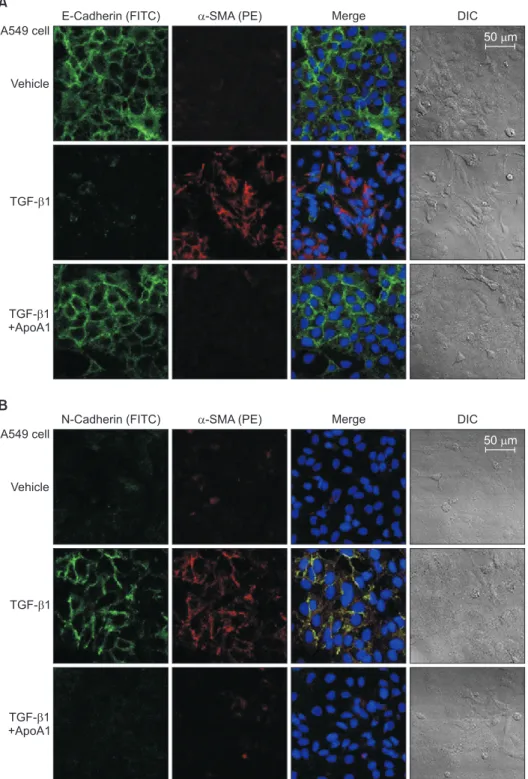
Figure 2. ApoA1 inhibits TGF-β1–in- duced EMT in A549 cells. A549 cells were treated with TGF-β1 (5 ng/mL) with or without ApoA1 (100 ng/mL) for 48 hours.
Expressions of E-cadherin (green) (A), N- cadherin (green) (B), and α-SMA (red) were analyzed by immunofluorescence staining. Treatment with TGF-β1 de- creased the expression of the epithelial marker, E-cadherin, and increased the expression of mesenchymal markers such as N-cadherin and α-SMA. ApoA1 inhibited TGF-β1–induced changes in EMT markers similar to control levels (A and B, ×400). ApoA1: apolipoprotein A1;
TGF-β1: transforming growth factor β1;
EMT: epithelial-to-mesenchymal transi- tion; α-SMA: α-smooth muscle actin;
DIC: differential interference contrast.
cadherin, E-cadherin, and α-smooth muscle actin (α-SMA).
Smad and non-Smad pathway related molecules and TGF-β1 receptor protein (type I [TβRI] and II [TβRII]) expression were measured by western blot in the cell.
2. Silica-induced lung fibrosis mouse model
Male ApoA1 overexpressing transgenic mice (6–8 weeks old) were treated with silica intratracheally following our previously described method
16. On day 0, the transgenic mice received 20 mg of sterile silica crystals (median diameter, 1–5 μm; Sigma-Aldrich, St. Louis, MO, USA) in endotoxin-free water in a total volume of 100 μL by intratracheal delivery.
ApoA1 overexpressing transgenic and ApoA1 non-overex- pressing wild type mice were housed and sacrificed on day 30.
Lung sections were subjected to immunofluorescence stain- ing with confocal microscopy (LSM 510 META; Carl Zeiss Microsystems).
3. Western blot analysis and immunofluorescence staining For western blot analysis, A549 cells were prepared by ex- tracting proteins with RIPA buffer (50 mM Tris-HCl [pH 7.4], 1% [V/V] NP-40, 150 mM NaCl, and 1 mM ethylenediamine- tetraacetic acid) supplemented with protease inhibitors (1 mM phenylmethylsulfonyl fluoride, 1 μM aprotinin, 1 μg/mL leupeptin, and 1 mM Na
3VO
4). Lung tissue was homogenized in RIPA buffer containing protease inhibitors. Equal amounts of proteins were resolved using 10% sodium dodecyl sulfate polyacrylamide gel electrophoresis and transferred onto nitrocellulose membranes (Amersham Pharmacia Biotech Inc., Piscataway, NJ, USA). After blocking with non-fat dried milk, the membranes were incubated with primary antibodies for 1 hour at room temperature followed by detection using horseradish peroxidase conjugated secondary antibodies.
Enhanced chemiluminescence detection was performed according to the manufacturer’s instructions (Boehringer Mannheim, Mannheim, Germany). The relative abundance of protein was determined by quantitative densitometry using Image J software (NIH, Bethesda, MD, USA). All western blot
A
TGF- 1 ApoA1 E-cadherin
N-cadherin
-SMA
-Actin
130 kDa
130 kDa
37 kDa
42 kDa 6 12 24 48 6 12 24 48 (hr)
(hr)
B
12 24 48 1.2
1.0
0.8
0.6
0.4
0.2 E-cadherin/-actin proteinexpressionratio
6 6 12 24 48
TGF- 1 ApoA1
(hr)
C
12 24 48 2.5
2.0
1.5
1.0
0.5 N-cadherin/-actin proteinexpressionratio
6 6 12 24 48
TGF- 1 ApoA1
(hr)
D
12 24 48 5
4
3
2
1
-SMA/-actin proteinexpressionratio
6 6 12 24 48
TGF- 1 ApoA1
* *
*
* * *
*
*
*
* *
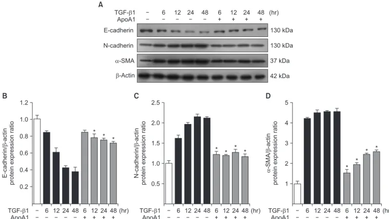
Figure 3. Effects of ApoA1 on EMT-related markers in A549 cells. A549 cells were incubated with TGF-β1 (5 ng/mL) in the absence of serum with or without ApoA1 (100 ng/mL) for up to 48 hours. Stimulation of cells by TGF-β1 down-regulated the epithelial marker, E-cadherin, and up-regulated the mesenchymal markers such as N-cadherin and α-SMA in a time dependent manner. β-Actin was used as a loading control (A). Densitometric analysis of band intensities for E-cadherin (B), N-cadherin (C), and α-SMA band (D). Each bar represents mean±standard error of at least three independent experiments. *p<0.05 versus same time of the TGF-β1–treated group. ApoA1: apolipoprotein A1; EMT:
epithelial-to-mesenchymal transition; TGF-β1: transforming growth factor β1; α-SMA: α-smooth muscle actin.
densitometry data were normalized to β-actin.
For immunofluorescence analysis, mouse lung tissue was incubated at 4
oC overnight with anti E-cadherin (1:500, Santa Cruz Biotechnology Inc., Santa Cruz, CA, USA) and N- cadherin antibodies (1:1,000, BD Bioscience, Bedford, MA, USA). Fluorescein isothiocyanate-conjugated donkey anti- goat antibodies (1:1,000, Santa Cruz Biotechnology Inc.) and goat anti-rabbit IgG-PE (1:1,000, Santa Cruz Biotechnology Inc.) were used as secondary antibodies for the localization of E-cadherin and N-cadherin in the mouse lung.
4. Antibodies
The antibodies used in the present study included anti- human E-cadherin (1:500, Santa Cruz Biotechnology Inc.), anti-human N-cadherin (1:1,000, BD Bioscience), anti-α-SMA
(1:300, Abcam, Cambridge, UK), anti–β-actin (1:5,000, Sigma- Aldrich), anti-Smad3, anti-pSmad3 (1:1,000, Cell Signaling Technology, Danvers, MA, USA), anti-ERK1/2, anti-pERK1/2 (1:1,000, Cell Signaling Technology), anti-p38 mitogen- activated protein kinase (MAPK), anti-pp38 MAPK (1:1,000, Cell Signaling Technology), anti-TβRI (1:1,000, Santa Cruz Biotechnology Inc.), and anti-TβRII (1:500, Abcam).
5. Statistical analysis
All data are expressed as mean±standard error of the mean.
Data were analyzed using the Kruskal-Wallis test followed by the Mann-Whitney U-test with Bonferroni correction for inter- group comparisons. A p-value less than 0.05 was deemed to indicate statistical significance.
A
E-Cadherin (FITC) Pro-SPC (PE) Merge DIC
WT Silica (D30)
ApoA1 TG +Silica
(D30) WT Silica (D30)
10 m
x400
B
N-Cadherin (FITC) Pro-SPC (PE) Merge DIC
WT Silica (D30)
ApoA1 TG +Silica
(D30) WT Silica (D30)
10 m
x400
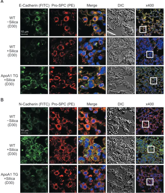
Figure 4. Overexpression of ApoA1 at- tenuates silica-induced EMT in the mouse lung. Immunofluorescence staining for E-cadherin (green) (A), N- cadherin (green) (B), and pro-SPC (red) in the mouse lung. Treatment with silica decreased the expression of the epithe- lial marker, E-cadherin, and increased the expression of the mesenchymal markers such as N-cadherin and α-SMA in the WT mouse lung. Overexpression of ApoA1 inhibited TGF-β1–induced changes in EMT markers similar to silica non-treated WT levels (first-fourth columns, ×1,000; fifth columns, ×400).
ApoA1: apolipoprotein A1; EMT: epi- thelial-to-mesenchymal transition; SPC:
surfactant protein C; α-SMA: α-smooth muscle actin; WT: wild type; TGF-β1:
transforming growth factor β1; DIC: dif-
ferential interference contrast; TG: trans-
genic.
Results
1. ApoA1 inhibits EMT in the TGF-β1–treated AEC and silica-induced lung fibrosis models
As previously reported, in response to TGF-β1, A549 cells were converted from an epithelial phenotype to a spindle-like mesenchymal phenotype, and treatment with ApoA1 restored the cells to the traditional polygonal epithelial shape (Figure 1). As expected, exposing the cells to TGF-β1 for 48 hours showed a reduced expression of the epithelial marker (E-cad- herin) with increased expression of the mesenchymal marker (N-cadherin and α-SMA). These phenotypic changes were inhibited by treatment of ApoA1 (Figures 2, 3). Silica-treated mouse lung showed decreased E-cadherin and increased N- cadherin expression in the alveolar epithelium, shown as the pro-surfactant protein C, and overexpression of ApoA1 inhib- ited the changes in the epithelial and mesenchymal markers in the alveolar epithelium (Figure 4). These findings demon- strate that ApoA1 effectively inhibits both TGF-β1–induced EMT in the AEC and silica-induced EMT in vivo.
2. ApoA1 inhibits TGF-β1–induced Smad-dependent and non-Smad signaling pathways
We determined whether ApoA1 interferes with the TGF-β1 signaling pathways. We examined whether the inhibitory ef- fects of ApoA1 against TGF-β1–induced EMT in A549 cells are mediated by changing the phosphorylated Smad2 or Smad3 expression, which have been demonstrated to be the major signal pathways in TGF-β1–induced EMT
17.
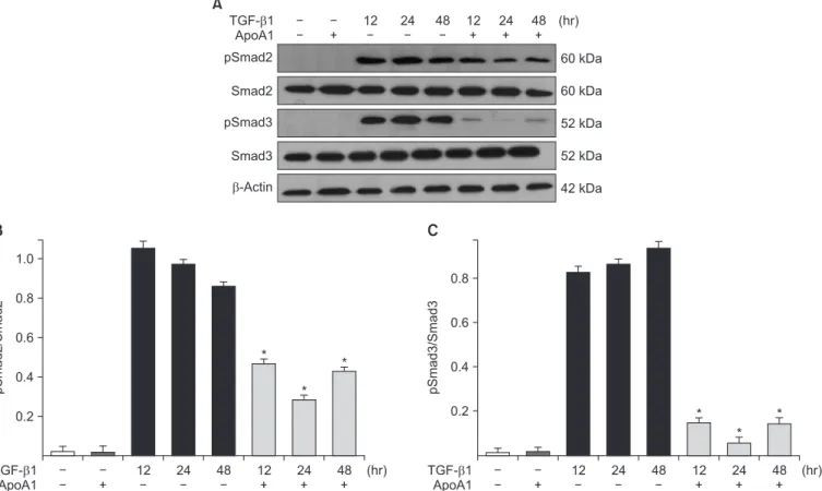
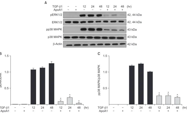
Phosphorylation of Smad2 and Smad3 in A549 cells was markedly increased in a time-dependent manner by TGF-β1 treatment. When the cells were treated with ApoA1, phos- phorylation of Smad2 and Smad3 were significantly reduced (Figure 5). This finding suggests that ApoA1 inhibits the Smad-dependent TGF-β1 signaling pathway. TGF-β1 also induced non-Smad responses, including ERK and p38 MAPK mediated signaling
18. ApoA1 also reduced the phosphoryla- tion of ERK and p38 MAPK induced by TGF-β1 (Figure 6). It appears that ApoA1 down-regulates both Smad-dependent and non-Smad signaling pathways induced by TGF-β1.
A
TGF- 1 ApoA1 pSmad2
Smad2
pSmad3
-Actin
60 kDa
60 kDa
52 kDa
52 kDa
42 kDa 12 24 48 12 24 48 (hr)
Smad3
B
1.0
0.8
0.6
0.4
0.2
pSmad2/Smad2
(hr)
12 24 48 12 24 48
TGF- 1 ApoA1
C
0.8
0.6
0.4
0.2
pSmad3/Smad3
(hr)
12 24 48 12 24 48
TGF- 1 ApoA1
*
*
*
*
*
*
Figure 5. ApoA1 down-regulates the TGF-β1–induced Smad dependent signaling pathway. A549 cells were incubated with TGF-β1 (5 ng/
mL) with or without ApoA1 (100 ng/mL) for 48 hours. (A) Phosphorylation of Smad2 and Smad3 occurred after TGF-β1 stimulation and co-
treatment with ApoA1 down-regulated the TGF-β1–induced Smad signaling pathway. β-Actin was used as a loading control. Densitometric
analysis of band intensities for phosphorylated Smad2 (B) and Smad3 (C). Each bar represents mean±standard error of at least three indepen-
dent experiments. *p<0.05 versus same time of the TGF-β1–treated group. ApoA1: apolipoprotein A1; TGF-β1: transforming growth factor β1.
3. ApoA1 down-regulates TGF-β1 receptor expression TGF-β1 signaling is initiated by the binding of TGF-β1 to TβRI and TβRII on the epithelial cell membrane
19. Increases in the TβRI and TβRII protein expression levels were observed in A549 cells upon TGF-β1 treatment, whereas the addition of ApoA1 significantly reduced the expression of these proteins (Figure 7).
Discussion
In this study, we have shown that ApoA1 effectively inhibits TGF-β1–induced EMT, both in vitro and in vivo. ApoA1 re- markably reduced TGF-β1–induced expression of mesenchy- mal markers such as α-SMA and N-cadherin and recovered the expression of the epithelial marker E-cadherin in AECs (Figures 2, 3). We also observed that ApoA1 overexpression inhibits silica-induced EMT in the mouse model (Figure 4).
In the currently accepted paradigm, the main pathogenesis of IPF is thought to be recurrent AEC injury that leads to aber-
rant activation of AEC, creating the accumulation of collagen producing fibroblasts and myofibroblasts
20,21. Although the significance and relative contribution of EMT to the source of activated fibroblasts and myofibroblasts has not been definite- ly established, EMT has been increasingly proposed as one of the main mechanisms of excessive fibroblast proliferation in lung fibrosis
22,23. For the induction of EMT in vitro and in vivo, TGF-β1 is regarded as the prototypical cytokine
24. TGF-β1–in- duced EMT starts with the activation of latent TGF-β1, which originally existed in the extracellular mileu, kept inactive by the latency-associated protein, and bound by the latent TGF binding protein. Then, TGF-β1 binds with the TβRII and TβRI, activating the receptor heterodimer and initiating the intracel- lular Smad and non-Smad signal pathways
24.
TGF-β1 signaling occurs via both Smad dependent and non-dependent pathways
25. The Smad dependent signal transduction system is the main process in the TGF-β1 sig- naling. When the extracellular TGF-β ligand binds to the cell membrane TGF-β receptor, the receptor-activated Smads (R- SMADS), Smad2 and 3, are phospholylated directly by the in- A
TGF- 1 ApoA1 pERK1/2
ERK1/2
pp38 MAPK
-Actin
42, 44 kDa
42, 44 kDa
43 kDa
43 kDa
42 kDa 12 24 48 12 24 48 (hr)
p38 MAPK
B
1.5
1.0
pERK/ERK 0.5
(hr)
12 24 48 12 24 48
TGF- 1 ApoA1
*
*
*
C
1.5
1.0
0.5
pp38MAPK/p38MAPK
(hr)
12 24 48 12 24 48
TGF- 1 ApoA1
* *
*
Figure 6. ApoA1 down-regulates the TGF-β1–induced non-Smad signaling pathways. A549 cells were incubated with TGF-β1 (5 ng/mL) with
or without ApoA1 (100 ng/mL) for 48 hours. (A) Phosphorylation of ERK1/2 and p38 MAPK occurred after TGF-β1 stimulation and co-treat-
ment with ApoA1 down–regulated the TGF-β1–induced non-Smad signaling pathways. β-Actin was used as a loading control. Densitometric
analysis of band intensities for phosphorylated ERK1/2 (B) and p38 MAPK (C). Each bar represents mean±standard error of at least three
independent experiments. *p<0.05 versus same time of the TGF-β1–treated group. ApoA1: apolipoprotein A1; TGF-β1: transforming growth
factor β1; MAPK: mitogen-activated protein kinase.
tracellular TβRI kinase domain, and then bind to the common mediator Smad (Co-SMAD), Smad4. These Smad proteins translocate into the nucleus, bind with Smad-binding ele- ments as a transcription factor complex, and activate TGF-β1 target genes (e.g., α-SMA and collagen), as well as inhibiting epithelial genes (e.g., E-cadherin) through the co-association of various transcription factors
17,24. For the full TGF-β1 re- sponse, phosphoinositide 3-kinase/Akt or MAPK-targeted transcription factors bind to the critical response element of the TGF-β target genes in the Smad independent pathway.
The ERK/MAPK cascade additionally phosphorylates R- SMADS, modifying Smad activity and enhances TGF-β1–
mediated collagen I synthesis
26. Besides such interaction with the Smad pathway in modulation and activation of transcrip- tion factors, non-Smad proteins are also involved in a variety of cellular responses, including cellular tight/adheren junction disassembly, and cytoskeletal rearrangement
24,25.
Each step in the TGF-β1–induced EMT signal pathway has been considered as a possible, effective therapeutic target for lung fibrosis
27. Several studies to identify the proper therapeu- tic target in the TGF-β1–induced EMT signal pathway have been completed or are in progress
22,28-31. However, currently, there is no therapeutic agent that directly inhibits TGF-β1, due to its complex function and the expected side effects of direct
TGF-β1 inhibition
28.
In this study, treatment of ApoA1 attenuated TGF-β1–in- duced Smad2 and Smad3 phosphorylation in A549 cell (Fig- ure 5). Moreover, ApoA1 also inhibited non-Smad signaling, particularly the ERK and MAPK pathways (Figure 6). Since both Smad-dependent and non-Smad pathways were inhib- ited by ApoA1 in our study, we evaluated further whether ApoA1 modulates TGF-β receptor expression. Rojas et al.
32reported that the TGF-β receptor levels regulate the specific- ity of the signaling pathway activation and the biologic effects of TGF-β1 and, in particular, that the TβRII expression levels are correlated with Smad signaling and MAPK-ERK signal- ing activation. We have shown that ApoA1 inhibits both TβRI and TβRII expression induced by TGF-β1 (Figure 7). The exact molecular mechanism by which ApoA1 modulates the expression of TGF-β receptors remains to be determined. We speculate that ApoA1 interferes with the binding or interac- tion between the TGF-β1 ligand and the TβRI-TβRII complex, resulting in down-regulation of downstream signaling mol- ecules.
We examined the anti-EMT activity of ApoA1 through the murine model of silica-induced lung fibrosis using a previ- ously reported method
16. ApoA1 transgenic mice and a silica-induced lung fibrosis model were used because even A
TGF- 1 ApoA1 T RI T RII
-Actin
53 kDa
63 kDa
42 kDa 12 24 48 12 24 48 (hr)
B
0.8
0.4
TRI/-actin
(hr)
12 24 48 12 24 48
TGF- 1 ApoA1
*
* *
C
1.2
0.8
0.4
(hr)
12 24 48 12 24 48
TGF- 1 ApoA1
*
* *
TRII/-actin
1.2