The Effect of Statin on
Epithelial-Mesenchymal Transition
in Peritoneal Mesothelial Cells
Tae Ik Chang
Department of Medicine
The Effect of Statin on
Epithelial-Mesenchymal Transition
in Peritoneal Mesothelial Cells
Tae Ik Chang
Department of Medicine
The Effect of Statin on
Epithelial-Mesenchymal Transition
in Peritoneal Mesothelial Cells
Directed by Professor Shin-Wook Kang
The Doctoral Dissertation
submitted to the Department of Medicine
the Graduate School of Yonsei University
in partial fulfillment of the requirements for the degree
of Doctor of Philosophy
Tae Ik Chang
This certifies that the Doctoral Dissertation
of Tae Ik Chang is approved.
---
Thesis Supervisor: Shin-Wook Kang
---
Thesis Committee Member#1: Hyeon Joo Jeong
---
Thesis Committee Member#2: Hyeong-Cheon Park
---
Thesis Committee Member#3: Hyunjin Noh
---
Thesis Committee Member#4: Seung Hyeok Han
The Graduate School
Yonsei University
ACKNOWLEDGEMENTS
My special gratitude goes to my mentor Prof. Shin-Wook
Kang who taught me nephrology and the right attitude for my
spiritual development and medical practice. Also, I wish to
express my appreciation to Prof. Hyeon Joo Jeong, Prof.
Hyeong-Cheon Park, Prof. Hyunjin Noh, and Prof. Seung
Hyeok Han who generously offered guidance and supports.
I really appreciate Dong Ki Kim, Hyun-Wook Kim, Jae
Hyun Chang, Jung Tak Park, Sung Jin Moon, Dong Hyung
Lee, Hyung Jung Oh, Dong Eun Yoo, Seung Jun Kim, and
Prof. Tae-Hyun Yoo for giving me big courage to the end. And
also give thanks to Sun Ha Lee, Bo Young Nam, Do Hee Kim,
Seung Jae Kwak, Hye-Young Kang, and Jisun Paeng. Without
their help, I could not dream of this thesis.
I would like to thank my parents and parents-in-law. They
have always encouraged me with love. Above all things, I
want to confess my love to my wife, Ye Jin Yang, who has
assisted me with devotion and has always been beside me with
great love. Finally, I send my gratitude separately to my lovely
daughter, Eun Chae, and my lovely son, Eun Heon.
<TABLE OF CONTENTS>
ABSTRACT ··· 1
I. INTRODUCTION ··· 4
II. MATERIALS AND METHODS ··· 7
1. Isolation of HPMCs ··· 7
2. HPMCs experiments ··· 7
3. Evaluation of small GTPase activation ··· 8
4. Animal studies ··· 9
5. Western blot analysis ··· 10
6. Immunofluorescence staining ··· 10
7. Immunohistochemical and Masson’s trichrome staining ··· 10
8. Statistical analysis ··· 12
III. RESULTS ··· 13
1. Cultured HPMCs studies ··· 13
A. Effects of simvastatin on EMT and fibronectin expression in
HPMCs ··· 13
B. Activation of small GTPases such as RhoA and Rac1 in HPMCs
··· 18
C. Involvement of isoprenoids of the mevalonate pathway in EMT
of HPMCs ··· 21
D. Effect of small GTPase inhibitors on EMT and fibronectin
expression in HPMCs ··· 23
2. Animal studies ··· 25
A. Effects of simvastatin on peritoneal EMT and ECM accumulation
in a PD rat model ··· 25
B. Immunohistochemical and Masson’s trichrome staining ··· 27
IV. DISCUSSION ··· 30
REFERENCES ··· 37
ABSTRACT (IN KOREAN) ··· 42
LIST OF FIGURES
Figure 1. MTT assay for cell viability ··· 15
Figure 2. Effects of simvastatin on EMT and fibronectin
expression in HPMCs ··· 16
Figure 3. Morphologic changes under an inverted
phase-contrast microscope in HPMCs exposed to 5.6 mM
glucose (NG), NG + mannitol (94.4 mM, NG + M), NG + 1
µM simvastatin (NG + statin), high glucose (100 mM, HG),
or HG + 1 µM simvastatin (HG + statin) ··· 17
Figure 4. RhoA1 and Rac1 protein expression in the
membrane and cytosol fractions of HPMCs exposed to 5.6
mM glucose (NG), NG + mannitol (94.4 mM, NG + M), NG
+ 1 µM simvastatin (NG + statin), high glucose (100 mM,
HG), or HG + 1 µM simvastatin (HG + statin) ··· 20
Figure 5. The protein expression of EMT markers and
fibronectin in HPMCs ··· 22
Figure 6. The protein expression of EMT markers and
fibronectin in HPMCs exposed to 5.6 mM glucose (NG), NG
with 1 µM Y27632 (Rho/ROCK inhibitor) (NG + Y27632),
NG with 1 µM EHT1864 (Rac1 inhibitor) (NG + EHT1864),
high glucose (100 mM, HG), HG with 1 µM Y27632 (HG +
Y27632) or HG with 1 µM EHT1864 (HG + EHT1864) ··· 24
Figure 7. The protein expression of EMT markers and ECM in
the peritoneum of control (C), C+ simvastatin (C + statin),
4.25% PDF instillation (PD), or 4.25% PDF + simvastatin
(PD + statin) rats ··· 26
Figure 8. Immunohistochemical staining of the peritoneum of
control (C), C+ simvastatin (C + statin), 4.25% PDF
instillation (PD), or 4.25% PDF + simvastatin (PD + statin)
rats ··· 28
Figure 9. Masson’s trichrome staining of the peritoneum of
control (C), C+ simvastatin (C + statin), 4.25% PDF
instillation (PD), or 4.25% PDF + simvastatin (PD + statin)
rats ··· 29
1
ABSTRACT
The Effect of Statin on Epithelial-Mesenchymal Transition
in Peritoneal Mesothelial Cells
Tae Ik Chang
Department of Medicine
The Graduate School, Yonsei University
(Directed by Professor Shin-Wook Kang)
Background: Statins have recently been highlighted due to their pleiotropic actions besides cholesterol-lowering effects. However, it is currently unknown whether statin therapy may inhibit peritoneal dialysis (PD)-related epithelial-mesenchymal transition (EMT).
Methods: In vitro, human peritoneal mesothelial cells (HPMCs) were exposed to 5.6 mM glucose (NG) or 100 mM glucose (HG) with or without simvastatin (1 µM). In vivo, PD catheters were inserted into 32 Sprague-Dawley rats, and
2
infused for 4 weeks. Eight rats from each group were treated with 5 mg/kg/day of simvastatin intraperitoneally. Changes in the protein expression of EMT markers such as E-cadherin, α-SMA, Snail, and fibronectin in HPMCs and the peritoneum were evaluated by Western blot analysis and immunofluorescence or immunohistochemical staining. I also explored whether activation of mevalonate pathway and its downstream small GTPases were involved in dialysis-related peritoneal EMT and could be inhibited by statin treatment. Results: Compared to NG cells, E-cadherin expression was significantly decreased, while α-SMA, Snail, and fibronectin expression were significantly increased in HPMCs exposed to HG, and these changes were abrogated by simvastatin (p<0.05). In addition, the cobblestone-like appearance of normal HPMCs was converted into a fibroblast-like morphology after HG treatment, which was reversed by simvastatin. These EMT-like changes were also observed in HPMCs treated with geranyl-geranyl pyrophosphate (5 µM). HG significantly increased the protein expression of RhoA and Rac1 in the membrane fractions, and these increases were ameliorated by simvastatin (p<0.05). In PD rats, E-cadherin in the peritoneum was significantly decreased, whereas α-SMA, Snail, and fibronectin expression were significantly increased (p<0.05) compared to C rats. The thickness of the mesothelial layer in the peritoneum were also significantly greater in PD rats than in C rats (p<0.05). These changes of the peritoneum in PD rats were significantly attenuated by simvastatin.
3
Conclusions: This study demonstrated that PD-related EMT was mediated via the mevalonate pathway, and statin treatment inhibited the EMT changes in HG-treated HPMCs and PDF-stimulated PD rats. These findings suggest that statins may be a promising therapeutic strategy for preservation of peritoneal membrane integrity in long-term PD patients.
---
Key words:
statin, peritoneal mesothelial cells, epithelial-mesenchymal transition4
The Effect of Statin on Epithelial-Mesenchymal Transition
in Peritoneal Mesothelial Cells
Tae Ik Chang
Department of Medicine
The Graduate School, Yonsei University
(Directed by Professor Shin-Wook Kang)
I. INTRODUCTION
Even though peritoneal dialysis (PD) is generally accepted as an established modality for the management of patients with end-stage renal disease (ESRD), a concern about peritoneal membrane failure has consistently been raised in long-term PD. Many factors are demonstrated to be involved in the development of peritoneal dysfunction. In particular, nonphysiologic nature of
5
PD solutions; high concentrations of glucose and lactate, low pH, and glucose degradation products is a major factor responsible for the deleterious effect on peritoneal membrane.1,2 These factors also induce chronic inflammation in the peritoneal cavity, which is often exacerbated by recurrent episodes of peritonitis, consequently leading to structural and functional alterations of the peritoneal
membrane.3
Peritoneal fibrosis (PF) is the ultimate form of peritoneal damage. It is characterized by the loss of peritoneal mesothelial cell (PMC) monolayer, submesothelial fibrosis, angiogenesis, and hyalinizing vasculopathy.3-6 In the past, resident stromal fibroblasts and inflammatory cells had been considered to
be the main cells responsible for PF.7,8 Recently, however, PMCs have emerged
as an active player in the alteration of the peritoneal membrane. After PD start, PMCs progressively lose their epithelial characteristics and acquire myofibroblast-like phenotype through the process of epithelial-mesenchymal
transition (EMT).5 EMT is a normal physiologic process during embryo
implantation, embryogenesis, or organ development, but it is also involved in various pathologic processes, including cancer metastasis and fibrotic
disorders.7 Indeed, EMT enables PMCs to gain migratory and invasive
capacities, thus they can intrude into the substromal stroma and produce extracellular matrix (ECM) components such as fibronectin and collagen, which ultimately lead to PF.8
6
reductase inhibitors or statins have recently been highlighted in numerous aspects due to their pleiotropic effects besides lipid-lowering property.9-11 Of note, one of the key actions of statins is the inhibition of the downstream products of the mevalonate pathway such as farnesyl pyrophosphate (FPP) and
geranyl-geranyl pyrophosphate (GGPP).12 In result, isoprenylation of small
RhoGTPases and Ras, the final products of this pathway, is inhibited by statins.13-15 Interestingly, previous studies have found that activation of small RhoGTPases such as RhoA, Rac1, and Cdc42 plays a key role in the process of EMT implicated in diverse renal diseases.16-19 In addition, a recent study by
Zhang et al.20 showed that RhoA/ROCK signaling pathway mediated EMT in
rat PMCs in response to transforming growth factor (TGF)-β1. These findings suggest that statins may reverse EMT-like changes through the inhibition of isoprenylation of small RhoGTPases. However, to my knowledge, this assumption has not yet been tested. In this study, therefore, I investigated the effect of statins on PD-related EMT both in vitro and in vivo.
7 II. MATERIALS AND METHODS
1. Isolation of human PMCs (HPMCs)
HPMCs were isolated according to the method described by Stylianou et al.21. Briefly, a piece of human omentum, obtained from consenting patients who underwent elective abdominal surgery, was washed three times with sterile phosphate-buffered saline (PBS) and incubated in 0.05% trypsin-0.02% ethylenediaminetetraacetic acid (EDTA) solution for 20 min at 37°C with continuous shaking. After incubation, the suspension containing free HPMCs was centrifuged at 100 × g for 10 min at 4°C. The cell pellet was then washed once and re-suspended in M199 medium supplemented with 10% fetal bovine serum (FBS), 100 U/ml penicillin, 100 mg/ml streptomycin, and 26 mM NaHCO3, and seeded onto culture dishes. The cells were grown in the same medium at 37°C in humidified 5% CO2 in air, and the medium was changed 24 hr after seeding, and then every 3 days.
2. HPMCs experiments
Subconfluent HPMCs were serum-restricted for 24 hr, and the medium was then changed to serum-free M199 medium containing normal glucose (5.6 mM, NG), NG + mannitol (94.4 mM, NG + M), NG + simvastatin (1 µM) (Sigma Chemical Co., St Louis, MO, USA), or high glucose (100 mM, HG) with or without simvastatin (1 µM). The dose of simvastatin used in the experiments
8
was determined using 3-(4,5-dimethylthiazol-2-yl)-2,5-diphenyltetrazolium bromide (MTT) cell viability assay. To explore whether isoprenoids of the mevalonate pathway were involved in peritoneal EMT, HPMCs were treated
with NG + GGPP (5 µM) (SigmaChemical Co.). HPMCs exposed to HG were
also treated with Rho/ROCK inhibitor (Y27632, 1 µM) (Sigma Chemical Co.) or Rac inhibitor (EHT1864, 1 µM) (R&D Systems, Minneapolis, MN, USA). At 72 hr after the media change, cells were harvested and conditioned media were collected.
3. Evaluation of small GTPase activation
To examine the activation of small GTPases, membrane and cytosol proteins were prepared separately and the expression of RhoA and Rac1 were determined in each fraction by Western blotting. Briefly, HPMCs treated as above were washed with cold PBS and lysed by freeze-thawing in ice-cold lysis buffer containing 50 mM HEPES (pH 7.4), 5 mM NaCl, 1 mM MgCl2, 2 mM
EDTA, 1 mM dithiothreitol, 10 mM sodium fluoride, 1 mM
phenylmethylsulfonyl fluoride, 10 μg/ml aprotonin, and 10 μg/ml leupeptin (Sigma Chemical Co.). The homogenates were centrifuged at 4°C and 100,000 × g for 30 min and the resulting supernatant (cytosolic fraction) was collected. The pellets were then homogenized in the same lysis buffer containing 2% Triton X-114 and centrifuged at 800 × g for 10 min at 4°C, and the supernatant was collected. This supernatant was referred to membrane fraction. Moreover,
9
the activity of Rho-kinase was determined by using the colorimetric G-LISA RhoA activation assay biochemical kit (Cytoskeleton, Denver, CO, USA)
according to manufacturer’s protocol as previously described.22
4. Animal studies
All animal studies were conducted under an approved protocol. Peritoneal access ports were inserted in 32 male Sprague-Dawley rats weighing 250-280 g, and 2 ml of saline with 1 IU/ml heparin was instilled intraperitoneally until wound healing. One week after surgery, 16 rats received a daily (once per day) 20 ml of saline instillation and 16 rats were instilled daily with 20 ml of 4.25%
peritoneal dialysis fluid (PDF, Dianeal®, Baxter Healthcare Ltd., Singapore) for
4 weeks. Eight rats from each group were treated with simvastatin (5 mg/kg per day) intraperitoneally, while 8 rats in each group were left untreated (control). After 4 weeks of PD, the abdomen was opened by a midline incision and the entire anterior abdominal wall was removed at the contralateral side to the tip of the implanted catheter. One fifth of the whole tissue adjacent to the liver was fixed in 10% neutral-buffered formalin for pathologic examination, while the parietal peritoneum dissected from the major part of the tissue was washed in ice-cold PBS, snap-frozen in liquid nitrogen, pulverized with a mortar and pestle while frozen, and suspended in SDS sample buffer [2% SDS, 10 mM Tris-HCl, pH 6.8, 10% (vol/vol) glycerol]. After a centrifugation at 16,000 × g for 15 min at 4°C, the supernatant was kept at -80°C until use.
10
5. Western blot analysis
The protein expression of E-cadherin (BD Biosciences, San Jose, CA, USA), Snail (Abcam, Cambridge, UK), α-SMA (Sigma Chemical Co.), fibronectin (DAKO, Glostrup, Denmark), RhoA (Santa Cruz Biotechnology, Inc., Santa Cruz, CA, USA), and Rac1 (Abcam) in HPMCs and peritoneal tissue were evaluated by Western blot as previously described.23 The band densities were measured using TINA image software (Raytest, Straubenhardt, Germany), and the changes in the optical densities of bands from the treated groups relative to NG cells or the peritoneum of control rats were used in the analysis.
6. Immunofluorescence staining
HPMCs grown on chamber slides were fixed in 4% paraformaldehyde for 10 min at 4°C, washed three times with PBS, and incubated with 1% BSA for 20 min at room temperature. For immunofluorescence staining, primary polyclonal antibodies to E-cadherin, Snail, α-SMA, RhoA, and Rac1 were diluted in 1:100 with antibody diluent (DAKO) and were applied for 3 hr at room temperature. After washing with PBS, Cy3 (red)- or Cy2 (green)-conjugated anti-rabbit IgG antibody (Research Diagnostics, Inc., Flanders, NJ, USA) was added for 60 min.
7. Immunohistochemical and Masson’s trichrome staining
11
processed in the standard manner, and 5 μm-thick sections of paraffin-embedded tissues were utilized for immunohistochemical staining. Slides were deparaffinized, hydrated in ethyl alcohol, and washed in tap water. Antigen retrieval was carried out in 10 mM sodium citrate buffer for 20 min using a Black & Decker vegetable steamer. Primary antibodies for E-cadherin, Snail, α-SMA, and fibronectin were diluted to the appropriate concentrations with 2% casein in bovine serum albumin (BSA) and then added to the slides, followed by an overnight incubation at 4°C. After washing, a secondary antibody was added for 20 min, and the slides were washed and incubated with a tertiary PAP complex for 20 min. Diaminobenzidine was added for 2 min and the slides were counterstained with hematoxylin. A semi-quantitative score of staining intensity was determined by examining at least 5 fields of the peritoneum in each section under × 400 magnification and by digital image analysis (MetaMorph version 4.6r5, Universal Imaging Corp., Downingtown, PA, USA). For Masson’s trichrome staining, 5 μm-thick sections of paraffin-embedded tissues were deparaffinized, hydrated in ethyl alcohol, washed in tap water, and re-fixed in Bouin’s solution at 56°C for 1 hr. After washing in running tap water for 10 min and staining with Weigert’s iron hematoxylin working solution for 10 min, the sections were stained with Biebrich scarlet-acid fuchsin solution for 15 min, followed by a 10-min wash. The slides were then differentiated in phosphomolybdic-phosphotungstic acid solution for 15 min, transferred to aniline blue solution and stained for 10 min,
12
and reacted with 1% acetic acid solution for 5 min. The thickness of the peritoneum, which was defined as the tissue between the mesothelial surface and the underlying muscle or parenchyma, was assessed as previously
described.24 Briefly, the maximal thickness of the peritoneum was measured in
three Masson’s trichrome-stained tissue sections per rats and five fields, the center of which included the area of maximal thickness, were examined under × 400 magnification. Areas and perimeter lengths of the peritoneum were obtained from drawn outlines and the average thickness was calculated from rectangular approximation based on the values for area and perimeter in each field of view.
8. Statistical analysis
All values are expressed as means standard errors of the mean (SEM). Statistical analyses were performed using the statistical package SPSS for Windows Ver. 11.0 (SPSS, Inc., Chicago, IL, USA). Results were analyzed using one-way ANOVA with a post hoc Bonferonni’s test for multiple comparisons. P-values < 0.05 were considered statistically significant.
13 III. RESULTS
1. Cultured HPMCs studies
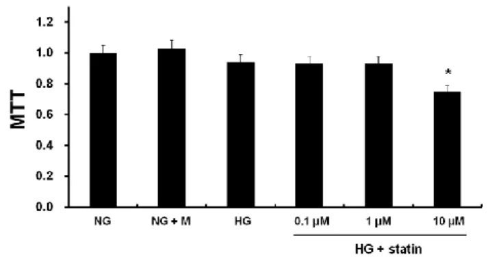
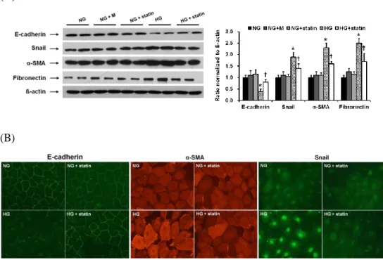
A. Effects of simvastatin on EMT and fibronectin expression in HPMCs As shown in Figure 1, MTT assay demonstrated that HPMCs remained viable at up to 1 µM of simvastatin, but the viability was decreased by 20% at 10 µM. Therefore, I determined to use the dose of 1 µM for the experiments. To evaluate the effects of statins on EMT in vitro, HPMCs were incubated for 72 hr with NG, NG + M, NG + simvastatin, or HG with or without simvastatin. E-cadherin protein expression was significantly lower, while the protein expression of Snail, α-SMA, and fibronectin were significantly higher in HG-stimulated HPMCs compared to NG cells (P < 0.05) (Fig. 2A). Furthermore, the changes in HPMCs exposed to HG were significantly abrogated by simvastatin treatment (P < 0.05) (Fig. 2A). These findings were corroborated by the immunofluorescence analysis. HPMCs cultured under HG medium showed a weak staining of E-cadherin, a strong signal intensity of α-SMA, and increased nuclear translocation of Snail, all of which were ameliorated by the administration of simvastatin (Fig. 2B). On the other hand, mannitol used as an osmotic control had no effect on EMT and fibronectin expression in HPMCs. In addition, the expression of EMT markers and fibronectin in NG cells was not affected by simvastatin.
14
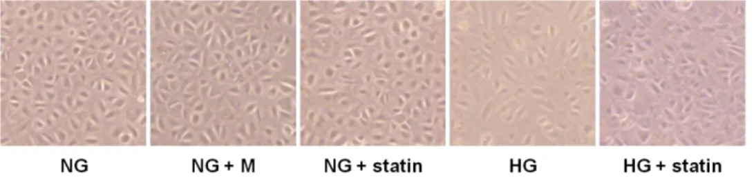
Moreover, I observed the morphologic changes of HPMCs under an inverted phase-contrast microscope. The cobblestone-like appearance of HPMCs was converted into a fibroblast-like morphology after HG treatment, which was reversed by simvastatin (Fig. 3).
15
Figure 1. MTT assay for cell viability. HPMCs were incubated for 72 hr with 5.6 mM glucose (NG), NG + mannitol (94.4 mM, NG + M), high glucose (100 mM, HG), or HG + 0.1 µM, 1 µM, or 10 µM simvastatin (HG + statin). Cell viability was maintained at up to 1 µM simvastatin, but was decreased by 20% at 10 µM. *; p<0.05 vs. 1 µM simvastatin.
16 (A)
(B)
Figure 2. Effects of simvastatin on EMT and fibronectin expression in HPMCs. (A) HPMCs were incubated for 72 hr with 5.6 mM glucose (NG), NG + mannitol (94.4 mM, NG + M), NG + 1 µM simvastatin (NG + statin), high glucose (100 mM, HG), or HG + 1 µM simvastatin (HG + statin) (A representative of five Western blots). E-cadherin protein expression was significantly lower, while the protein expression of Snail, α-SMA, and fibronectin were significantly higher in HG-stimulated HPMCs compared to NG cells, and these changes were significantly attenuated by simvastatin. *; p<0.05 vs. NG, †; p<0.05 vs. HG. (B) Compared to NG cells, HPMCs cultured under HG medium showed a weak staining of E-cadherin, a strong signal intensity of α-SMA, and increased nuclear translocation of Snail, all of which were ameliorated by the administration of simvastatin (× 400).
17
Figure 3. Morphologic changes under an inverted phase-contrast microscope in HPMCs exposed to 5.6 mM glucose (NG), NG + mannitol (94.4 mM, NG + M), NG + 1 µM simvastatin (NG + statin), high glucose (100 mM, HG), or HG + 1 µM simvastatin (HG + statin). The cobblestone-like appearance of HPMCs was converted into a fibroblast-like morphology 72 hr after HG treatment, which was reversed by simvastatin (× 40).
18
B. Activation of small GTPases such as RhoA and Rac1 in HPMCs
Posttranslational modification of Rho proteins by geranyl-geranlyation is essential for their membrane location and activity. Thus, the assessment of these proteins in the membrane fraction of the cells can reflect their degree of
prenylation through the mevalonate pathway.25,26 Since inhibiting isoprenylation
of the mevalonate pathway products was the main mechanism of statins, I evaluated the membrane-associated protein expression of RhoA and Rac1 in HPMCs after separation of the membrane and cytosol fractions by Western blot analysis. Compared to NG cells, the protein expression of RhoA and Rac1 were significantly increased in the membrane fraction of HG-stimulated HPMCs (P < 0.05), and simvastatin significantly abrogated the increases in RhoA and Rac1 expression in the membrane fraction of HPMCs exposed to HG (P < 0.05) (Fig. 4A). Furthermore, the immunofluorescence study revealed that HG provoked the translocation of RhoA and Rac1 from the cytosol to the membrane fraction, and simvastatin treatment inhibited this translocation of RhoA and Rac1 induced by HG (Fig. 4B). In addition, the levels of Rho kinase were significantly increased in HG-treated HPMCs than in NG cells (P < 0.05), and these changes were significantly ameliorated by simvastatin (P < 0.05) (Fig. 4C).
19 (A)
(B)
20
Figure 4. RhoA1 and Rac1 protein expression in the membrane and cytosol fractions of HPMCs exposed to 5.6 mM glucose (NG), NG + mannitol (94.4 mM, NG + M), NG + 1 µM simvastatin (NG + statin), high glucose (100 mM, HG), or HG + 1 µM simvastatin (HG + statin). (A) The protein expression of RhoA and Rac1 were significantly increased in the membrane fraction of HG-stimulated HPMCs compared to NG cells, and simvastatin significantly attenuated the increases in RhoA and Rac1 expression in the membrane fraction of HPMCs exposed to HG. *; p<0.05 vs. NG, †; p<0.05 vs. HG. (B) Immunofluorescence study revealed that HG provoked the translocation of RhoA and Rac1 from the cytosol to the membrane fraction, and simvastatin treatment inhibited this translocation of RhoA and Rac1 induced by HG (× 40). (C) The levels of Rho kinase were significantly increased in HG-treated HPMCs than in NG cells, and these changes were significantly abrogated by simvastatin. *; p<0.05 vs. NG, †; p<0.05 vs. HG.
21
C. Involvement of isoprenoids of the mevalonate pathway in EMT of HPMCs
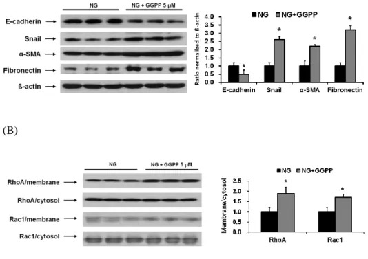
To evaluate whether isoprenoids of the mevalonate pathway were involved in peritoneal EMT, HPMCs were incubated with 5 µM GGPP for 72 hr. The administration of GGPP significantly decreased E-cadherin protein expression and significantly increased the protein expression of Snail, α-SMA, and fibronectin in HPMCs (P < 0.05) (Fig. 5A). The protein expression of RhoA and Rac1 were also significantly increased in the membrane fraction of HPMCs exposed to GGPP (P < 0.05) (Fig. 5B).
22 (A)
(B)
Figure 5. The protein expression of EMT markers and fibronectin in HPMCs. HPMCs were incubated with 5.6 mM glucose (NG) and NG + 5 µM GGPP (NG + GGPP) for 72 hr. (A) GGPP treatment significantly decreased E-cadherin expression and significantly increased the protein expression of Snail, α-SMA, and fibronectin in HPMCs (A representative of five Western blots). *; p<0.05 vs. NG. (B) The protein expression of RhoA and Rac1 were significantly increased in the membrane fraction of HPMCs exposed to GGPP. *; p<0.05 vs. NG.
23
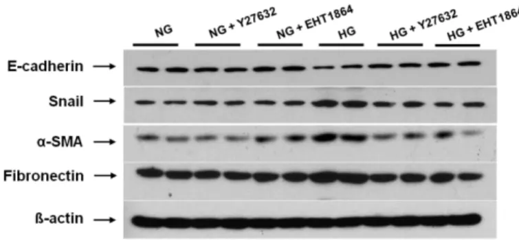
D. Effect of small GTPase inhibitors on EMT and fibronectin expression in HPMCs
I added Rho/ROCK inhibitor (Y27632) and Rac inhibitor (EHT1864) to HG-stimulated HPMCs, and determined the changes in EMT markers and fibronectin expression. The administration of these two small GTPase inhibitors significantly ameliorated the changes in EMT markers and fibronectin expression in HPMCs cultured under HG medium (P < 0.05) (Fig. 6).
24
Figure 6. The protein expression of EMT markers and fibronectin in HPMCs exposed to 5.6 mM glucose (NG), NG with 1 µM Y27632 (Rho/ROCK inhibitor) (NG + Y27632), NG with 1 µM EHT1864 (Rac inhibitor) (NG + EHT1864), high glucose (100 mM, HG), HG with 1 µM Y27632 (HG + Y27632), or HG with 1 µM EHT1864 (HG + EHT1864). The administration of Y27632 and EHT1864 significantly attenuated the changes in EMT markers and fibronectin expression in HPMCs cultured under HG medium. *; p<0.05 vs. NG, †; p<0.05 vs. HG, ‡; p<0.05 vs. HG.
25 2
. Animal studies
A. Effects of simvastatin on peritoneal EMT and ECM accumulation in a PD rat model
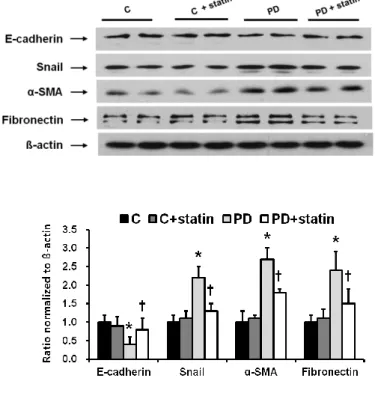
Finally, the effects of simvastatin on peritoneal EMT and ECM accumulation were explored in a PD rat model. E-cadherin protein expression was significantly lower, while Snail, α-SMA, and fibronectin protein expression were significantly higher in rats treated with 4.25% PDF compared to control rats (P < 0.01), and these changes were significantly abrogated by simvastatin treatment (P < 0.05) (Fig. 7).
26
Figure 7. The protein expression of EMT markers and ECM in the peritoneum of control (C), C+ simvastatin (C + statin), 4.25% PDF instillation (PD), or 4.25% PDF + simvastatin (PD + statin) rats. E-cadherin protein expression was significantly lower, while Snail, α-SMA, and fibronectin protein expression were significantly higher in rats treated with 4.25% PDF compared to control rats, and these changes were significantly ameliorated by simvastatin. *; p<0.05 vs. C, †; p<0.05 vs. PD.
27
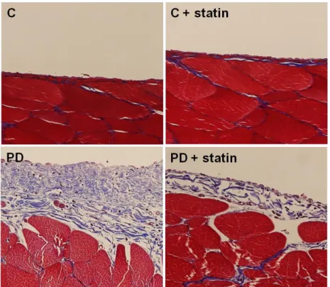
B. Immunohistochemical and Masson’s trichrome staining
Immunohistochemical staining of the peritoneum also revealed that EMT markers and fibronectin protein expression were significantly higher in rats treated with 4.25% PDF relative to control rats, and simvastatin significantly attenuated EMT and ECM accumulation in PD rats (Fig. 8). Moreover, Masson’s trichrome staining found that submesothelial layer was significantly thicker and peritoneal fibrosis was more extensive in PD rats with 4.25% PDF than control rats, and these changes were significantly abrogated by simvastatin treatment (Fig. 9).
28
Figure 8. Immunohistochemical staining of the peritoneum of control (C), C+ simvastatin (C + statin), 4.25% PDF instillation (PD), or 4.25% PDF + simvastatin (PD + statin) rats. The intensity of E-cadherin staining was significantly lower, while Snail, α-SMA, and fibronectin staining intensities were significantly higher in PD rats compared to C rats, and simvastatin significantly ameliorated these changes in PD rats (× 200).
29
Figure 9. Masson’s trichrome staining of the peritoneum of control (C), C+ simvastatin (C + statin), 4.25% PDF instillation (PD), or 4.25% PDF + simvastatin (PD + statin) rats. Peritoneal fibrosis assessed by Masson’s trichrome staining was significantly more extensive in PD rats with 4.25% PDF than C rats, and these changes were significantly attenuated by simvastatin treatment (× 200).
30 IV. DISCUSSION
PF is one of the most serious complications of long-term PD, leading to membrane failure. Even though resident peritoneal fibroblasts and infiltrating inflammatory cells have been considered to play a key role in the development PF, EMT of PMCs is recently highlighted as another potential mechanism responsible for PF.5-8 In this study, I show for the first time that statin treatment abrogates PD-related EMT of HPMCs and ECM accumulation in a PD rat model. In addition, I demonstrate that these beneficial effects of statin are mediated, at least in part, by inhibiting isoprenylation of small RhoGTPases such as RhoA and Rac1.
Besides a physiologic role of EMT in embryogenesis or organ development, it also plays a pathologic role in cancer metastasis and fibrotic disorders.7 A number of recent studies have found that PMCs also undergo EMT during
PD.5,27-29 In particular, Yanez-Mo et al.5 showed that PMCs underwent a
transition from an epithelial phenotype to a mesenchymal phenotype soon after PD was initiated along with a decrease in the expression of cytokeratins and E-cadherin, suggesting that these cells indeed acquire structural changes during PD. Consistent with these findings, in my study, E-cadherin expression was significantly decreased, while Snail, a-SMA, and fibronectin expression were significantly increased in HPMCs exposed to HG and in the peritoneum of rats instilled with 4.25% PDF. Furthermore, the cobblestone-like appearance of
31
normal HPMCs was converted into a fibroblast-like morphology after HG treatment. These findings support previous evidence of EMT of PMCs under pathologic conditions.
HMG-CoA reductase inhibitors or statins are potent inhibitors of cholesterol biosynthesis and have emerged as the leading lipid-lowering agents. However, it has been acknowledged that the beneficial effects of statins are not mediated solely by their lipid-lowering property but also through effects separate from inhibiting cholesterol synthesis, known as “pleiotropic” effects.9-11 In fact, statins exert these effects by preventing the synthesis of other important isoprenoids of cholesterol biosynthetic pathway, such as FPP and GGPP that are
downstream of the mevalonate pathway.12 These intermediates play key roles in
the post-translational modification of many proteins, including small GTP binding proteins; the family of Ras, Rho, Rap, and Rab GTPase, by serving as
lipid attachments through a process known as “prenylation”.13-15 Isoprenylation
of these proteins permits the covalent attachment, subcellular localization, and intracellular trafficking of membrane-associated proteins.13-15 Therefore, small G proteins are anchored into the cell membrane if they are prenylated, while they remain in the cytoplasm when prenylation is inhibited. In general, modification of FPP is necessary for localization of Ras, whereas GGPP is
required for Rho, Rap, and Rab family proteins.12 By inhibiting the synthesis of
mevalonate products, statins prevent isoprenylation of small GTPases, leading to suppression of these signal molecules.14 To support this notion, I clearly
32
demonstrated that the expression of RhoA and Rac1 protein in the membrane fraction were increased in cultured HPMCs exposed to HG, and these increases were ameliorated by statin treatment.
In addition to pivotal roles of small RhoGTPases in the regulation of cell shape, adhesion, migration, secretion, and proliferation,12-14 several recent studies have found that small RhoGTPases such as RhoA, Rac1, and Cdc42 exert a direct effect on EMT in a number of cell types including renal,16-20 lens,30 bronchial,31 and mammary epithelial cells.32 Bhowmick et al.32 showed that a RhoA-dependent mechanism was responsible for TGF-β1-induced mammary epithelial EMT. Moreover, some investigators demonstrated that overexpression of active RhoA reduced E-cadherin expression and increased
mesenchymal cell markers,16 while transfection of RhoA dominant-negative
vector or ROCK inhibition with Y-27632 or fasudil inhibited EMT provoked by angiotensin II in renal tubular epithelial cells,19 indicating that RhoA may be directly involved in renal tubular epithelial EMT. Furthermore, in contrast to the putative roles of Rac1 and Cdc42, which are believed to be involved in the
establishment and maintenance of epithelial intercellular adhesions,33-35
activation of these proteins can also induce EMT accompanied by breakdown of cell-cell adhesion and rearrangement of the actin cytoskeleton.36-38 Similar to these cells, EMT-like changes caused by small GTPases can occur in PMCs. A recent study by Zhang et al.20 found that activation of RhoA in rat PMCs by TGF-β1 up-regulated α-SMA, vimentin, and collagen expression and
33
down-regulated E-cadherin expression, suggesting that RhoA/ROCK signaling pathway mediated EMT in rat PMCs in response to TGF-β1. Based on these findings, it is surmised that small GTPases such as RhoA, Cdc42, and Rac1 may be involved in EMT.
Because statins have inhibitory effect on the synthesis of isoprenoid intermediates, it can be presumed that statins may reverse EMT-like changes through inhibiting isoprenylation of small RhoGTPases. This assumption was verified in the current study. I showed for the first time that statin treatment attenuated HG- or PD-induced EMT and ECM accumulation in HPMCs in vitro and in vivo. In addition, I provided the underlying mechanisms of the effect of statins against EMT. The present study revealed that HG increased membrane translocation of RhoA and Rac1 and enhanced Rho-kinase activity in cultured HPMCs. Moreover, HG-induced changes in EMT markers were reversed by Rho and Rac inhibitors. Taken together, it was suggested that HG increased isoprenylation of small GTPases, and these proteins played a role in HG-induced EMT of HPMCs. Furthermore, GGPP-treated HPMCs lost epithelial marker and acquired mesenchymal markers, indicating that isoprenoid intermediates were directly involved in EMT of HPMCs. All these findings support evidence that statins can inhibit HG-induced EMT in HPMCs, at least in part, through inhibiting isoprenalytion process and subsequently leading to inactivation of RhoA and Rac1.
34
inhibiting small GTPases, it is possible that statins may exhibit this favorable effect via other mechanisms. In fact, EMT can be induced by a variety of cytokines or growth factors including TGF-β,28 angiotensin II,39 fibroblast
growth factor-2,40 epidermal growth factor,41 and platelet-derived growth
factor.42 Furthermore, it can be triggered by inflammation or oxidative stress,43
and statins have been reported to abrogate some of these stimuli such as inflammation, oxidative stress,44-46 connective tissue growth factor,47 or TGF- β.48
However, there is lack of evidence elucidating whether statins can inhibit EMT by reducing these triggering factors. On the other hand, my in vivo experiment demonstrated that alteration of EMT markers and increased ECM accumulation in a PD rat model were not completely ameliorated by statin treatment. Based on these findings, it is implied that peritoneal EMT is a complex process which is engaged by a wide spectrum of factors other than RhoA and Rac1 activation. Therefore, the results of the current study should be interpreted with caution, but provide another potential mechanism of pleiotropic effects of statins with respect to inhibiting EMT.
In conclusion, the present study found that PD-related EMT was mediated through activation of the mevalonate pathway and statin treatment attenuated EMT changes in HG-stimulated HPMCs and 4.25%-PDF-instilled PD rats. These findings suggest that statins may be a promising therapeutic strategy for preservation of peritoneal membrane integrity in long-term PD patients.
35 V. CONCLUSION
In this study, I investigated the effects of simvastatin on EMT and fibronectin expression and whether isoprenoids of the mevalonate pathway were involved in HG-induced EMT in cultured HPMCs. The effects of simvastatin on peritoneal EMT and PF were also examined in an animal model of PD.
1. Compared to NG cells, E-cadherin expression was significantly decreased, while Snail, α-SMA, and fibronectin expression were significantly increased in HPMCs exposed to HG, and these changes were abrogated by simvastatin. 2. The cobblestone-like appearance of normal PMCs was converted into a fibroblast-like morphology after HG treatment, which was reversed by simvastatin treatment.
3. HG significantly increased the protein expression of RhoA and Rac1 in the membrane fraction of HPMCs, and these increases were significantly ameliorated by simvastatin.
4. The activity of Rho kinase was significantly increased in HG-stimulated HPMCs than in NG cells, and this increment was significantly inhibited by simvastatin treatment.
5. The administration of GGPP significantly reduced E-cadherin protein expression and significantly increased the protein expression of Snail, α-SMA, and fibronectin in cultured HPMCs.
36
6. Administration of Rho/ROCK inhibitor (Y27632) or Rac inhibitor (EHT1864) significantly attenuated the increase in E-cadherin protein expression and the decreases in Snail, α-SMA, and fibronectin protein expression in HPMCs cultured under HG medium.
7. Western blot and immunohistochemical staining revealed that Snail, α-SMA, and fibronectin protein expression were significantly higher, while E-cadherin protein expression was significantly lower in the peritoneum of rats treated with 4.25% PDF compared to control rats, and these changes were significantly abrogated simvastatin.
8. Submesothelial layer of the peritoneum assessed by Masson’s trichrome staining was significantly thicker in rats treated with 4.25% PDF relative to control rats.
9. Simvastatin significantly ameliorated EMT and ECM accumulation and significantly reduced submesothelial thickness in PD rats.
In conclusion, PD-related EMT was mediated through activation of the mevalonate pathway and statin treatment attenuated EMT changes in HG-treated HPMCs and 4.25%-PDF-instilled PD rats. These findings suggest that statins may be a promising therapeutic strategy for preservation of peritoneal membrane integrity in long-term PD patients.
37 REFERENCES
1. Topley N. Membrane longevity in peritoneal dialysis: impact of
infection and bio-incompatible solutions. Adv Ren Replace Ther 1998;5:179–84.
2. Higuchi C, Nishimura H, Sanaka T. Biocompatibility of peritoneal
dialysis fluid and influence of compositions on peritoneal fibrosis. Ther Apher Dial 2006;10:372–9.
3. Margetts PJ, Bonniaud P. Basic mechanisms and clinical implications of
peritoneal fibrosis. Perit Dial Int 2003;23:530–41.
4. Dobbie JW. Pathogenesis of peritoneal fibrosing syndromes (sclerosing
peritonitis) in peritoneal dialysis. Perit Dial Int 1992;12:14–27.
5. Yanez-Mo M, Lara-Pezzi E, Selgas R, Ramirez-Huesca M,
Dominguez-Jimenez C, Jimenez-Heffernan JA, et al. Peritoneal dialysis and epithelial-to-mesenchymal transition of mesothelial cells. N Engl J Med 2003;348:403–13.
6. Mateijsen MA, van der Wal AC, Hendriks PM, Zweers MM, Mulder J,
Struijk DG, et al. Vascular and interstitial changes in the peritoneum of CAPD patients with peritoneal sclerosis. Perit Dial Int 1999;19:517–25.
7. Kalluri R, Weinberg RA. The basics of epithelial-mesenchymal
transition. J Clin Invest 2009;119:1420–8.
8. Aroeira LS, Aguilera A, Sanchez-Tomero JA, Bajo MA, del Peso G,
Jimenez-Heffernan JA, et al. Epithelial to mesenchymal transition and peritoneal membrane failure in peritoneal dialysis patients: pathologic significance and potential therapeutic interventions. J Am Soc Nephrol 2007;18:2004–13.
9. Fried LF. Effects of HMG-CoA reductase inhibitors (statins) on
progression of kidney disease. Kidney Int 2008;74:571–6.
10. Wang CY, Liu PY, Liao JK. Pleiotropic effects of statin therapy:
molecular mechanisms and clinical results. Trends Mol Med 2008;14:37–44.
38
11. Liao JK. Isoprenoids as mediators of the biological effects of statins. J
Clin Invest 2002;110:285–8.
12. Goldstein JL, Brown MS. Regulation of the mevalonate pathway.
Nature 1990;343:425–30.
13. Van Aelst L, D'Souza-Schorey C. Rho GTPases and signaling networks.
Genes Dev 1997;11:2295–322.
14. Hall A. Rho GTPases and the actin cytoskeleton. Science
1998;279:509–14.
15. Rikitake Y, Liao JK. Rho GTPases, statins, and nitric oxide. Circ Res
2005;97:1232–5.
16. Patel S, Takagi KI, Suzuki J, Imaizumi A, Kimura T, Mason RM, et al.
RhoGTPase activation is a key step in renal epithelial mesenchymal transdifferentiation. J Am Soc Nephrol 2005;16:1977–84.
17. Patel S, Mason RM, Suzuki J, Imaizumi A, Kamimura T, Zhang Z.
Inhibitory effect of statins on renal epithelial-to-mesenchymal transition. Am J Nephrol 2006;26:381–7.
18. Masszi A, Di Ciano C, Sirokmany G, Arthur WT, Rotstein OD, Wang J,
et al. Central role for Rho in TGF-beta1-induced alpha-smooth muscle actin expression during epithelial-mesenchymal transition. Am J Physiol Renal Physiol 2003;284:F911–24.
19. Rodrigues-Diez R, Carvajal-Gonzalez G, Sanchez-Lopez E,
Rodriguez-Vita J, Rodrigues Diez R, Selgas R, et al. Pharmacological modulation of epithelial mesenchymal transition caused by angiotensin II. Role of ROCK and MAPK pathways. Pharm Res 2008;25:2447–61.
20. Zhang H, Liu X, Liu Y, Yi B, Yu X. Epithelial-mesenchymal transition
of rat peritoneal mesothelial cells via Rhoa/Rock pathway. In Vitro Cell Dev Biol Anim 2011;47:165–72.
21. Stylianou E, Jenner LA, Davies M, Coles GA, Williams JD. Isolation,
culture and characterization of human peritoneal mesothelial cells. Kidney Int 1990;37:1563–70.
22. Ramseyer VD, Hong NJ, Garvin JL. Tumor necrosis factor α decreases
39
the thick ascending limb. Hypertension 2012;59:1145–50.
23. Nam BY, Paeng J, Kim SH, Lee SH, Kim DH, Kang HY, et al. The
MCP-1/CCR2 axis in podocytes is involved in apoptosis induced by diabetic conditions. Apoptosis 2012;17:1–13.
24. Musi B, Braide M, Carlsson O, Wieslander A, Albrektsson A, Ketteler
M, et al. Biocompatibility of peritoneal dialysis fluids: long-term exposure of nonuremic rats. Perit Dial Int 2004;24:37–47.
25. Laufs U, Liao JK. Direct vascular effects of HMG-CoA reductase
inhibitors. Trends Cardiovasc Med 2000;10:143–8.
26. Blanco-Colio LM, Villa A, Ortego M, Hernandez-Presa MA, Pascual A,
Plaza JJ, et al. 3-Hydroxy-3-methyl-glutaryl coenzyme A reductase inhibitors, atorvastatin and simvastatin, induce apoptosis of vascular smooth muscle cells by downregulation of Bcl-2 expression and Rho A prenylation. Atherosclerosis 2002;161:17–26.
27. Aroeira LS, Aguilera A, Selgas R, Ramirez-Huesca M, Perez-Lozano
ML, Cirugeda A, et al. Mesenchymal conversion of mesothelial cells as a mechanism responsible for high solute transport rate in peritoneal dialysis: role of vascular endothelial growth factor. Am J Kidney Dis 2005;46:938–48.
28. Margetts PJ, Bonniaud P, Liu L, Hoff CM, Holmes CJ, West-Mays JA,
et al. Transient overexpression of TGF-β1 induces epithelial mesenchymal transition in the rodent peritoneum. J Am Soc Nephrol 2005;16:425–36.
29. Lee SH, Kang HY, Kim KS, Nam BY, Paeng J, Kim S, et al. The
monocyte chemoattractant protein-1 (MCP-1)/CCR2 system is involved in peritoneal dialysis-related epithelial-mesenchymal transition of peritoneal mesothelial cells. Lab Invest 2012;92:1698–711.
30. Cho HJ, Yoo J. Rho activation is required for transforming growth
factor-beta-induced epithelial-mesenchymal transition in lens epithelial cells. Cell Biol Int 2007;31:1225–30.
31. Hu YB, Li X, Liang GN, Deng ZH, Jiang HY, Zhou JH. Roles of
Rho/Rock Signaling Pathway in Silica-induced Epithelial-mesenchymal Transition in Human Bronchial Epithelial Cells. Biomed Environ Sci 2013;26:571–6.
40
32. Bhowmick NA, Ghiassi M, Bakin A, Aakre M, Lundquist CA, Engel
ME, et al. Transforming growth factor-beta1 mediates epithelial to
mesenchymal transdifferentiation through a RhoA-dependent
mechanism. Mol Biol Cell 2001;12:27–36.
33. Braga VM, Machesky LM, Hall A, Hotchin NA. The small GTPases
Rho and Rac are required for the establishment of cadherin-dependent cell-cell contacts. J Cell Biol 1997;137:1421–31.
34. Hordijk PL, ten Klooster JP, van der Kammen RA, Michiels F, Oomen
LC, Collard JG. Inhibition of invasion of epithelial cells by Tiam1-Rac signaling. Science 1997;278:1464–6.
35. Takaishi K, Sasaki T, Kotani H, Nishioka H, Takai Y. Regulation of
cell-cell adhesion by rac and rho small G proteins in MDCK cells. J Cell Biol 1997;139:1047–59.
36. Keely PJ, Westwick JK, Whitehead IP, Der CJ, Parise LV. Cdc42 and
Rac1 induce integrin-mediated cell motility and invasiveness through PI(3)K. Nature 1997;390:632–6.
37. Royal I, Lamarche-Vane N, Lamorte L, Kaibuchi K, Park M. Activation
of Cdc42, Rac PAK, and Rho-kinase in response to hepatocyte growth factor differentially regulates epithelial cell colony spreading and dissociation. Mol Biol Cell 2000;11:1709–25.
38. Zondag GCM, Evers EE, ten Klooster JP, Janssen L, van der Kammen
RA, Collard JG. Oncogenic Ras downregulates Rac activity, which leads to increased Rho activity and epithelial-mesenchymal transition. J Cell Biol 2000;149:775–81.
39. Kiribayashi K, Masaki T, Naito T, Ogawa T, Ito T, Yorioka N, et al.
Angiotensin II induces fibronectin expression in human peritoneal mesothelial cells via ERK1/2 and p38 MAPK. Kidney Int 2005;67:1126–35.
40. Strutz F, Zeisberg M, Ziyadeh FN, Yang CQ, Kalluri R, Muller GA, et
al. Role of basic fibroblast growth factor-2 in epithelial-mesenchymal transformation. Kidney Int 2002;61:1714–28.
41. Zhuang S, Yan Y, Han J, Schnellmann RG. p38 kinase-mediated
41
dedifferentiation of renal epithelial cells after oxidant injury. J Biol Chem 2005;280:21036–42.
42. Floege J, Eitner F, Alpers CE. A new look at platelet-derived growth
factor in renal disease. J Am Soc Nephrol 2008;19:12–23.
43. Lee HB, Ha H. Mechanisms of epithelial-mesenchymal transition of
peritoneal mesothelial cells during peritoneal dialysis. J Korean Med Sci 2007;22:943–5.
44. Yoshimura A, Inui K, Nemoto T, Uda S, Sugenoya Y, Watanabe S, et al.
Simvastatin suppresses glomerular cell proliferation and macrophage infiltration in rats with mesangial proliferative nephritis. J Am Soc Nephrol 1998;9:2027–39.
45. Usui H, Shikata K, Matsuda M, Okada S, Ogawa D, Yamashita T, et al.
HMG-CoA reductase inhibitor ameliorates diabetic nephropathy by its pleiotropic effects in rats. Nephrol Dial Transplant 2003;18:265–72.
46. Vecchione C, Gentile MT, Aretini A, Marino G, Poulet R, Maffei A, et
al. A novel mechanism of action for statins against diabetes-induced oxidative stress. Diabetologia 2007;50:874–80.
47. Heusinger-Ribeiro J, Fischer B, Goppelt-Struebe M. Differential effects
of simvastatin on mesangial cells. Kidney Int 2004;66:187–95.
48. Vieira JM Jr, Mantovani E, Rodrigues LT, Delle H, Noronha IL,
Fujihara CK, et al. Simvastatin attenuates renal inflammation, tubular transdifferentiation and interstitial fibrosis in rats with unilateral ureteral obstruction. Nephrol Dial Transplant 2005;20:1582–91.
42
ABSTRACT (IN KOREAN)
실험적 복막투석 모형에서 스타틴의
상피-중간엽 이행 억제효과에 관한 연구
<지도교수 강 신 욱>
연세대학교 대학원 의학과
장 태 익
배경: 복막을 이루고 있는 중피세포는 복막투석을 하면서 상피세포의 성격을 소실하고, 중간엽세포로 이행하는 상피-중간엽 이행의 변화를 보이며, 궁극적으로는 복막섬유화를 이루게 된다. 최근 여러 연구들을 통하여 복막 중피세포의 상피-중간엽 이행 유발인자들이 보고되었다. 그러나 아직까지 이를 억제할 수 있는 치료적인 방법을 제시한 연구들은 드물다. 이에 본 연구자는 스타틴이 복막투석에 의한 상피-중간엽 이행의 변화를 억제 할 수 있는지 알아보고자 하였다. 방법: 생체 외 실험은 복강 수술을 시행한 환자에서 얻은 복막을 처리하여 복막 중피세포를 얻은 후 정상 포도당군 (5.6 mM), 정상 포도당+만니톨군 (94.4 mM), 정상 포도당+심바스타틴 처치군 (1 µM), 고포도당군 (100 mM), 고포도당+심바스타틴 처치군 (1 µM) 으로43
나누어 72시간 동안 배양 후 세포 형태 변화 및 상피-중간엽 이행 표식자를 Western blot으로 확인하였다. 또한 스타틴의 상피-중간엽 이행 억제 기전을 규명하고자, RhoA, Rac1 단백 발현 변화를
관찰하였고, mevalonate pathway 대사 산물인 geranyl-geranyl
pyrophosphate (GGPP, 5 µM)를 처리 후 상피-중간엽 이행 변화가 나타나는지도 관찰하였다. 생체 내 실험으로는 32마리의 Sprague-Dawley 백서에 복막관을 삽입 후, 16마리는 생리식염수 20 ml를 1일 1회 주입하는 대조군, 그리고 16마리는 4.25% 투석액 20 ml를 1일 1회 주입하는 복막투석군으로 나누었으며, 각 군에서 8마리씩은 심바스타틴을 피하로 1일에 5 mg/kg의 용량으로 4주간 주사하였다. 복막의 비후 및 섬유화는 Masson’s trichrome 염색을 통해, 그리고 상피-중간엽 이행 여부는 면역조직화학 염색을 통해 관찰 하였다. 결과: 고포도당으로 처리한 복막 중피세포와 고농도 포도당을 이용한 복막투석 백서의 복막에서 정상군에 비해 E-cadherin 단백이 의미있게 감소하였으며, α-SMA, Snail, fibronectin은 유의하게 증가하였다. 또한, 이와 같은 상피-중간엽 이행의 변화는 심바스타틴에 의해 의의있게 억제되었고, 복막투석군에서 관찰되었던 복막의 비후 및 섬유화도 심바스타틴 전처치로 의미있게 완화되었다. 한편, 고포도당은 복막
44 중피세포의 세포막내 RhoA와 Rac1의 단백 발현을 유의하게 증가시켰고, 심바스타틴은 이를 의미있게 감소시켜, 고포도당으로 유발된 상피-중간엽 이행에 미치는 심바스타틴의 보호효과는 mevalonate pathway의 억제에 기인함을 확인 하였다. 결론: 이상의 결과를 종합해 볼 때, 고포당으로 자극한 복막 중피세포와 복막투석 백서에서 유발된 상피-중간엽 이행은 mevalonate pathway의 활성화에 기인하며, 스타틴을 통한 상피-중간엽 이행 발생의 보호 효과는 이의 억제와 연관이 있을 것으로 생각된다. --- 핵심되는 말: 스타틴, 복막 중피세포, 상피-중간엽 이행