D I A B E T E S & M E T A B O L I S M J O U R N A L D I A B E T E S & M E T A B O L I S M J O U R N A L
This is an Open Access article distributed under the terms of the Creative Commons Attribution Non-Commercial License (http://creativecommons.org/licenses/by-nc/4.0/) which permits unrestricted non-commercial use, distribution, and reproduction in any medium, provided the original work is properly cited.
Fibroblast Growth Factor 21 Attenuates Diabetes-
Induced Renal Fibrosis by Negatively Regulating TGF- β-p53-Smad2/3-Mediated Epithelial-to-Mesenchymal Transition via Activation of AKT
Sundong Lin1,2,3,*, Lechu Yu1,*, Yongqing Ni1, Lulu He1,2,3, Xiaolu Weng1,2, Xuemian Lu1, Chi Zhang1,2,3
1Ruian Center of Chinese-American Research Institute for Diabetic Complications, The Third Affiliated Hospital of Wenzhou Medical University, Wenzhou,
2Chinese-American Research Institute for Diabetic Complications, Wenzhou Medical University, Wenzhou,
3School of Pharmaceutical Science, Wenzhou Medical University, Wenzhou, China
Background: Epithelial-to-mesenchymal transition (EMT) is required for renal fibrosis, which is a characteristic of diabetic ne- phropathy (DN). Our previous study demonstrated that fibroblast growth factor 21 (FGF21) prevented DN associated with the suppressing renal connective tissue growth factor expression, a key marker of renal fibrosis. Therefore, the effects of FGF21 on re- nal fibrosis in a DN mouse model and the underlying mechanisms were investigated in this study.
Methods: Type 1 diabetes mellitus was induced in C57BL/6J mice by intraperitoneal injections of multiple low doses of strepto- zotocin. Then, diabetic and non-diabetic mice were treated with or without FGF21 in the presence of pifithrin-α (p53 inhibitor) or 10-[4´-(N,N-Diethylamino)butyl]-2-chlorophenoxazine hydrochloride (10-DEBC) hydrochloride (Akt inhibitor) for 4 months.
Results: DN was diagnosed by renal dysfunction, hypertrophy, tubulointerstitial lesions, and glomerulosclerosis associated with severe fibrosis, all of which were prevented by FGF21. FGF21 also suppressed the diabetes-induced renal EMT in DN mice by neg- atively regulating transforming growth factor beta (TGF-β)-induced nuclear translocation of Smad2/3, which is required for the transcription of multiple fibrotic genes. The mechanistic studies showed that FGF21 attenuated nuclear translocation of Smad2/3 by inhibiting renal activity of its conjugated protein p53, which carries Smad2/3 into the nucleus. Moreover pifithrin-α inhibited the FGF21-induced preventive effects on the renal EMT and subsequent renal fibrosis in DN mice. In addition, 10-DEBC also blocked FGF21-induced inhibition of renal p53 activity by phosphorylation of mouse double minute-2 homolog (MDM2).
Conclusion: FGF21 prevents renal fibrosis via negative regulation of the TGF-β/Smad2/3-mediated EMT process by activation of the Akt/MDM2/p53 signaling pathway.
Keywords: Epithelial-mesenchymal transition; Fibroblast growth factor 21; Fibrosis; Kidney; Transforming growth factor beta;
Tumor suppressor protein p53
Corresponding authors: Chi Zhang https://orcid.org/0000-0002-3717-7665 Chinese-American Research Institute for Diabetic Complications, Wenzhou Medical University, Wenzhou 325035, China
E-mail: [email protected]
Xuemian Lu https://orcid.org/0000-0002-2277-0052
Ruian Center of Chinese-American Research Institute for Diabetic Complications, Wenzhou Medical University, Wenzhou 325200, China
E-mail: [email protected]
INTRODUCTION
Diabetic nephropathy (DN) is one of the most severe compli-
cations of diabetes [1]. Approximately 40% of patients with DN develop end-stage renal disease (ESRD) [2]. Progression of DN is characterized by gradual renal fibrosis, which is at- https://doi.org/10.4093/dmj.2018.0235
pISSN 2233-6079 · eISSN 2233-6087
tributed to the excessive accumulation of the extracellular ma- trix (ECM) manifested by the overexpression of matrix pro- teins, including connective tissue growth factor (CTGF), fibro- nectin 1 (FN1), and collagen [3]. In diabetes, ECM deposition in the glomerulus and tubular-interstitial compartment is mainly responsible for tubulointerstitial fibrosis and glomeru- losclerosis [4].
Strong evidence has demonstrated that ECM deposition is primarily induced by the epithelial-to-mesenchymal transition (EMT), a process in which epithelial cells transdifferentiate into motile mesenchymal cells [5]. Epithelial cells play impor- tant roles in maintaining normal renal structure and function.
However, during the EMT, epithelial cells gradually lose apical- basal polarity and intracellular adhesions, along with acquisi- tion of the mesenchymal cell phenotype such as migratory and invasive abilities. This process is also characterized by the de- creased expression of specific epithelial proteins, including E- cadherin and zonula occludens-1 (ZO-1), followed by the in- creased expression of mesenchymal-specific proteins, includ- ing α-smooth muscle actin (α-SMA), vimentin, and laminin [6]. Moreover, accumulating studies have confirmed that the inhibition of the EMT in the kidney significantly suppresses the progression of renal fibrosis and subsequent DN [5].
Mechanistic studies have indicated that the transforming growth factor beta (TGF-β)-p53-Smad2/3 pathway plays a critical role in mediating the EMT and renal fibrosis [7,8]. Sus- tained high-glucose levels induce the renal expression of TGF-β, which activates Smad 2/3 by phosphorylation. Then, phosphorylated Smad2/3 translocates into the nuclei from the cytosol and functions as transcription factors that induce the expression of multiple fibrotic genes, including CTGF, TGF-β, and plasminogen activator inhibitor-1 (PAI-1) [7,9]. During the above-mentioned process, p53 is required for Smad2/3 nu- clear translocation. Activated p53 assembles and binds to TGF-β-activated Smad2/3 and forms a complex that is able to translocate across the nuclear membrane [7,9]. Therefore, sup- pression of the EMT by inhibiting the p53-mediated Smad2/3 nuclear translocation may be an effective strategy for prevent- ing diabetes-induced renal fibrosis and subsequent DN.
Fibroblast growth factor 21 (FGF21) is an important meta- bolic regulator that induces preventive effects against diabetes and its complications [10]. Our previous study indicated that the administration of FGF21 attenuated type 1 or type 2 diabe- tes mellitus-induced early-stage renal apoptosis and late-stage DN [11]. Although we found that FGF21 significantly sup-
pressed CTGF expression in the diabetic kidney, CTGF is just one of multiple markers of renal fibrosis at the molecular level.
It is not sufficient to conclude that FGF21 induces anti-fibrotic effects in the kidney of DN mice. If so, whether its anti-fibrotic effects are attributed to the suppression of the EMT is still un- clear. Additionally, strong evidence has shown that FGF21 is a negative regulator of p53 [12]. Wang et al. [13] reported that FGF21 represses cerebrovascular aging by improving mito- chondrial biogenesis and inhibiting the p53 signaling pathway.
Another study demonstrated that FGF21 suppresses cisplatin- induced kidney tubular cell injury by suppressing p53 induc- tion and activity [12]. In contrast, a decrease of endogenous FGF21 in the liver is associated with aberrant p53 and TGF-β/
Smad signaling during hepatocellular carcinoma development [14]. Moreover, increasing evidence has demonstrated that p53 activity is negatively regulated by the activation of Akt, which is the classic downstream target enzyme of FGF21 [15].
In this study, we mainly focused on test the hypothesized that FGF21 prevents renal fibrosis and subsequent DN by the activation of Akt-mediated suppression of the TGF-β-p53- Smad2/3-induced renal EMT.
METHODS
Ethics statement
The experimental protocol was approved by the Committee on the Ethics of Animal Experiments of Wenzhou Medical Uni- versity (IRB number: SYXK2015-0009, Zhejiang, China). The animals were killed under anesthesia induced by intraperito- neal injection of 1.2% 2,2,2-tribromoethanol (avertin) (Sigma- Aldrich, St. Louis, MO, USA) at a dose of 20 μL/g body weight, and all efforts were made to minimize their suffering.
Animal model and treatment
Type 1 diabetes mellitus was induced in wild type (WT) and FGF21 knockout (FGF21-KO) mice (8 weeks old, male) with the background of C57BL/6J by intraperitoneal injection of streptozotocin at a dose of 50 mg/kg/day for 5 consecutive days, while age-matched control mice received injections of the same volume of sodium citrate buffer [16]. After hyperglycemia was stably diagnosed, diabetic mice (WT) were treated with or without FGF21 (produced in our laboratory, 100 μg/kg/day) in the presence of pifithrin-α (PF-α) (intraperitoneally adminis- tered at 1.1 mg/kg five times a week; Sigma-Aldrich) [10,11] or 10-[4´-(N,N-Diethylamino)butyl]-2-chlorophenoxazine hy-
drochloride (10-DEBC) (intraperitoneally administered at 1 mg/kg seven times a week; Sigma-Aldrich) for 4 months. Same volume of phosphate-buffered saline was used as vehicle. The grouping is as following: control (Con) group (n=8), FGF21 group (n=8), DN group (n=8), DN/FGF21 group (n=8), DN/
PF group (n=8), DN/DEBC group (n=8), DN/FGF21/PF group (n=8), and DN/FGF21/DEBC group (n=8). The tech- nical route was shown in Supplementary Fig. 1.
Measurements of renal function
Four months after FGF21 and/or PF-α treatment, 24-hour urine collection was done from mice housed in metabolic cag- es, and the sera were collected after the mice were killed. Blood urea nitrogen (BioAssay Systems, Hayward, CA, USA), urine protein (BioAssay Systems), urine microalbumin (Bethyl Lab- oratories, Montgomery, TX, USA), and serum creatinine (Bio- Assay Systems) were measured according to the manufactur- ers’ instructions. The urinary albumin-to-creatinine ratio was expressed as urine albumin/urine creatinine (µg/mg).
Histological staining
The renal tissue of mice was fixed in 10% formalin at room temperature for 48 hours. After dehydration, the tissue blocks were embedded in paraffin and cut into 4-mm-thick blocks, followed by hematoxylin and eosin (H&E) staining for general morphological examination, periodic acid–Schiff (PAS) stain- ing to evaluate glomerulosclerosis, and Sirius red staining and Masson trichrome staining to evaluate collagen accumulation, which represent fibrosis [17,18].
Nuclei isolation
The nuclei of renal cells were isolated using the Nuclei Isolation Kit (NUC-201; Sigma-Aldrich) as previously described [19].
Briefly, 50 mg renal tissue was homogenized for 50 seconds in 300 mL cold lysis buffer containing 1 mL dithiothreitol and 0.1% Triton X-100. Then, 600 mL of cold 1.8 M Cushion Solu- tion was added to the lysis solution. The mixture was trans- ferred to a new tube pre-loaded with 300 mL of 1.8 M Sucrose Cushion Solution followed by centrifugation at 30,000 ×g for 45 minutes. The supernatant containing the cytoplasmic com- ponent was saved for later analysis. Nuclei were visible as a thin pellet at the bottom of the tube.
RNA isolation and quantitative polymerase chain reaction Total RNA was isolated from kidney tissue using TRIzol re-
agent (Invitrogen, Carlsbad, CA, USA). The concentration of total mRNA in each sample was quantified with the Nanodrop 2000 Spectrometer (Thermo Fisher Scientific, San Jose, CA, USA). The mRNA samples were reverse transcribed into cDNA using the High-Capacity cDNA Reverse Transcription Kit (PE Applied Biosystems, Foster City, CA, USA). Quantita- tive polymerase chain reaction was conducted in triplicate us- ing the SYBR GREEN PCR master mix (Invitrogen) on the Stratagene MX3000p Thermocycler (Agilent StrataGene, Santa Clara, CA, USA). The amount of mRNA was calculated by the comparative computed tomography (CT) method, which de- pends on the ratio of the amount of target genes to the refer- ence gene β-actin.
Western blot analysis
Renal tissues were homogenized in lysis buffer (Santa Cruz Bio- technology, Santa Cruz, CA, USA), followed by centrifugation at 12,000 ×g at 4°C to collect the supernatant. Equal amounts of protein from each sample were separated on 10% sodium dodec- yl sulfate polyacrylamide gel electrophoresis and transferred to nitrocellulose membranes. After blocking in non-fat milk for 1 hour at room temperature, membranes were incubated overnight at 4°C with the following primary antibodies: CTGF (1:1,000), FN1 (1:1,000), PAI-1 (1:1,000), collagen I (Col I, 1:1,000), colla- gen II (Col II, 1:1,000), collagen IV (Col IV, 1:1,000), E-cadherin (1:1,000), phosphorylated cadherin (1:1,000), nephrin (1:1,000), ZO-1 (1:1,000), α-SMA (1:1,000), vimentin (1:1,000), laminin (1:1,000), TGF-β (1:1,000), p-Smad2 (1:1,000), total Smad2 (t- Smad2, 1:1,000), p-Smad3 (1:1,000), t-Smad3 (1:1,000), p-Smad7 (1:1,000), t-Smad7 (1:1,000), snail (1:1,000), twist (1:1,000), slug (1:1,000), phosphorylated mouse double minute-2 homolog (p- MDM2, 1:1,000), total MDM2 (1:1,000), lamin B (1:1,000), and β-actin (1:1,000) (all purchased from Abcam, Cambridge, MA, USA), p-p53 (1:1,000) and t-P53 (1:1,000) were purchased from Cell Signaling Technology (Danvers, MA, USA). After three washes in Tris-buffered saline containing 0.05% Tween 20, the membranes were incubated with horseradish peroxidase-conju- gated secondary antibodies for 1 hour at room temperature. An- tigen-antibody complexes were visualized using an enhanced chemiluminescence kit (Amersham, Piscataway, NJ, USA), and the intensity of the protein bands was quantified using Quantity One software (Bio-Rad, Hercules, CA, USA).
Immunoprecipitation
Immunoprecipitation (IP) was performed with kidney tissues
(30 mg from each mouse) lysed in IP buffer using the Pierce Co-IP Kit (Pierce Biotechnology Ltd., Rockford, IL, USA) as previously described [20,21]. Immunocomplexes were deter- mined using specific antibodies as indicated.
Statistical analysis
Data were collected from eight mice per group, and are pre- sented as the mean±standard deviation. One-way analysis of variance was used to determine general differences, followed by the post hoc Tukey’s test to determine the difference between groups. Origin 7.5 software (OriginLab Corp., Northampton, MA, USA) was used for laboratory data analysis and graphing.
Statistical significance was considered P<0.05.
RESULTS
FGF21 suppresses renal hypertrophy and maintains renal function
Renal hypertrophy and dysfunction are the key symptoms of DN, which were examined to evaluate the preventive effects of FGF21 on DN in this study. We found that the ratio of kidney weight to tibia length significantly increased, indicating renal hypertrophy in DN mice (Supplementary Table 1). Meanwhile, renal function was also impaired in 4-month-old DN mice, as characterized by an increase of urine volume, urine protein content, blood creatinine level, urine microalbumin level, blood urine nitrogen level, and the ratio of urinary albumin/urinary creatinine (Supplementary Table 1). However, the parameters representing renal hypertrophy and renal dysfunction were strongly suppressed by administration of FGF21 (Supplemen- tary Table 1), indicating that FGF21 treatment induces preven- tive effects on DN. Additionally, FGF21 treatment slightly de- creased blood glucose level and glycosylated hemoglobin in type 1 diabetes mellitus mice (Supplementary Table 1).
FGF21 prevents renal morphological changes, glomerulosclerosis, and fibrosis in the diabetic kidney Generally, diabetes-induced renal dysfunction is associated with renal pathological changes. H&E staining (Supplementa- ry Fig. 2A) and PAS staining (Supplementary Fig. 2B and E) showed that pathological changes were obvious in the diabetic kidney and included tubular atrophy, tubulointerstitial lesions, glomerular expansion, glomerulosclerosis, mesangial cell pro- liferation, mesangial matrix expansion, and capillary collapse, all of which were suppressed by FGF21 treatment for 4 months
(Supplementary Fig. 2A, B, and E). In addition, Sirius red staining (Supplementary Fig. 2C and F) and Masson staining (Supplementary Fig. 2D and G) showed clear collagen accu- mulation in both the glomeruli and tubule, representing glo- merular and tubulointerstitial fibrosis, respectively. In contrast, diabetes-induced renal fibrosis was prevented by 4 months of FGF21 treatment (Supplementary Fig. 2 C, D, F, and G).
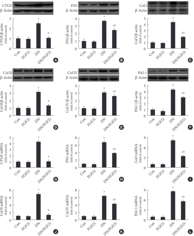
FGF21 prevents diabetic renal fibrosis associated with suppression of renal ECM accumulation
Since the ECM is the predominant contributor to diabetic renal fibrosis, the markers represented ECM were examined in this study. The expression of CTGF, a strong inducer of ECM syn- thesis, was significantly increased in the diabetic kidney (Fig.
1A). Meanwhile, the expression of multiple classic matrix pro- teins in the diabetic kidneys, including FN1 (Fig. 1B), collagen of types I (Fig. 1C), II (Fig. 1D), and IV (Fig. 1E), were signifi- cantly increased. Additionally, the expression of PAI-1, consid- ered both an inflammatory and fibrotic marker, was strongly increased (Fig. 1F). Furthermore, the quantitative polymerase chain reaction assay showed that the mRNA levels of these ECM markers above were also increased in the diabetic kidney (Fig. 1G-L), indicating that the upregulated ECM markers were derived from the kidney rather than transferred into the kidney in an endocrine manner. However, FGF21 treatment for 4 months notably inhibited the increased ECM mRNA and pro- tein expression in the diabetic kidney (Fig. 1G-L), indicating that FGF21-induced anti-fibrotic effect might be attributed to their suppression of the ECM in the diabetic kidney.
FGF21 prevents the EMT in the diabetic kidney
Next, we examined the effects of FGF21 on the EMT, which is the predominant inducer of ECM accumulation and fibrosis in the diabetic kidney. To this end, the protein expression levels of EMT biomarkers in the renal cortex were detected by West- ern blotting. The results showed that the EMT was significantly enhanced in the diabetic kidney, as characterized by a decrease in the expression of epithelial cells markers, including E-cad- herin (Supplementary Fig. 3A and B) and ZO-1 (Supplemen- tary Fig. 3A and C), and an increase in the expression of mes- enchymal cells markers, including α-SMA (Supplementary Fig. 3A and D), vimentin (Supplementary Fig. 3A and E), and laminin (Supplementary Fig. 3A and F). However, the EMT process above was prevented by FGF21 treatment (Supple- mentary Fig. 3A-F). Next, we found that the FGF21-induced
Fig. 1. Effects of fibroblast growth factor 21 (FGF21) supplement on renal extracellular matrix (ECM) accumulation in diabetic nephropathy (DN) mice. Since ECM leads to renal fibrosis, Western blotting was used to examine the expressions of the markers of ECM and fibrosis in the kidneys, including (A) connective tissue growth factor (CTGF), (B) fibronectin 1 (FN1), (C) collagen I (Col I), (D) collagen II (Col II), (E) collagen IV (Col IV), and (F) plasminogen activator inhibitor-1 (PAI-1). The mRNA level of multiple markers of EMC accumulation and renal fibrosis including (G) CTGF, (H) FN1, (I) Col I, (J) Col II, (K) Col IV, (L) and PAI-1 in the kidney with or without FGF21 treatment were examined by quantitative polymerase chain reaction. Data are pre- sented as the mean±standard deviation (n=8/group). aP<0.05 vs. the control (Con) group, bP<0.05 vs. DN group.
3 2 1 0
4 3 2 1 0
5 4 3 2 1 0
8 6 4 2 0
4 3 2 1 0
4 3 2 1 0
8 6 4 2 0
8 6 4 2 0
5 4 3 2 1 0
5 4 3 2 1 0
9 6 3 0
9 6 3 0 CTGF/β-actin (fold of control)Col II/β-actin (fold of control)CTGF mRNA (fold of control)Col II mRNA (fold of control) FN1/β-actin (fold of control)Col IV/β-actin (fold of control)FN1 mRNA (fold of control)Col IV mRNA (fold of control) Col I/β-actin (fold of control)PAI-1/β-actin (fold of control)Col I mRNA (fold of control)PAI-1 mRNA (fold of control)
Con
Con
Con
Con
Con
Con
Con
Con
Con
Con
Con
Con DN/FGF21
DN/FGF21
DN/FGF21
DN/FGF21
DN/FGF21
DN/FGF21
DN/FGF21
DN/FGF21
DN/FGF21
DN/FGF21
DN/FGF21
DN/FGF21 DN
DN
DN
DN
DN
DN
DN
DN
DN
DN
DN
DN FGF21
FGF21
FGF21
FGF21
FGF21
FGF21
FGF21
FGF21
FGF21
FGF21
FGF21
FGF21 β-ActinCTGF
Col II β-Actin
β-ActinFN1
Col IV β-Actin
Col I β-Actin
PAI-1 β-Actin A
D
G
J
B
E
H
K
C
F
I
L
a
a
a
a
a
a
a
a
a
a
a
a b
b
b
b
a,b
a,b
a,b
a,b
a,b
a,b
a,b
a,b
preventive effects on EMT in the diabetic kidney were associ- ated with the inhibition of snail (Supplementary Fig. 3A and G), twist (Supplementary Fig. 3A and H), and slug (Supple- mentary Fig. 3A and I), which are negative transcriptional reg- ulators of E-cadherin. Generally, studies related to EMT focus on tubular cells; however, in this study, we found that FGF21 treatment also preserved the renal expression of P-cadherin
(Supplementary Fig. 3A and J), which is derived from podo- cytes in the diabetic kidney. Therefore, we also detected the ex- pression of nephrin, a biomarker of podocytes that are classi- fied as epithelial cells in the glomeruli. Nephrin expression was strongly inhibited in the diabetic kidney (Supplementary Fig.
3A and K), indicating that the EMT occurs in both the tubules and glomeruli of the diabetic kidney. Although under healthy
Fig. 2. Effects of fibroblast growth factor 21 (FGF21) supplement on the formation and nuclear translocation of p53/Smad2/3 complex. Western blot analysis was used to examine the expression of (A) renal transforming growth factor beta (TGF-β) and phosphorylation of (B) Smad2, (C) Smad3, and (D) Smad7 as well as (E) renal p53 phosphorylation. (F, G) Immunoprecipitation (IP) was used to examine the protein-protein interaction between p53 and Smad2/3 with or without FGF21 treatment. The nucle- ar translocation of p53/Smad2/3 complex was examined by changes in the expression of nuclear (H) p53, (I) Smad2, (J) and Smad3, as determined by Western blot analysis. Data are presented as the mean±standard deviation (n=8/group). aP<0.05 vs.
the control (Con) group, bP<0.05 vs. diabetic nephropathy (DN) group.
54 32 10
4 3 2 1 0
8 6 4 2 0
6 4 2 0
6 4 2 0 4
3 2 1 0
4 3 2 1 0
1.2 0.8 0.4
-β1/β-actinTGF (fold of control) n-p53/c-p53p-p53/t-p53 l)rontld o(fof col)ntf cold o(foro n-Smad2/c-Smad2 ntl)ro(fof cold o n-Smad3/c-Smad3 ntrol)f cold o(fo 0
p-Smad2/t-Smad2 (fold of control) p-Smad3/t-Smad3 (fold of control) p-Smad7/t-Smad7 (fold of control)
Con
Con
Con Con Con
Con Con
Con Con Con
DN/FGF21
DN/FGF21
DN/FGF21 DN/FGF21 DN/FGF21
DN/FGF21 DN/FGF21
DN/FGF21 DN/FGF21 DN/FGF21
DN
DN
DN DN DN
DN DN
DN DN DN
FGF21
FGF21
FGF21 FGF21 FGF21
FGF21 FGF21
FGF21 FGF21 FGF21
TGF-β1 β-Actin
p-p53 t-p53
n-p53 Lamin B C-p53 β-Actin
n-Smad2 Lamin B C-Smad2 β-Actin
n-Smad3 Lamin B C-Smad3 β-Actin p-Smad2
t-Smad2 p-Smad3
t-Smad3 p-Smad7
t-Smad7 13
42
55 55
55 55
50 50
A
E
H I J
F G
B C D
a
a
a a
a
a a
a a
a,b
a,b a,b a,b
a a
a
IP: p-p53
Blot: p-Smad2 IP: p-p53
Blot: p-Smad3
p-p53 p-p53
p-Smad2 p-Smad3
p-p53 p-p53
Input Input
a
Fig. 3. Effect of pifithrin-α (PF-α) on fibroblast growth factor 21 (FGF21)-induced suppression of renal p53 phosphorylation and interaction of p53/Smad2/3. (A) The phosphorylation and expression of renal p53 in the presence of FGF21 and/or PF-α were ex- amined by Western blotting. (B, C) The protein-protein interaction between p53 and Smad2/3 in the presence of FGF21 and/or PF-α was examined by immunoprecipitation (IP). Western blot analysis was used to examine the activity and (D) expression of renal AKT and the expression of the extracellular matrix (ECM) and fibrotic markers in the kidneys including (E, F) connective tissue growth factor (CTGF), (E, G) fibronectin 1 (FN1), (E, H) plasminogen activator inhibitor-1 (PAI-1), (E, I) collagen I (Col I), (E, J) collagen II (Col II), and (E, K) collagen IV (Col IV). Data are presented as the mean±standard deviation (n=8/group).
aP<0.05 vs. the diabetic nephropathy (DN) group.
1.2 0.8 0.4 0
1.2 0.8 0.4 0
1.2 0.8 0.4 0
1.2 0.8 0.4 0
1.2 0.8 0.4 0 1.2
0.8 0.4 0 1.2
0.8 0.4 0
p-p53/t-p53 (fold of control)p-p53/t-p53 (fold of control) CTGF/β-actin (fold of control)FN1/β-actin (fold of control) Col IV/β-actin (fold of control)
Col II/β-actin (fold of control)
Col I/β-actin (fold of control)
DN
DN
DN
DN
DN DN
DN
DN DN/FGF21/P DN
F
DN/FGF21/P F
DN/P F
DN/P F
DN/P F
DN/P F
DN/P F DN/P
F DN/P
F DN/FGF21/P
F
DN/FGF21/P F
DN/FGF21/P F
DN/FGF21/P F
DN/FGF21/P F DN/FGF21/P
F DN/FGF21/P
F
DN/P F
DN/P F
DN/FGF21
DN/FGF21
DN/FGF21
DN/FGF21
DN/FGF21 DN/FGF21
DN/FGF21
DN/FGF21 DN/FGF21
p-p53 t-p53
p-AKT
t-AKT
β-Actin
A
D
F
G
K J
I
E
B C
a
a
a
a
a a
a a
a
a
a
a a
a a
a
a
a
a a
a
1.2 0.8 0.4 0 PAI-1/β-actin (fold of control)
DN DN/P
F DN/FGF21/P
F DN/FGF21
H
a a
a
IP: p-p53
Blot: p-Smad2 IP: p-p53
Blot: p-Smad3
p-p53 p-p53
p-Smad2 p-Smad3
p-p53 p-p53
Input Input
CTGF β-Actin FN1 β-Actin PAI-1 β-Actin Col I β-Actin Col II β-Actin Col IV β-Actin
condition, FGF21 treatment had no impact on EMT, which were notably suppressed under diabetic condition (Supple- mentary Fig. 3).
FGF21 negatively regulates Smad2/3 nuclear translocation and renal fibrosis by inhibiting p53 activity
In this study, we found that diabetes strongly increased the ex-
pression of PAI-1, a classic marker of TGF-β signaling, at both the mRNA and protein levels. Importantly, TGF-β/Smad2/3 signaling is the predominant regulatory pathway of the EMT in the kidney. Western blot analyses showed that TGF-β ex- pression was significantly upregulated in diabetic conditions (Fig. 2A), followed by activity (phosphorylation) enhancement of Smad2 and Smad3 in the diabetic kidney (Fig. 2B and C).
Opposite changing pattern of the phosphorylation renal Smad7, negative regulator of Smad2/3 (Fig. 2D) was observed in diabetic mice. Unexpected, FGF21 treatment had no impact on the expression of TGF-β (Fig. 2A) and phosphorylation of Smad2/3/7 in the diabetic kidney (Fig. 2B-D), indicating that FGF21 does not affect TGF-β/Smad2/3 signaling transduction.
Strong evidence has demonstrated that p53 is a key kinase that regulates TGF-β-Smad2/3 by binding with Smad2/3. This study showed that diabetes induced p53 phosphorylation in the diabetic kidney (Fig. 2E). Meanwhile, the binding amount of phosphorylated p53 to phosphorylated Smad2/3 signifi- cantly increased in the diabetic kidney (Fig. 2F and G). How- ever, FGF21 treatment for 4 months not only suppressed p53 phosphorylation (Fig. 2E), but also attenuated the binding amount of p-p53 and p-Smad2/3 (Fig. 2F and G). Next, we evaluated the nuclear translocation of the p53/Smad2/3 com- plex by detecting the ratio of nuclear content to cytosol content of both p53 and Smad2/3, which were significantly increased in the diabetic kidney (Fig. 2H-J). However, translocation of the complex was inhibited in the presence of FGF21 (Fig. 2H- J). In order to further confirmed the negative regulating effect of FGF21 on renal p53 activity and its nuclear translocation with Smad2/3, reverse experiments was performed in FGF21- KO mice compared with WT mice. The results showed that compared with WT diabetic mice FGF21 deletion further en- hanced diabetes-induced renal p53 phosphorylation (Supple- mentary Fig. 4A) and its nuclear translocation (Supplementary Fig. 4B). Similarly, enhanced nuclear translocation of Smad2/3 were also observed in the diabetic kidney of FGF21-KO mice (Supplementary Fig. 4C and D), indicating that endogenous FGF21 negatively regulated p53-Smad pathway in the diabetic kidney.
To determine the reason for the decreased interaction be- tween p53 and Smad2/3 in the FGF21-treated diabetic kidney, PF-α (p53 specific inhibitor) was applied. The results showed that FGF21 or PF-α significantly suppressed p53 phosphoryla- tion (Fig. 3A) and the protein-protein interaction between p53 and Smad2/3 (Fig. 3B and C). However, within treatment of
PF-α, FGF21 failed to decrease p53 phosphorylation and the binding amount to Smad2/3 (Fig. 3), indicating that the FGF21-mediated decrease in the interaction between p53 and Smad2/3 was purely due to inhibition of the activity of p53 rather than suppressing the binding ability of p53 to Smad2/3.
Additionally, we found inhibition of p53 had no effect on FGF21-induced renal AKT activity (Fig. 3D), indicating AKT is not the downstream target of p53. Moreover, diabetes-in- duced renal fibrosis and ECM accumulation were notably pre- vented at varying degrees by FGF21 or PF-α treatment, respec- tively, accompanied by the decreased expression of fibrotic markers and ECM markers, including CTGF (Fig. 3E and F), FN1 (Fig. 3E and G), PAI-1 (Fig. 3E and H), Col I (Fig. 3E and I), Col II (Fig. 3E and J), and Col IV (Fig. 3E and K). With PF-α treatment, FGF21 failed to further enhance the preven- tion of fibrosis and ECM accumulation in the diabetic kidney (Fig. 3E-K), indicating that FGF21 prevented diabetes-induced EMT in the kidney via inhibition of renal p53 activity.
FGF21 prevents the renal EMT via inhibition of p53 activity in DN mice
Since we found that FGF21 inhibited the TGF-β/Smad2/3 pathway by attenuating p53-mediated Smad2/3 nuclear trans- location, we next examined the role of p53 in the FGF21-in- duced preventive effects of the EMT in the diabetic kidney. The results showed that diabetes-induced EMT in the kidney was notably prevented by FGF21 or PF-α treatment at varying de- grees, with characteristics of increased expression of epithelial cell markers, including E-cadherin (Supplementary Fig. 5A and B), P-cadherin (Supplementary Fig. 5A and C), nephrin (Supplementary Fig. 5A and D), and ZO-1 (Supplementary Fig. 5A and E), and the decreased expression of α-SMA (Sup- plementary Fig. 5A and F), vimentin (Supplementary Fig. 5A and G), and laminin (Supplementary Fig. 5A and H). As ex- pected, with PF-α treatment, FGF21 failed to further attenuate renal EMT in DN mice (Supplementary Fig. 5), indicating that FGF21 suppresses diabetes-induced EMT in the diabetic kid- ney via inhibition of renal p53 activity.
AKT mediates FGF21-induced suppression of p53 activity and its binding to Smad2/3 by upregulating MDM2 expression
To identify the role of Akt in FGF21-induced negative regula- tion of p53 activity in the diabetic kidney, a specific AKT in- hibitor, 10-DEBC, was applied. The results showed that FGF21
Fig. 4. Effects of 10-[4´-(N,N-Diethylamino)butyl]-2-chlorophenoxazine hydrochloride (10-DEBC) on fibroblast growth factor 21 (FGF21)-induced suppression of renal p53 phosphorylation and complex formation of p53/Smad2/3. The phosphorylation and expression of renal (A, B) AKT, (A, C) p53, and (A, D) mouse double minute-2 homolog (MDM2) in the presence of FGF21 and/or 10-DEBC were examined by Western blotting. (E, F) The protein-protein interaction between p53 and Smad2/3 in the presence of FGF21 and/or 10-DEBC was examined by immunoprecipitation (IP). Data are presented as the mean±standard de- viation (n=8/group). DEBC=10-DEBC (10-DEBC hydrochloride). aP<0.05 vs. the diabetic nephropathy (DN) group.
3
2
1
0
3
2
1
0 2.5
2.0 1.5 1.0 0.5 0
p-AKT/t-AKT (fold of control)MDM2/β-actin (fold of control)
p-p53/t-p53 (fold of control)
DN
DN DN
DN/FGF21/D EBC
DN/FGF21/D EBC DN/FGF21/D
EBC
DN/D EBC
DN/D EBC DN/D
EBC
DN/FGF21
DN/FGF21 DN/FGF21
p-AKT t-AKT p-p53 t-p53 MDM2 β-Actin
B
D C
A
a
a a
a
a a
a
a
a
DN DN/FGF21/D DN
EBC
DN/FGF21/D EBC DN/D
EBC
DN/D EBC
DN/FGF21 DN/FGF21
E F
IP: p-p53
Blot: p-Smad2 IP: p-p53
Blot: p-Smad3
p-p53 p-p53
p-Smad2 p-Smad3
p-p53 p-p53
Input Input
enhanced, but DEBC suppressed AKT phosphorylation rather than its expression in the diabetic kidney (Fig. 4A and B), indi- cating that renal AKT inhibition model was successfully estab- lished. Additionally, FGF21 notably decreased, but 10-DEBC increased p53 phosphorylation in the diabetic kidney (Fig. 4A and C). However, the converse was observed with renal MDM2 in DN mice (Fig. 4A and D), suggesting that FGF21 suppresses renal p53 phosphorylation by increasing AKT-mediated MDM2 activation. Additionally, inhibition of Akt enhanced
formation of the p53/Smad2/3 complex (Fig. 4E and F). Inter- estingly, FGF21 treatment failed to induce inhibitory effects on p53 phosphorylation and subsequent binding to Smad2/3 in the presence of 10-DEBC (Fig. 4), indicating that Akt is the key enzyme that negatively regulates p53/Smad2/3 signaling.
FGF21 prevents diabetes-induced EMT and subsequent fibrosis in the kidney via activation of Akt
The results showed that 10-DEBC treatment enhanced diabe-
Fig. 5. Effects of 10-[4´-(N,N-Diethylamino)butyl]-2-chlorophenoxazine hydrochloride (10-DEBC) on fibroblast growth factor 21 (FGF21)-induced prevention of the epithelial-to-mesenchymal transition (EMT) in the diabetic kidney. Western blot analysis was used to examine the markers of EMT and extracellular matrix (ECM) in the kidneys, including (A) E-cadherin, (B) P-cad- herin, (C) zonula occludens-1 (ZO-1), (D) α-smooth muscle actin (α-SMA), (E) vimentin, and (F) laminin, (G) connective tissue growth factor (CTGF), (H) fibronectin 1 (FN1), (I) plasminogen activator inhibitor-1 (PAI-1), (J) collagen I (Col I), (K) collagen II (Col II), and (L) collagen IV (Col IV). Data are presented as the mean±standard deviation (n=8/group). aP<0.05 vs. the dia- betic nephropathy (DN) group.
2.5 2.0 1.5 1.0 0.5 0
2.0 1.5 1.0 0.5 0
1.5 1.0 0.5 0
2.5 2.0 1.5 1.0 0.5 0
3 2 1 0
1.5 1.0 0.5 0
1.8 1.2 0.6 0
3 2 1 0
4 3 2 1 0
4 3 2 1 0
2.0 1.5 1.0 0.5 0
2.0 1.5 1.0 0.5 0 E-cadherin/β-actin (fold of control)α-SMA/β-actin (fold of control)CTGF/β-actin (fold of control)Col I/β-actin (fold of control) P-cadherin/β-actin (fold of control)Vimentin/β-actin (fold of control)FN1/β-actin (fold of control)Col II/β-actin (fold of control) ZO-1/β-actin (fold of control)Laminin/β-actin (fold of control)PAI-1/β-actin (fold of control)Col IV/β-actin (fold of control)
DN
DN
DN
DN
DN
DN
DN
DN
DN
DN
DN
DN DN/FGF21/D
EBC
DN/FGF21/D EBC
DN/FGF21/D EBC
DN/FGF21/D EBC
DN/FGF21/D EBC
DN/FGF21/D EBC
DN/FGF21/D EBC
DN/FGF21/D EBC
DN/FGF21/D EBC
DN/FGF21/D EBC
DN/FGF21/D EBC
DN/FGF21/D EBC DN/D
EBC
DN/D EBC
DN/D EBC
DN/D EBC
DN/D EBC
DN/D EBC
DN/D EBC
DN/D EBC
DN/D EBC
DN/D EBC
DN/D EBC
DN/D EBC DN/FGF21
DN/FGF21
DN/FGF21
DN/FGF21
DN/FGF21
DN/FGF21
DN/FGF21
DN/FGF21
DN/FGF21
DN/FGF21
DN/FGF21
DN/FGF21 E-cadherin
β-Actin
α-SMA β-Actin
CTGF β-Actin
Col I β-Actin
P-cadherin β-Actin
Vimentin β-Actin
FN1 β-Actin
Col II β-Actin
ZO-1 β-Actin
Laminin β-Actin
PAI-1 β-Actin
Col IV β-Actin A
D
G
J
B
E
H
K
C
F
I
L
a
a
a
a
a
a
a
a
a
a
a
a a
a
a
a
a
a
a
a
a
a
a
a a
a
a
a
a
a
a
a
a
a
a
a
tes-induced renal EMT by further decreasing the expression of epithelial cells markers, including E-cadherin (Fig. 5A), P-cad- herin (Fig. 5B), and ZO-1 (Fig. 5C), as well as further increas- ing the expression of mesenchymal cell markers, including α-SMA (Fig. 5D), vimentin (Fig. 5E), and laminin (Fig. 5F).
Moreover, 10-DEBC treatment also enhanced diabetes-in- duced renal fibrosis, characterized by a further increase in the expression of multiple fibrotic and EMC markers, including CTGF (Fig. 5G), FN1 (Fig. 5H), PAI-1 (Fig. 5I), Col I (Fig. 5J), Col II (Fig. 5K), and Col IV (Fig. 5L). As expected, FGF21 was incapable of inducing preventive effects on renal EMT and fi- brosis in the diabetic kidney in the presence of 10-DEBC (Fig.
5), indicating that Akt plays a key role in mediating FGF21-in- duced anti-fibrotic effects in the diabetic kidney.
DISCUSSION
FGF21 is a member of FGF family, which induces beneficial effects in multiple organs in an endocrine manner [22]. In- creasing clinical studies have demonstrated that circulating FGF21 levels are dramatically increased in patients with vari- ous kidney diseases, including chronic kidney disease (CKD) [23,24], kidney transplantation [25,26], ESRD [27], early-stage diabetic kidney disease [28], acute renal dysfunction [29], and long-term peritoneal dialysis [30], indicating that an increase in FGF21 levels above baseline is a stress response to induce renal protection. This hypothesis was confirmed by a subse- quent study, which showed that the administration of FGF21 maintained renal function in mice with CKD [31]. Kim et al.
[32] reported that FGF21 improved insulin resistance and ameliorated renal injury in spontaneous type 2 diabetes melli- tus (db/db) mice. Our previous studies also indicated that FGF21 supplement prevents, but FGF21 deletion enhances DN progression [11,17]. Importantly, we found that FGF21 supplement suppresses renal CTGF expression in the diabetic kidney [27,33]. However, CTGF is only one marker of fibrosis;
thus, the effects of FGF21 on renal CTGF expression are not enough to identify the anti-fibrotic effects of FGF21 in the kid- ney of DN mice, which were evaluated in this study.
Four months after diabetes was diagnosed, the symptoms of DN were clearly observed. Meanwhile, fibrosis and it-induced glomerulosclerosis also obviously appeared in tubular and glo- merular areas. The above findings indicate that fibrosis devel- oped in both the tubules and glomerulus. Moreover, renal fi- brosis was further confirmed by the increased expression of
multiple fibrosis and ECM markers in the diabetic kidney.
However, all of the above-mentioned symptoms were marked- ly prevented by the administration of FGF21, indicating that FGF21 induced anti-fibrotic effects in the kidney of DN mice.
Although FGF21 is considered as metabolic regulator, in the present study only a slight glucose-lowering effect (approxi- mately 10%) of FGF21 with the dose of 100 μg/kg was found in type 1 diabetes mellitus mice, which may not be the predomi- nant contributor to the strong anti-fibrotic effect of FGF21 in the diabetic kidney indicating other mechanism must exist. Strong evidence has indicated that the EMT plays a significant role in renal fibrosis and ECM accumulation, which is a hallmark of DN [5]. The EMT can be divided into three different types ac- cording to the different biological circumstances in which they occur and the associated consequences [34,35]. Among them, the type 2 EMT is implicated in wound healing, organ fibrosis, and tissue regeneration, and induces the generation of activated mesenchymal cells, which stimulate ECM accumulation ulti- mately leading to tissue damage [36,37]. The EMT occurs in the diabetic kidney and is classified as type 2 EMT, which is consid- ered the inducer of diabetic renal fibrosis [34].
In this study, we also confirmed the enhanced EMT in the diabetic kidney with characteristics of decreased expression of epithelial cells markers and increased expressions of mesen- chymal cells markers. Normally, the EMT in the kidney refers to tubular EMT [8], but the EMT in glomeruli also plays im- portant roles in inducing podocyte loss and glomerular dam- age [38,39], which was observed in this study. Importantly, the expression of all of the above-mentioned EMT markers was oppositely regulated by the administration of FGF21, indicat- ing that FGF21 induced preventive effects on the EMT in both the tubular and glomerular areas of DN mice. Next, we focused on determining the underlying mechanisms of FGF21-in- duced suppression on the EMT in the diabetic kidney. EMT- induced renal fibrosis is predominantly mediated by the TGF-β/Smad2/3 pathway [40,41]. In this study, we confirmed activated TGF-β/Smad2/3 in the diabetic kidney, characterized by increased renal TGF-β expression and phosphorylation of its downstream Smad2/3. Unexpectedly, FGF21 supplement inhibited TGF-β/Smad2/3-mediated EMT via the suppression of Smad2/3 nuclear translocation rather than inhibiting the ex- pression or activity of TGF-β/Smad2/3. Importantly, we found that renal p53 is a key target of FGF21 that decreases activated Smad2/3 nuclear translocation. Increasing studies have report- ed that p53 plays an important role in regulating the TGF-β/
Smad2/3 pathway [42,43]. Activated p53 recognizes and binds to Smad2/3 phosphorylated by TGF-β to form a complex that is able to translocate into the nucleus and acts as a transcription factor of multiple fibrotic genes [42,43]. Moreover, in vitro and in vivo studies have demonstrated that FGF21 can induce in- hibitory effects on p53 activity [13,44,45]. Similar effects were also identified in this study, in that FGF21 supplement sup- pressed renal p53 phosphorylation in the diabetic kidney, which contributed to less activated p53 binding to phosphory- lated Smad2/3 to form the transcriptional complex. We also found that FGF21 supplement failed to reduce the amount and nuclear translocation of p53-Smad2/3 complex in the presence of PF-α, suggesting that suppression of p53 is required in FGF21-induced negative regulation of Smad2/3 nuclear trans- location. Next, we mainly focused on determining whether FGF21-induced downregulation of Smad2/3 nuclear translo- cation via inhibition of p53 contributes to the suppression of EMT and subsequent renal fibrosis. We found that FGF21 sup- plement was incapable of inducing preventive effects on diabe- tes-induced renal EMT process and fibrosis in DN mice in the presence of PF-α, suggesting that FGF21 negatively regulated the EMT and subsequent fibrosis in the diabetic kidney by in- hibition of the p53-mediated TGF-β/Smad2/3 pathway.
Finally, we explored the underlying mechanisms of FGF21- induced negative regulation of renal p53 activity in the DN mice. Akt, an effector of phosphoinositide 3-kinase, is a serine/
threonine protein kinase that regulates a variety of cellular functions and induces multiple beneficial effects in DN [46, 47]. AKT negatively regulates p53 activity by phosphorylating MDM2, a p53 negative regulator [48]. Then, activated MDM2 translocates into the nucleus and assembles phosphorylated p53. MDM2 carries p53 out of the nucleus for degradation [49,50]. In this study, we found that FGF21 supplement in- creased the phosphorylation of renal MDM2. However, this effect was not observed in the presence of 10-DEBC, indicat- ing that AKT is required for FGF21-induced activation of MDM2. Moreover, 10-DEBC also blocked FGF21-induced suppression of renal p53 and subsequent EMT and fibrosis in the diabetic kidney.
In summary, we confirmed that FGF21 attenuates DN asso- ciated with the prevention of diabetes-induced renal ECM ac- cumulation and fibrosis. Mechanistic studies indicated that FGF21 suppressed renal fibrosis in the diabetic kidney by neg- atively regulating TGF-β-p53-Smad2/3-mediated EMT via ac- tivation of Akt.
SUPPLEMENTARY MATERIALS
Supplementary materials related to this article can be found online at https://doi.org/10.4093/dmj.2018.0235.
CONFLICTS OF INTEREST
No potential conflict of interest relevant to this article was re- ported.
AUTHOR CONTRIBUTIONS
Conception or design: X.L., C.Z.
Acquisition, analysis, or interpretation of data: S.L., L.Y., Y.N., L.H., X.W., X.L., C.Z.
Drafting the work or revising: X.L., C.Z.
Final approval of the manuscript: S.L., L.Y., Y.N., L.H., X.W., X.L., C.Z.
ORCID
Sundong Lin https://orcid.org/0000-0002-9640-061X Lechu Yu https://orcid.org/0000-0002-0560-4028 Xuemian Lu https://orcid.org/0000-0002-2277-0052 Chi Zhang https://orcid.org/0000-0002-3717-7665
ACKNOWLEDGMENTS
This work was supported by grants from the Medical and Healthy Technological Grant of Zhejiang Province (Nos.
2015KYB236 to Chi Zhang and 2018KY769 to Lechu Yu), the National Science Foundation of China (No. 81670767 to Chi Zhang and 81700732 to Lechu Yu), and the Project for Selected Overseas Chinese supported by Zhejiang Technology Founda- tion to Chi Zhang. The funding institutions had no role in the study design, data collection and analysis, decision to publish, or preparation of the manuscript.
REFERENCES
1. Reidy K, Kang HM, Hostetter T, Susztak K. Molecular mecha- nisms of diabetic kidney disease. J Clin Invest 2014;124:2333- 40.
2. Gallagher H, Suckling RJ. Diabetic nephropathy: where are we on the journey from pathophysiology to treatment? Diabetes
Obes Metab 2016;18:641-7.
3. Jha JC, Banal C, Okabe J, Gray SP, Hettige T, Chow BSM, Thal- las-Bonke V, De Vos L, Holterman CE, Coughlan MT, Power DA, Skene A, Ekinci EI, Cooper ME, Touyz RM, Kennedy CR, Jandeleit-Dahm K. NADPH oxidase Nox5 accelerates renal in- jury in diabetic nephropathy. Diabetes 2017;66:2691-703.
4. Pontrelli P, Conserva F, Papale M, Oranger A, Barozzino M, Vocino G, Rocchetti MT, Gigante M, Castellano G, Rossini M, Simone S, Laviola L, Giorgino F, Grandaliano G, Di Paolo S, Gesualdo L. Lysine 63 ubiquitination is involved in the pro- gression of tubular damage in diabetic nephropathy. FASEB J 2017;31:308-19.
5. Zhao Y, Yin Z, Li H, Fan J, Yang S, Chen C, Wang DW. MiR- 30c protects diabetic nephropathy by suppressing epithelial-to- mesenchymal transition in db/db mice. Aging Cell 2017;16:
387-400.
6. Thiery JP, Acloque H, Huang RY, Nieto MA. Epithelial-mesen- chymal transitions in development and disease. Cell 2009;139:
871-90.
7. Higgins SP, Tang Y, Higgins CE, Mian B, Zhang W, Czekay RP, Samarakoon R, Conti DJ, Higgins PJ. TGF-β1/p53 signaling in renal fibrogenesis. Cell Signal 2018;43:1-10.
8. Ma J, Zhang L, Hao J, Li N, Tang J, Hao L. Up-regulation of mi- croRNA-93 inhibits TGF-β1-induced EMT and renal fibro- genesis by down-regulation of Orai1. J Pharmacol Sci 2018;
136:218-27.
9. Piccolo S. P53 regulation orchestrates the TGF-beta response.
Cell 2008;133:767-9.
10. Yang H, Feng A, Lin S, Yu L, Lin X, Yan X, Lu X, Zhang C. Fi- broblast growth factor-21 prevents diabetic cardiomyopathy via AMPK-mediated antioxidation and lipid-lowering effects in the heart. Cell Death Dis 2018;9:227.
11. Zhang C, Shao M, Yang H, Chen L, Yu L, Cong W, Tian H, Zhang F, Cheng P, Jin L, Tan Y, Li X, Cai L, Lu X. Attenuation of hyperlipidemia- and diabetes-induced early-stage apoptosis and late-stage renal dysfunction via administration of fibro- blast growth factor-21 is associated with suppression of renal inflammation. PLoS One 2013;8:e82275.
12. Li F, Liu Z, Tang C, Cai J, Dong Z. FGF21 is induced in cisplatin nephrotoxicity to protect against kidney tubular cell injury.
FASEB J 2018;32:3423-33.
13. Wang XM, Xiao H, Liu LL, Cheng D, Li XJ, Si LY. FGF21 re- presses cerebrovascular aging via improving mitochondrial biogenesis and inhibiting p53 signaling pathway in an AMPK- dependent manner. Exp Cell Res 2016;346:147-56.
14. Zhang Q, Li Y, Liang T, Lu X, Liu X, Zhang C, Jiang X, Martin RC, Cheng M, Cai L. Loss of FGF21 in diabetic mouse during hepatocellular carcinogenetic transformation. Am J Cancer Res 2015;5:1762-74.
15. Guo D, Xiao L, Hu H, Liu M, Yang L, Lin X. FGF21 protects human umbilical vein endothelial cells against high glucose- induced apoptosis via PI3K/Akt/Fox3a signaling pathway. J Diabetes Complications 2018;32:729-36.
16. Zhang C, Zhang L, Chen S, Feng B, Lu X, Bai Y, Liang G, Tan Y, Shao M, Skibba M, Jin L, Li X, Chakrabarti S, Cai L. The pre- vention of diabetic cardiomyopathy by non-mitogenic acidic fibroblast growth factor is probably mediated by the suppres- sion of oxidative stress and damage. PLoS One 2013;8:e82287.
17. Shao M, Yu L, Zhang F, Lu X, Li X, Cheng P, Lin X, He L, Jin S, Tan Y, Yang H, Zhang C, Cai L. Additive protection by LDR and FGF21 treatment against diabetic nephropathy in type 2 diabetes model. Am J Physiol Endocrinol Metab 2015;309:
E45-54.
18. Shao M, Lu X, Cong W, Xing X, Tan Y, Li Y, Li X, Jin L, Wang X, Dong J, Jin S, Zhang C, Cai L. Multiple low-dose radiation pre- vents type 2 diabetes-induced renal damage through attenua- tion of dyslipidemia and insulin resistance and subsequent re- nal inflammation and oxidative stress. PLoS One 2014;9:
e92574.
19. Zhang C, Lu X, Tan Y, Li B, Miao X, Jin L, Shi X, Zhang X, Miao L, Li X, Cai L. Diabetes-induced hepatic pathogenic damage, inflammation, oxidative stress, and insulin resistance was exacerbated in zinc deficient mouse model. PLoS One 2012;7:e49257.
20. Gu J, Wang B, Liu Y, Zhong L, Tang Y, Guo H, Jiang T, Wang L, Li Y, Cai L. Murine double minute 2 siRNA and wild-type p53 gene therapy interact positively with zinc on prostate tumours in vitro and in vivo. Eur J Cancer 2014;50:1184-94.
21. Zhang Z, Wang S, Zhou S, Yan X, Wang Y, Chen J, Mellen N, Kong M, Gu J, Tan Y, Zheng Y, Cai L. Sulforaphane prevents the development of cardiomyopathy in type 2 diabetic mice probably by reversing oxidative stress-induced inhibition of LKB1/AMPK pathway. J Mol Cell Cardiol 2014;77:42-52.
22. Zhang F, Yu L, Lin X, Cheng P, He L, Li X, Lu X, Tan Y, Yang H, Cai L, Zhang C. Minireview: roles of fibroblast growth factors 19 and 21 in metabolic regulation and chronic diseases. Mol Endocrinol 2015;29:1400-13.
23. Anuwatmatee S, Tang S, Wu BJ, Rye KA, Ong KL. Fibroblast growth factor 21 in chronic kidney disease. Clin Chim Acta 2019;489:196-202.


![Fig. 4. Effects of 10-[4´-(N,N-Diethylamino)butyl]-2-chlorophenoxazine hydrochloride (10-DEBC) on fibroblast growth factor 21 (FGF21)-induced suppression of renal p53 phosphorylation and complex formation of p53/Smad2/3](https://thumb-ap.123doks.com/thumbv2/123dokinfo/5220608.123338/9.892.159.753.139.756/effects-diethylamino-chlorophenoxazine-hydrochloride-fibroblast-suppression-phosphorylation-formation.webp)
![Fig. 5. Effects of 10-[4´-(N,N-Diethylamino)butyl]-2-chlorophenoxazine hydrochloride (10-DEBC) on fibroblast growth factor 21 (FGF21)-induced prevention of the epithelial-to-mesenchymal transition (EMT) in the diabetic kidney](https://thumb-ap.123doks.com/thumbv2/123dokinfo/5220608.123338/10.892.127.763.137.958/diethylamino-chlorophenoxazine-hydrochloride-fibroblast-prevention-epithelial-mesenchymal-transition.webp)
