• 검색 결과가 없습니다.

Luteolin Induces Apoptosis via Mitochondrial Pathway and Inhibits Invasion and Migration of Oral Squamous Cell Carcinoma by Suppressing Epithelial-Mesenchymal Transition Induced Transcription Factors

N/A
N/A
Protected

Academic year: 2021

Share "Luteolin Induces Apoptosis via Mitochondrial Pathway and Inhibits Invasion and Migration of Oral Squamous Cell Carcinoma by Suppressing Epithelial-Mesenchymal Transition Induced Transcription Factors"

Copied!
8
0
0

로드 중.... (전체 텍스트 보기)

전체 글

(1)

http://dx.doi.org/10.11620/IJOB.2018.43.2.069 pISSN 1226-7155, eISSN 2287-6618

Oral squamous cell carcinoma (OSCC) is the most common type of oral malignancy. Numerous therapies have been proposed for its cure. Research is continually being conducted to develop new forms of treatment as current therapies are associated with numerous side-effects. Luteolin, a common dietary flavonoid, has been demonstrated to possess strong anti-cancer activity against various human cancer cell lines.

Nevertheless, research into luteolin-based anticancer activity against oral cancer remains scarce. Thus, the objective of this study was to assess the effect of luteolin as an anti-cancer agent.

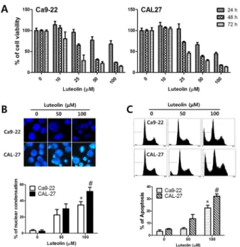
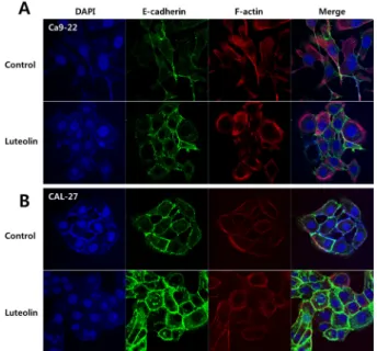
After treatment with luteolin, Ca9-22 and CAL-27 oral cancer cells showed condensed nuclei and enhanced apoptotic rate with evidence of mitochondria-mediated apoptosis. Epithelial- mesenchymal transition (EMT) is closely related to tumor migration and invasion. Luteolin suppressed cancer cell invasion and migration in the current study. Elevated

expression of E-cadherin, an adherens junction protein, was evident in both cell lines after luteolin treatment. Luteolin also significantly inhibited transcription factors (i.e., N-cadherin, Slug, Snail, Twist, and ZEB-1) that regulated expression of tumor suppressors such as E-cadherin based on Western blot analysis and quantitative PCR. Thus, luteolin could induce mitochondrial apoptosis and inhibit cancer cell invasion and migration by suppressing EMT-induced transcription factors.

Key words: OSCC, luteolin, apoptosis, EMT, E-cadherin

Introduction

Oral squamous cell carcinoma (OSCC) is the most common type of malignant tumor in the oral cavity [1]. It is caused by exposure to various types of carcinogen (i.e., chemical, physical, or microbial) and results in DNA mutation [2]. A combination of surgery, radiation therapy, and chemotherapy is commonly proposed as treatment for OSCC [3]. Research is continually being conducted into new forms of treatment as the current therapies are associated with low survival rates and adverse effects [4, 5].

Flavonoids are polyphenolic compounds that are found in various fruits, vegetables, and medicinal plants [6, 7]. Luteolin (3’,4’,5,7-tetrahydroxy flavone) is a common dietary flavonoid

Luteolin Induces Apoptosis via Mitochondrial Pathway and Inhibits Invasion and Migration of Oral Squamous Cell Carcinoma by Suppressing

Epithelial-Mesenchymal Transition Induced Transcription Factors

Bong-Soo Park

1,2

, Jong-Jin Kil

1

, Hae-Mi Kang

1,2

, Su-Bin Yu

1,2

, Dan-Bi Park

1,2

, Jin-A Park

1,2

and In-Ryoung Kim

1*

1

Department of Oral Anatomy, School of Dentistry, Pusan National University, Busandaehak-ro, 49, Mulguem-eup, Yangsan-si, Gyeongsangnam-do, 50612, South Korea

2

BK21 PLUS Project, School of Dentistry, Pusan National University, Busandaehak-ro, 49, Mulguem-eup, Yangsan-si, Gyeongsangnam-do, 50612, South Korea

(received May 28, 2018; revised June 12, 2018; accepted June 14, 2018)

*Correspondence to: In-Ryoung Kim, Department of Oral Anatomy, School of Dentistry, Pusan National University, Yangsan, 50612, Korea.

Tel: 82-51-510-8552, Fax.: 82-51-510-8241 E-mail: [email protected]

ORCID : 0000-0003-0232-0385

This is an Open-Access article distributed under the terms of the Creative Commons Attribution Non-Commercial License (http://creati- vecommons.org/licenses/by-nc/3.0) which permits unrestricted non- commercial use, distribution, and reproduction in any medium, pro- vided the original work is properly cited.

69

(2)

that is present in grapefruit, oranges, parsley, celery, sweet bell peppers, and broccoli [8, 9]. It has been shown to have strong anti-cancer activity against different human cancer cell lines [10-12]. Its anticancer properties have been demonstrated to inhibit cell growth and metastasis-associated properties in A431 cells [13], induce apoptosis in human myeloid leukemia [14], and cause G2/M phase cell cycle arrest in prostate cancer cells [15]. Tjioe et al. reported that luteolin impacts positively on DNA damage and suggested its use as an adjuvant reagent in oral cancer therapy [16]. However, research into luteolin-based anti-cancer activity against oral cancer remains scarce.

Apoptosis is regulated by a number of genes and plays an important role in morphogenesis, homeostasis, and cancer regression [17]. It was suggested recently that apoptosis is influential in a number of diseases. Cancer appears to relate closely to cell death, in particular. Therefore, it is feasible to study the anti-cancer capacity of natural compounds in order to develop anti-cancer drugs with less side-effects than those associated with the current cancer treatment options [18].

Epithelial-mesenchymal transition (EMT) is a well-organized process that evolves into cancer metastasis [19]. A significant correlation has been identified between cancer metastasis and stem cell characteristics in research on EMT [20], the latter of which is characterized by the loss of epithelial properties and the procurement of mesenchymal properties [21]. Cancer cell proliferation is closely related to acquisition of the EMT phenotype, which equips the tumor cells with the ability to penetrate the surrounding tissue and the cancer cells to migrate from distant sites [22]. These EMT characteristics are associated with OSCC progression and the development of an oral epithelium [23].

EMT-related changes generally occur in the early stages of the development of OSCC. The ability to identify the genes involved and their products is important in elucidating the transition process [24, 25]. EMT is often responsible for causing a reduction in the sensitivity of apoptotic-induced cancer and is known to be refractory to a variety of therapies, including chemotherapy and targeted drugs [26].

However, the molecular mechanisms by which luteolin affects apoptosis and EMT, and increases the invasiveness and metastatic potential of OSCC, remain unclear. Thus, the current study objective was to evaluate whether or not luteolin was capable of inducing apoptosis and suppressing EMT, and therefore of reducing motility in and the invasiveness of human OSCC cell lines.

Materials and Methods

Materials

Commercially obtained reagents, i.e., luteolin (3-[4,5- dimethylthiazol-2-yl]), 2, 5-diphenyl tetrazolium bromide (thiazolyl blue tetrazolium blue[MTT]), Hoechst 33342, propidium iodide, protease inhibitor, and RNase A, were obtained from Sigma-Aldrich (St Louis, USA). Fetal bovine serum (FBS) and Dulbecco’s Modified Eagle Medium (DMEM) were purchased from HyClone (Logan, USA). Penicillin- streptomycin was acquired from Gibco-BRL (Rockville, USA).

Radioimmunoprecipitation assay (RIPA) buffer and antibodies (caspase-3, poly (ADP-ribose) polymerase[PARP], E-cadherin, Slug, and Snail were sourced from Cell Signaling Technology (Beverly, USA). N-cadherin, cytochrome c, and β-actin were obtained from Santa Cruz Biotechnology (Santa Cruz, USA).

Mouse anti-rabbit immunoglobulin (Ig) G and rabbit anti-mouse IgG antibodies were purchased from Enzo Biochem (Farmingdale, USA). Rhodamin-phalloidin and MitoTracker

®

DeepRed were acquired from Thermo Fisher Scientific (Waltham, USA).

Cell culture and luteolin treatment

Human oral squamous carcinoma cell lines, Ca9-22 and CAL-27, were purchased from ATCC (Rockville, USA). The cells were maintained in DMEM supplemented with 10%

FBS and 1% antibiotics at 37°C in 5% CO

2

. Luteolin (100 mM) was dissolved in distilled water and kept frozen at -20°C until use. Prior to the luteolin treatment, the cells were grown to 80% confluence and then exposed to luteolin at different concentrations (0–100 μM) and time points (24–

72 hours).

MTT assay

The cell viability of luteolin was assessed using an MTT assay. Ca9-22 and CAL-27 cell lines (1 × 10

4

cells/100 μl medium) were seeded in a 96-well culture plateand treated with different concentrations of luteolin (0–100 μM) for 24–

72 hours. Thereafter, the existing medium was removed and treated with 100 μl of MTT working solution (500 μg/mL) and incubated at 37°C for 4 hours. The MTT solution was then removed and 100 μl DMSO was added to each well.

The absorbance of each well was measured using an ELISA

reader (Tecan; Männedorf, Switzerland) at 620 nm.

(3)

Hoechst staining

The Ca9-22 and CAL-27 cells (2 × 10

5

cells/chamber) were seeded in a 4-well Invitrogen

®

Lab-Tek II Chambered Coverglass (Thermo Fisher Scientific; Waltham, USA) and incubated overnight. The next day, both cell lines were treated with 50 and 100 μM of luteolin for 24 hours. The cells were washed with phosphate-buffered saline (PBS) and fixed in a 4% paraformaldehyde solution for 10 minutes. After fixation, the cells were washed again and stained with 1 μg/ml Hoechst 33342 for 10 minutes in the dark. Finally, the slides were mounted with 100% glycerol. Each sample was analyzed using fluorescence microscopy (Axioskop

®

) (CarlZeiss; Göttingen, Germany).

Determination of apoptosis using flow cytometry The Ca9-22 and CAL-27 cells were treated with luteolin (0 μ M, 50 μM, and 100 μM) for 24 hours. Thereafter, they were harvested, washed twice with PBS containing 1% bovine serum albumin (BSA), fixed in 70% (ice-cold) ethanol, and stored overnight at 4°C. The next day, the fixed cells were washed twice with PBS containing 1% BSA and re-suspended in a PBS containing 1 μg/ml propidium iodide and 50 μg/ml RNase A, and then incubated at 4°C for 30 minutes. The stained cells were measured with a Cytomics

®

FC500 Flow Cytometry System (Beckman Coulter; Indianapolis, USA). The data were analyzed using CXP

®

version 2.2.

Western blot analysis

The Ca9-22 and CAL-27 (2 × 10

6

) cells were seeded in a 100 mm culture dish and incubated overnight. Both cell lines were treated with various concentrations of luteolin (10–100 μM) for 24 hours. Thereafter, the cells were harvested and centrifuged at 3000 rpm for five minutes. The cell pellets were lysed in RIPA buffer at 4°C for an hour. The total cell lysate (10 μg of protein) was loaded onto the 10% sodium dodecyl sulfate-polyacrylamide gel electrophoresis and transferred onto a polyvinyl difluoride membrane. The membranes were blocked with 5% non-fat dry milk (a lyophilized blocking buffer) for an hour and incubated with the respective primary antibodies (1:1000) for an hour at room temperature, then washed twice with PBS. A secondary antibody (1:5000) was applied and washed in the same manner as the primary one. The protein bands were exposed to gel imaging using an Alphalmager

®

HP System (Alpha Innotech; Santa Clara, USA). Caspase-3, PARP, E-cadherin, N-cadherin, Slug, Snail, and β-actin were selected

as the primary antibodies. Anti-rabbit and anti-mouse IgG antibodies were used as these secondary antibodies.

Invasion assay

The Ca9-22 and CAL-27 cell lines (2 × 10

5

cells) were plated in the top chamber with a Corning

®

Matrigel

®

-coated membrane (Corning Costar; Cambridge, USA) and treated with luteolin (0 μM and 25 μM). The upper transwell chamber was filled with a serum-free medium and the lower one with 800 μl of medium comprising 10% FBS for 48 hours at 37°C in 5% CO

2

. The cells were fixed in methanol, stained with hematoxylin and eosin for 30 minutes, and placed in a glycerol-based mounting medium.

Scratch migration assay

The Ca9-22 and CAL-27 cell lines were plated in 6-well plates seeded at a density of 5 × 10

5

cell seach. Following overnight incubation, each well under went a scratch procedure with a yellow tip. The cells were rinsed with PBS and treated with luteolin (0 μM and 25 μM) for 48 hours at 37°C in 5%

CO

2

. The distance above the scratch area (the migration area) was determined using Image J

®

software.

Quantitative PCR

The Ca9-22 and CAL-27 (2 × 10

6

) cell lines were seeded in a 100 mm culture dish and incubated overnight. The cell lines were treated with luteolin (25 μM) for 24 hours.

Quantitative PCR analysis was performed using an Applied Biosystems

®

7500 Fast Real-Time PCR System (Applied Biosystems; Foster City, USA) using Sequence Detection System

®

software version 2.0.1, as described elsewhere [27].

The primer sequences for Twist and ZEB-1 were as follows:

Twist, forward 5’-TCAAGAGGTCGTGCCAATCA-3’, reverse 5’-TTGCAGGCCAGTTTGATCCC-3’; ZEB-1, forward 5’-GG ATCAGGTCGTCGGTCT-3’, reverse 5’-TTTGTAACGTTATT GCGCCG-3’; glyceraldehyde 3-phosphate dehydrogenase (GAPDH), forward 5’-GAAGGTGAAGGTCGGAGT-3’ and reverse 5’-GAAGATGGTGATGGGATTTC-3’. The relative mRNA levels were normalized by using GAPDH.

Statistical analysis

Data analysis was performed using GraphPad Prism

®

version

5.0. Statistical significance between the groups was determined

using one-way analysis of variance and Dunnett’s multiple

comparison test. A p-value of < 0.05 was considered to denote

(4)

statistical significance. All the data were reported as mean ±  standard error of the mean.

Results

Evidence of apoptosis following luteolin treatment of OSCC

Concentration- and time-dependent cell growth inhibition and cell viability of the Ca9-22 and CAL-27 cells was demonstrated following treatment with luteolin (0–100 μM). Survival of ≤ 50% was associated with luteolin treatment of ≥ 50 μM (Figure 1A). Nuclear condensation in the OSCC cells was evaluated.

Condensed and fragmented nuclei were observed in the group treated with 50 μM and 100 μM of luteolin, compared to the typically round nuclei seen in the control group (Figure 1B).

When the apoptosis ratio for both cell lines was compared

Figure 1. Cell viability was suppressed and DNA fragmentation in the Ca9-22 and CAL-27 cell lines was induced following treatment with luteolin (0-100 μM). (A) Cell viability was determined using an MTT assay. Various concentrations of luteolin (0 μM, 10 μM, 25 μM, 50 μM, and 100 μM) were applied for 24–72 hours to treat both cell lines. (B) The nuclei of both cells were subject to hoechst staining and visualized by fluorescence microscope. Both cell lines were treated with luteolin (0 μM, 50 μM, and 100 μM) for 24 hours. The graph reflects the quantification of the condensed nuclei. (C) Flow cytometry was used to determine the apoptotic rate in both cell lines treated with luteolin (0 μM, 50 μM, and 100 μM) (*p< 0.05 vs. untreated cells [Ca9-22] and #p< 0.05 vs. untreated cells [CAL-27]).

with that for the control group, the degree of apoptosis following treatment with luteolin was determined to be 8% and 24% in the CA9-22 line at concentrations of 50 μM and 100 μM, respectively; and 15% and 32% in the CAL-27 line at concentrations of 50 μM and 100 μM, respectively (Figure 1C).

When cells are stimulated, triggering apoptosis, cytochrome c protein is released from the mitochondria into the cytosol [28]. Treatment with 50 μM of luteolin significantly increased the translocation and expression of cytochrome c into the cytosol, compared to that observed in the control group for both Ca9-22 and CAL-27 cell lines (Figure 2A and B). The association of PARP and caspase-3 with the mitochondrial- intrinsic pathway of apoptosis is well known [29]. PARP and caspase-3 expression was assessed using Western blot analysis.

Figure 2. Luteolin induced apoptosis in the Ca9-22 and CAL-27

cell lines by activating the mitochondria-mediated caspase

pathway. (A and B) Cytochrome c was released from the

mitochondria following treatment with luteolin (50 μM) and was

visualized by confocal microscope. (C) Both the cell lines were

treated with various concentration of luteolin (0 μM, 10 μM, 25

μ M, and 50 μM) for 24 hours. The expression of protein was then

determined using Western blot analysis.

(5)

PARP and caspase-3 were shown to be downregulated dose- dependently when the cells were treated with luteolin (0–100 μ M). Cleaved caspase-3 and cleaved PARP were strongly upregulated at concentrations of ≥100 μM luteolin in the Ca9-22 cell line and at a concentration of 50 μM in the CAL-27 line (Figure 2C). These data suggest that there is evidence of apoptosis and the degradation of mitochondrial dysfunction following the treatment of OSCC with luteolin, leading to a decrease in caspase-3 and PARP levels.

EMT pathway suppression and the inhibition of OSCC-related invasion and migration

A migration and invasion assay, immunofluorescence, Western blot analysis, and quantitative PCR were performed to elucidate the suppression effect of EMT due to luteolin in the Ca9-22 and CAL-27 cell lines. Both cell lines were treated with luteolin (0 μM and 25 μM) for 48 hours when conducting the invasion and migration assays. A marked decrease in cell invasion was observed in these cells treated with luteolin (0 μM and 25 μM) compared to the control group (Figure 3A).

Cell migration was also inhibited by the luteolin treatment (Figure 3B).

There is a close correlation between EMT and tumor migration and invasion [22]. EMT leads to the onset of metastasis by inducing the loss of cell adhesion, owing to the downregulation of the epithelial marker, E-cadherin, and

Figure 3. Luteolin inhibited the migratory and invasive capability of the Ca9-22 and CAL-27 cells. Both cells were treated with luteolin (25 μM) for 48 hours. (A) Cell invasion in a transwell chamber as depicted on a slide. (B) Migration in a 6-well plate (determined using a scratch migration assay) was visualized with light microscopy (*p< 0.05 and **p< 0.01 vs.

untreated cells [Ca9-22] and #p< 0.05 and ## p< 0.01 vs.

untreated cells [CAL-27]).

increased expression of the mesenchymal marker, N-cadherin [30]. Immunofluorescence was carried out to evaluate the effects of luteolin on E-cadherin, a marker for the adherens junction.

E-cadherin was shown to be highly expressed in both the Ca9-22 and CAL-27 cell lines in the luteolin treatment group (25 μM for 24 hours), compared to the control group (Figure 4A and B). Luteolin was demonstrated to significantly increase

Figure 4. Luteolin treatment activated E-cadherin expression in the Ca9-22 and CAL-27 cells. Both cell lines were treated with luteolin (25 μM) for 24 hours and visualized by confocal microscope.

Figure 5. Luteolin treatment increased the expression of the epithelial marker, E-cadherin, and decreased the expression of the mesenchymal marker, N-cadherin, in the Ca9-22 and CAL27 cells.

(A) Protein expression was determined using Western blot analysis. Both cell lines were treated with luteolin at various concentrations (0 μM, 10 μM, 25 μM, and 50 μM) for 24 hours.

(B) The inhibitory capacity of E-cadherin transcription suppressor,

i.e., Twist and ZEB-1, was investigated using real-time PCR. Both

cell lines were treated with luteolin (0 μM and 25 μM) for 24 hours

(*p< 0.05 vs. control cells [Ca9-22] and #p< 0.05 vs. control cells

[CAL-27]).

(6)

E-cadherin, and to decrease N-cadherin, Slug, and Snail levels dose-dependently, following Western blot analysis (Figure 5A).

Several factors have been identified as transcription suppressors of E-cadherin, including Snail, Slug, Twist, and ZEB-1 [31]. Quantitative PCR was performed to further characterize the mRNA expression levels of EMT-related genes.

Luteolin (25 μM) decreased the mRNA expression of Twist and ZEB-1 in the Ca9-22 and CAL-27 cell lines (Figure 5B).

These results suggest that luteolin inhibits OSCC migration and invasion by increasing and decreasing the epithelial and mesenchymal markers, respectively.

Discussion

Low five-year survival is associated with OSCC, although it is less common worldwide than other tumors [32, 33]. It has been reported in many studies that natural plant compounds have biologically advantageous properties that can be used to counter various types of cancer [34-36]. Luteolin, a plant-derived flavonoid, exhibits anti-oxidative, anti-inflammatory, anti- allergic, and anti-cancer properties [37-39]. The biological mechanism of luteolin-induced apoptosis in OSCC cell lines has not been well researched nor elucidated.

Thus, the effects of luteolin on the viability of Ca9-22 and CAL-27 cells was investigated using an MTT assay in the current study. Luteolin was shown to produce a dose- and time-dependent reduction of tumor migration and invasion rate.

In particular, the survival rate of the CAL-27 cells was seen to be lower than that of the Ca9-22 cells. This phenomenon was similar to the results obtained from the application of methanolic extracts of Cryptocarya concinna. Hance roots (MECCrt) to Ca9-22 and CAL-27 cell lines, where the relative survival rate of the Ca9-22 cells at indicated concentrations of MECCrt was higher than that for the CAL-27 cells. The apoptosis rate as lower for the former [40]. However, the difference between the cells was not and has still not been explained. Further research in this area is warranted.

During apoptosis, cells exhibit specific morphological changes, such as chromatin condensation, cell volume reduction, and DNA fragmentation [41]. In the current study, morphological changes in the OSCC cells (including chromatin condensation and fragmentation) were observed when Hoechst 33258 staining was used. The nuclei in the Ca9-22 and CAL27 cells treated with luteolin were shown to be affected by

morphological changes (i.e., condensation and fragmentation).

Apoptosis, through the mitochondrial signaling pathway, plays an important role in regulating apoptosis in response to various stimuli [42]. When cells undergo apoptosis, mitochondrial outer membrane permeability increases, triggering the release of cytochrome c into the cytosol [43]. The released cytochrome c binds to the Apaf-1/caspase-9 apoptosome, which subsequently results in a cascade of caspase activation [44].

Caspase-3 is the primary executioner caspase. It cleaves to various intracellular substrates, destroying normal cell function and causing apoptosis. It is also known to cleave to PARP [45].

In the current study, the effect of luteolin-induced apoptosis on the mitochondrial signaling pathway was investigated.

Luteolin was found to release cytochrome c from the mitochondria, which led to PARP cleavage by caspases in both the Ca9-22 and CAL-27 cell lines. These findings clearly suggest that luteolin induces apoptosis by activating the mitochondrial pathway and caspase cascades in OSCC cells.

EMT is an important determinant of migration within a tumor [46]. It is influential in tumor progression and is associated with significant changes, including the downregulation of E-cadherin and the translocation of β-catenin [47, 48]. Although several mechanisms affect E-cadherin loss in EMT, methylation and the transcriptional suppression of the E-cadherin promoter is predominant in most carcinomas [49].

Several factors have been described as transcription suppressors of the E-cadherin gene, including Snail, Slug, Twist, and ZEB-1, and when the cells undergo EMT, these transcription factors are upregulated in the process [31, 47, 50].

The inhibition of EMT by natural compounds has been explored in numerous studies and is thought to be achieved through the downregulation of ZEB-1, Slug, and Snail expression, as these are EMT-inducing transcription factors which slow migration and invasion in various cancer cells [51-53]. In the current study, luteolin significantly increased E-cadherin, and decreased cell migration, invasion, and the influence of EMT-inducing transcription factors, such as Snail, Slug, Twist, and ZEB-1, in both cell lines.

Conclusion

The inhibitory effect of luteolin, achieved via the mitochondrial

apoptosis pathway, was demonstrated in this study, confirming

that it is able to inhibit cell invasion and migration through

(7)

the suppression of EMT-induced transcription factors. More research is needed, including animal experiments, but luteolin could be considered a possibility of novel cancer prevention agent.

Acknowledgements

This work was supported by a 2-Year Research Grant of Pusan National University

Conflict of interest

All authors declare there are no conflicts of interest.

References

1. Ishida T, Hijioka H, Kume K, Miyawaki A, Nakamura N.

Notch signaling induces EMT in OSCC cell lines in a hypoxic environment. Oncol Lett 2013;6:1201-1206.

2. Scully C, Bagan J. Oral squamous cell carcinoma overview.

Oral Oncol 2009;45:301-308.

3. Li C, Yazawa K, Kondo S, Mukudai Y, Sato D, Kurihara Y, Kamatani T, Shintani S. The root bark of Paeonia moutan is a potential anticancer agent in human oral squamous cell carcinoma cells. Anticancer research 2012;32:2625-2630.

4. Petersen PE, Bourgeois D, Bratthall D, Ogawa H. Oral health information systems-towards measuring progress in oral health promotion and disease prevention. Bulletin of the World Health Organization 2005;83:686-693.

5. Reid BC, Rozier RG. Continuity of care and early diagnosis of head and neck cancer. Oral oncology 2006;42:510-516.

6. Birt DF, Hendrich S, Wang W. Dietary agents in cancer prevention: flavonoids and isoflavonoids. Pharmacology &

therapeutics 2001;90:157-177.

7. Ross JA, Kasum CM. Dietary flavonoids: bioavailability, metabolic effects, and safety. Annual review of nutrition 2002;22:19-34.

8. Miean KH, Mohamed S. Flavonoid (myricetin, quercetin, kaempferol, luteolin, and apigenin) content of edible tropical plants. Journal of agricultural and food chemistry 2001;49:

3106-3112.

9. Sun T, Xu Z, Wu CT, Janes M, Prinyawiwatkul W, No H.

Antioxidant activities of different colored sweet bell peppers (Capsicum annuum L.). Journal of Food Science 2007;72.

10. Shi RX, Ong CN, Shen HM. Protein kinase C inhibition and x-linked inhibitor of apoptosis protein degradation contribute to the sensitization effect of luteolin on tumor necrosis factor-related apoptosis-inducing ligand-induced apoptosis in cancer cells. Cancer Res 2005;65:7815-7823.

11. Lim DY, Jeong Y, Tyner AL, Park JH. Induction of cell cycle

arrest and apoptosis in HT-29 human colon cancer cells by the dietary compound luteolin. Am J Physiol Gastrointest Liver Physiol 2007;292:G66-75.

12. Fang J, Zhou Q, Shi XL, Jiang BH. Luteolin inhibits insulin- like growth factor 1 receptor signaling in prostate cancer cells. Carcinogenesis 2007;28:713-723.

13. Huang YT, Hwang JJ, Lee PP, Ke FC, Huang JH, Huang CJ, Kandaswami C, Middleton E, Lee MT. Effects of luteolin and quercetin, inhibitors of tyrosine kinase, on cell growth and metastasis‐associated properties in A431 cells overexpressing epidermal growth factor receptor. British journal of pharmacology 1999;128:999-1010.

14. Ko W, Kang T, Lee S, Kim Y, Lee B. Effects of luteolin on the inhibition of proliferation and induction of apoptosis in human myeloid leukaemia cells. Phytotherapy Research 2002;16:295-298.

15. Haddad A, Venkateswaran V, Viswanathan L, Teahan S, Fleshner N, Klotz L. Novel antiproliferative flavonoids induce cell cycle arrest in human prostate cancer cell lines.

Prostate cancer and prostatic diseases 2006;9:68.

16. Tjioe KC, Tostes Oliveira D, Gavard J. Luteolin impacts on the DNA damage pathway in oral squamous cell carcinoma.

Nutrition and cancer 2016;68:838-847.

17. Loro LL, Vintermyr OK, Liavaag PG, Jonsson R, Johannessen AC. Oral squamous cell carcinoma is associated with decreased bcl-2/bax expression ratio and increased apoptosis.

Human pathology 1999;30:1097-1105.

18. Ahn G, Lee W, Kim K-N, Lee J-H, Heo S-J, Kang N, Lee S-H, Ahn C-B, Jeon Y-J. A sulfated polysaccharide of Ecklonia cava inhibits the growth of colon cancer cells by inducing apoptosis. EXCLI journal 2015;14:294.

19. Dia VP, Pangloli P. Epithelial-to-Mesenchymal Transition in Paclitaxel-Resistant Ovarian Cancer Cells Is Downregulated by Luteolin. J Cell Physiol 2017;232:391-401.

20. Weinberg RA. Twisted epithelial–mesenchymal transition blocks senescence. Nature Cell Biology 2008;10:1021.

21. Hennessy BT, Gonzalez-Angulo A-M, Stemke-Hale K, Gilcrease MZ, Krishnamurthy S, Lee J-S, Fridlyand J, Sahin A, Agarwal R, Joy C. Characterization of a naturally occurring breast cancer subset enriched in epithelial-to- mesenchymal transition and stem cell characteristics. Cancer research 2009;69:4116-4124.

22. Kong D, Li Y, Wang Z, Sarkar F. Cancer Stem Cells and Epithelial-to-Mesenchymal Transition (EMT)-Phenotypic Cells: Are They Cousins or Twins? Cancers 2011;3:716.

23. da Silva SD, Morand GB, Alobaid FA, Hier MP, Mlynarek AM, Alaoui-Jamali MA, Kowalski LP. Epithelial- mesenchymal transition (EMT) markers have prognostic impact in multiple primary oral squamous cell carcinoma.

Clinical & Experimental Metastasis 2015;32: 55-63.

24. Zhou J-P, Gao Z-L, Zhou M-L, He M-Y, Xu X-H, Tao D-T, Yang C-C, Liu L-K. Snail interacts with Id2 in the regulation of TNF-α-induced cancer cell invasion and migration in OSCC. American journal of cancer research 2015;5:1680.

25. Yu C-C, Chen P-N, Peng C-Y, Yu C-H, Chou M-Y.

Suppression of miR-204 enables oral squamous cell

(8)

carcinomas to promote cancer stemness, EMT traits, and lymph node metastasis. Oncotarget 2016;7:20180.

26. Singh A, Settleman J. EMT, cancer stem cells and drug resistance: an emerging axis of evil in the war on cancer.

Oncogene 2010;29:4741.

27. Kim IR, Kim SE, Baek HS, Kim BJ, Kim CH, Chung IK, Park BS, Shin SH. The role of kaempferol-induced autophagy on differentiation and mineralization of osteoblastic MC3T3-E1 cells. BMC complementary and alternative medicine 2016;

16:333.

28. Sun J, Shi X, Li S, Piao F. 2,5-hexanedione induces bone marrow mesenchymal stem cell apoptosis via inhibition of Akt/Bad signal pathway. J Cell Biochem 2017.

29. Morales J, Li L, Fattah FJ, Dong Y, Bey EA, Patel M, Gao J, Boothman DA. Review of poly (ADP-ribose) polymerase (PARP) mechanisms of action and rationale for targeting in cancer and other diseases. Critical Reviews™ in Eukaryotic Gene Expression 2014;24.

30. Torres MP, Rachagani S, Souchek JJ, Mallya K, Johansson SL, Batra SK. Novel pancreatic cancer cell lines derived from genetically engineered mouse models of spontaneous pancreatic adenocarcinoma: applications in diagnosis and therapy. PLoS One 2013;8:e80580.

31. Kojc N, Zidar N, Gale N, Poljak M, Fujs Komlos K, Cardesa A, Hofler H, Becker KF. Transcription factors Snail, Slug, Twist, and SIP1 in spindle cell carcinoma of the head and neck. Virchows Arch 2009;454:549-555.

32. Wu J-Y, Yi C, Chung H-R, Wang D-J, Chang W-C, Lee S-Y, Lin C-T, Yang Y-C, Yang W-CV. Potential biomarkers in saliva for oral squamous cell carcinoma. Oral oncology 2010;

46:226-231.

33. Marocchio LS, Lima J, Sperandio FF, Corrêa L, Sousa SOd.

Oral squamous cell carcinoma: an analysis of 1,564 cases showing advances in early detection. Journal of oral science 2010;52:267-273.

34. Rates SMK. Plants as source of drugs. Toxicon 2001;39:

603-613.

35. Kumar S, Pandey AK. Chemistry and biological activities of flavonoids: an overview. The Scientific World Journal 2013;

2013.

36. Balunas MJ, Kinghorn AD. Drug discovery from medicinal plants. Life sciences 2005;78:431-441.

37. Ueda H, Yamazaki C, Yamazaki M. Luteolin as an anti- inflammatory and anti-allergic constituent of Perilla frutescens. Biological and Pharmaceutical Bulletin 2002;25:

1197-1202.

38. Seelinger G, Merfort I, Schempp CM. Anti-oxidant, anti- inflammatory and anti-allergic activities of luteolin. Planta medica 2008;74:1667-1677.

39. Seelinger G, Merfort I, Wölfle U, Schempp CM. Anti- carcinogenic effects of the flavonoid luteolin. Molecules 2008;13:2628-2651.

40. Huang HW, Chung YA, Chang HS, Tang JY, Chen IS, Chang HW. Antiproliferative effects of methanolic extracts of Cryptocarya concinna Hance roots on oral cancer Ca9-22 and CAL 27 cell lines involving apoptosis, ROS induction, and

mitochondrial depolarization. ScientificWorldJournal 2014;

2014:180462.

41. Cohen G, Sun X, Snowden R, Dinsdale D, Skilleter D. Key morphological features of apoptosis may occur in the absence of internucleosomal DNA fragmentation. Biochemical Journal 1992;286:331-334.

42. Tait SW, Green DR. Mitochondria and cell death: outer membrane permeabilization and beyond. Nature reviews Molecular cell biology 2010;11:621-632.

43. Kang MH, Reynolds CP. Bcl-2 Inhibitors: Targeting Mitochondrial Apoptotic Pathways in Cancer Therapy.

Clinical Cancer Research 2009;15:1126-1132.

44. Kim H-E, Jiang X, Du F, Wang X. PHAPI, CAS, and Hsp70 Promote Apoptosome Formation by Preventing Apaf-1 Aggregation and Enhancing Nucleotide Exchange on Apaf-1. Molecular Cell 2008;30:239-247.

45. Kim CH, Ko AR, Lee SY, Jeon HM, Kim SM, Park HG, Han SI, Kang HS. Hypoxia switches glucose depletion-induced necrosis to phosphoinositide 3-kinase/Akt-dependent apoptosis in A549 lung adenocarcinoma cells. International journal of oncology 2010;36:117.

46. Biddle A, Mackenzie IC. Cancer stem cells and EMT in carcinoma. Cancer and Metastasis Reviews 2012;31:285-293.

47. Wells A, Yates C, Shepard CR. E-cadherin as an indicator of mesenchymal to epithelial reverting transitions during the metastatic seeding of disseminated carcinomas. Clin Exp Metastasis 2008;25:621-628.

48. Shin NR, Jeong EH, Choi CI, Moon HJ, Kwon CH, Chu IS, Kim GH, Jeon TY, Kim DH, Lee JH. Overexpression of Snail is associated with lymph node metastasis and poor prognosis in patients with gastric cancer. BMC cancer 2012;12:1.

49. Bolós V, Peinado H, Pérez-Moreno MA, Fraga MF, Esteller M, Cano A. The transcription factor Slug represses E- cadherin expression and induces epithelial to mesenchymal transitions: a comparison with Snail and E47 repressors.

Journal of cell science 2003;116:499-511.

50. Zeisberg M, Neilson EG. Biomarkers for epithelial- mesenchymal transitions. The Journal of clinical investigation 2009;119:1429-1437.

51. Shankar S, Nall D, Tang S-N, Meeker D, Passarini J, Sharma J, Srivastava RK. Resveratrol inhibits pancreatic cancer stem cell characteristics in human and Kras G12D transgenic mice by inhibiting pluripotency maintaining factors and epithelial- mesenchymal transition. PLoS One 2011;6:e16530.

52. Tania M, Khan MA, Fu J. Epithelial to mesenchymal transition inducing transcription factors and metastatic cancer. Tumor Biology 2014;35:7335-7342.

53. Lu Q, Ji X-J, Zhou Y-X, Yao X-Q, Liu Y-Q, Zhang F, Yin X-X.

Quercetin inhibits the mTORC1/p70S6K signaling- mediated

renal tubular epithelial-mesenchymal transition and renal

fibrosis in diabetic nephropathy. Pharmacological research

2015;99:237-247.

수치

Figure 2. Luteolin induced apoptosis in the Ca9-22 and CAL-27  cell lines by activating the mitochondria-mediated caspase  pathway
Figure 3. Luteolin inhibited the migratory and invasive  capability of the Ca9-22 and CAL-27 cells

참조

관련 문서