105
©The Korean Society of Food Science and Technology
EMT 억제를 통한 멜리틴의 폐암세포 이동 및 침투 억제 효과
조현지
1,2·정윤정
1,2·김문현
1,2·정일경
3·강동욱
4·장영채
1,2,*
1대구가톨릭대학교 의용생체공학연구소, 2대구가톨릭대학교 의과대학 세포생물학교실, 3대구가톨릭대학교 생명공학과, 4대구가톨릭대학교 제약산업공학과
Melittin inhibits cell migration and invasion via blocking of
the epithelial-mesenchymal transition (EMT) in lung cancer cells
Hyun-Ji Cho1,2, Yun-Jeong Jeong1,2, Mun-Hyeon Kim1,2, Il-Kyung Chung3, Dong Wook Kang4, and Young-Chae Chang1,2,*
1Research Institute of Biomedical Engineering,
2Department of Medicine, Catholic University of Daegu School of Medicine, 3Department of Biotechnology, Catholic University of Daegu,
4Department of pharmaceutical science and technology, Catholic University of Daegu
Abstract Melittin is the main component of apitoxin (bee venom) that has been reported to have anti-inflammatory and anti-cancer effects. Herein, we demonstrated that inhibition of epithelial-mesenchymal transition (EMT) by melittin causes suppression of cancer cell migration and invasion. Melittin significantly suppressed the epidermal growth factor (EGF)-induced cell migration and invasion in lung cancer cells. Moreover, melittin up-regulated the expression of epithelial marker protein, E-cadherin, and down-regulated the expression of EMT related proteins, vimentin and fibronectin. Mechanistic studies revealed that melittin markedly suppressed the expression of EMT mediated transcription factors, ZEB2, Slug, and Snail. The EGF-induced phosphorylation of AKT, mTOR, P70S6K, and 4EBP1 was also inhibited by melittin, but not that of ERK and JNK. Therefore, the inhibitory effect of melittin on migration and invasion of lung cancer cells may be associated with the inhibition of EMT via blocking of the AKT-mTOR-P70S6K-4EBP1 pathway. Keywords: melittin, epithelial-mesenchymal transition (EMT), migration, invasion, lung cancer
서
론
암세포 이동과 침투는 암세포 전이에 중요한 현상으로 EMT (epithelial-mesenchymal transition)나 세포 외 기질(extracellular matrix, ECM) 분해와 같은 여러 메커니즘에 의해 유발된다(1,2). EMT는 ‘상피세포의 간엽세포로 이행’을 뜻하는 것으로 상피세포 가 양극성이나 세포간 결합능력을 잃고 이동성이나 침투성을 얻 어 간엽세포가 되는 것을 의미하며, 세포분열 초기발달 동안 정 상세포 분화단계에서 나타나는 현상으로 처음 발견되었다(3). EMT 는 세포의 정상발달과 암세포 전이에 모두 관련하는 것으로 알 려지고 있다(4). 정상발달에서 EMT는 세포분화와 발달을 자극하 여 조직을 형성하거나 특화된 세포로 분열된다(5). 그러나 암세 포에서 나타나는 EMT의 경우 세포간 결합과 관련된 이카드헤린 (E-cadherin)이나 바이멘틴(vimentin), 피브로넥틴(fibronectin)와 같 은 유전자 발현이 비정상적으로 감소 혹은 증가하여 세포간 결 합이 저하되고 세포 결합 분해 효소 활성이 증가하여 세포 외 기 질 분해가 증가된다(6). 결과적으로 암세포 이동과 침투가 원활 하게 되어 암전이가 유발된다. 특히 폐암 세포에서 EMT 활성이 암세포 전이 공격성을 증가시키고, Rac3 유전자가 p38 MAPK 메 커니즘을 통해 세포의 이동과 침투를 조절하며(7), ING5 (inhibitor of growth, ING; tumor suppressor family) 유전자 억제로 EGFR/ PI3K/AKT와 IL-6/STAT3 메커니즘을 통해 EMT가 활성화 된다는 연구결과가 발표되었다(8). 따라서 폐암세포의 이동과 침투를 억 제하기 위해서는 EMT의 억제와 관련된 연구가 요구된다. 봉독은 꿀벌(Apis mellifera)의 독성성분으로써 퇴행성, 류머티 스 관절염, 피부질환 등에 예로부터 사용되어온 성분으로 뉴질랜 드에서는 벌꿀과 섞어 기능성 식품으로 판매되고 있다(9,10). 봉 독은 여러 생리활성 물질로 이루어져 있으며, 특히 정제 봉독의 50%를 이루고 있는 단일물질인 멜리틴은 항염증(11), 항암활성 (1), 섬유화 억제(12), 간세포와 연골세포 보호(13,14) 등 다양한 분야에서 그 효과가 확인되고 있다. 우리 연구에서도 멜리틴이 PMA로 유도된 자궁경부암 세포 침투를 NF-κB와 AP-1 의존적 으로 억제하는 것을 확인하였다(15). 또한 멜리틴이 EGF (epider-mal growth factor)로 유도된 유방암세포 운동성과 침투를 PI3K/ AKT/mTOR 의존적으로 억제하였다(1). 이처럼 멜리틴은 다양한 암세포에서 항암활성에 대한 연구가 진행되고 있다. 그러나 폐암 세포의 이동과 침투에 주요하게 작용하는 현상인 EMT에 대한 멜리틴의 효과 연구는 아직 수행되지 않았다. 따라서 우리 연구 *Corresponding author: Young-Chae Chang, Ph.D., Department of
Cell Biology, Catholic University of Daegu School of Medicine, Daegu 42472, Korea
Tel: 82-53-650-4848 Fax: 82-53-650-4849 E-mail: [email protected]
Received October 19, 2017; revised December 22, 2017; accepted December 26, 2017
조건에서 배양하였다. 멜리틴은 Sigma-Aldrich (St. Louis, MO, USA) 에서 구입하여 살균 증류수에 녹여 사용하였다.
세포 생존율 측정
멜리틴 농도에 따른 세포 생존율을 측정하기 위해 세포(1×10⁴
cells/well)를 96 well plate에 분주하여 24시간 안정화 시킨 후, 멜 리틴을 농도 별로 처리하여 24시간 배양하였다. 24시간 후 MTT 용액(Roche Molecular Biochemicals, Indianapolis, IN, USA)을 처 리하여 37oC, 5% 이산화탄소 조건에서 4시간 동안 반응 후
ELISA 판독기 (Molecular Devices, Sunnyvale, CA, USA)로 545 nm에서 흡광도를 측정하였다.
웨스턴 블롯 분석
세포 내 단백질을 추출하기 위하여 total lysis buffer (50 mM Tris, 150 mM 염화소듐, 에틸렌다이아민테트라아세트산 (ethylene-diaminetetraacetic acid, EDTA), 1 mM 다이싸이오트레이톨(dithio-threitol, DTT), 0.5% 노니데트(nonidet) P-40, 100 mM 페닐메틸설 포닐 플루오라인드(phenylmethylsulfonyl fluoride, PMSF), 20 mM 아프로티닌(aprotinin), 20 mM 류펩틴(leupeptin, pH 8.0))를 넣고 4oC에서 30분간 용해 후, 12000 rpm에서 10분간 원심 분리하여 단백질을 추출하였다. 추출한 단백질은 정량 후 도데실황산소듐 (SDS)-폴리아크릴아마이드 젤에 전기 영동하고 나이트로셀룰로스 막(Whatman, GE healthcare Corp., Fairfield, CT, USA)에 이동 시켰다. 항체는 이카드헤린, 바이멘틴, ZEB2, Slug, Snail, p-AKT, AKT, mTOR, mTOR, p70S6K, 4EBP1, FAK, JNK, p-ERK, β-actin (Santa Cruz Biotechnology Inc., Dallas, TX, USA) 를 사용하였다. 결과 확인은 chemil luminoesence (ECL, Amer-sham Life Science Corp., Arlington Heights, IL, USA)을 처리하 여 발광된 정도를 처리 이미지 시스템(Bio-rad, Hercules, CA, USA)을 이용하여 결과를 확인하였다.
역전사 효소 중합 효소 연쇄 반응 (RT-PCR) 분석
세포의 RNA를 추출하기 위해 TRlzol (Invitrogen Co.)를 사용 하였으며, 동량의 total RNA와 oligo (dT), RT premix kit (Bion-eer Inc., Daejeon, Republic of Korea)을 사용하여 cDNA를 합성 하였다. 중합 효소 연쇄 반응 (PCR) 방법으로 증폭한 cDNA을 1.5% 아가로스 젤로 전기 영동하여, EtBr로 염색한 후 gel-Doc system (Bio-Rad)을 이용하여 결과를 확인하였다. 실험에 사용한 프라이머는 피브로넥틴 (forward 5'-GAGCTATTCCCTGCAC-CTGA-3', reverse 3'-CGTGCAAGGCAACCACACT-5')과 β-actin (forward 5'-AGGGTGTGATGGGTATGGG-3', reverse 3'-CAGGA TCTTCATGAGGTAGTC-5')이다. 지를 담아 24시간 동안 배양하였다. 24시간 후 이주용기 막 하 위 표면에 침투한 세포를 H&E 염색법으로 염색하였다. 현미경 을 이용하여 결과를 이미지화하고 침투된 세포 수를 확인하여 결 과를 분석하였다. 액틴 필라멘트(F-actin) 염색 세포를 차가운 4% 폼알데하이드를 이용하여 4oC에서, 20분 동 안 고착시킨 후 0.2% triton X-100 (Sigma-Aldrich)을 녹인 PBS 버퍼로 5분간 씻는다. 1 μg/mL FITC conjugated phalloidin (Sigma-Aldrich)을 이용하여 30 분간 세포를 염색한다. 염색된 액 틴 필라멘트는 Nikon TE300 digital inverted microscope를 이용하 여 이미지화 하였다.
통계적 분석
모든 실험은 독립적으로 3회 반복 시행하고 실험결과는 평균 ±표준평균오차로 표기하였으며, 통계처리는 GraphPad Prism 5 program (GraphPad Software, Inc., La Jolla, CA, USA)을 이용하 여 p값이 0.05 미만일 경우(p<0.05) 통계적인 유의성이 있다고 판 정하였다. 일원배치 분산분석(one-way analysis of varience)을 한 후, 각 시료 간의 통계적 유의성은 Newman-Keuls 다중비교 검정 을 수행하였다.
결과 및 고찰
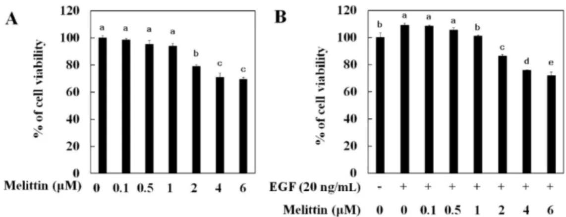
멜리틴에 의한 폐암세포 이동과 침투 억제 효과 멜리틴이 폐암세포 생존에 미치는 영향을 확인하기 위해 MTT 를 수행하였다. 멜리틴 0.1, 0.5, 1, 2, 4, 6 μM을 24 시간 처리하 여 약물에 대한 폐암세포 생존율을 확인한 결과 멜리틴 2-4 μM 처리에 농도의존적으로 세포 생존율이 20, 30, 35% 정도 각각 감 소하였다(Fig. 1A). 암세포는 여러 가지 사이토카인(cytokine)에 의 해 세포의 이동이 증가되며, 그 중 EGF 또한 EMT를 유도하여 세 포의 이동을 증가시킨다고 알려져 있다(16). 이에 EGF에 의해 EMT 형질로 변화된 세포에서 멜리틴의 약물 독성을 알아보기 위해 EGF 와 멜리틴을 같이 처리하여 폐암세포에 대한 멜리틴의 약물 독성 을 확인하였다. 그 결과 멜리틴 단독 처리와 유사하게 2-4 μM 농 도에서 세포 생존율이 18, 30, 35% 정도 각각 감소하였다(Fig. 1B). 따라서 멜리틴의 약물 독성으로 인해 암세포 이동과 침투, EMT 억제 효과가 나타날 수 있으므로 향후 실험은 20% 미만의 세포 독성을 나타내는 2 μM 농도를 최고농도로 설정하여 수행하였다. EGF는 암세포를 자극하여 세포의 이동과 침투를 유도하고 암 세포 성장과 증식을 증가시키는 것으로 알려져 있으며(16), 이전 에 도출된 연구결과에서도 EGF에 의해 증가된 암세포의 이동과침투가 멜리틴에 의해 억제되었다(1). 따라서 EGF 처리에 의해 증가된 암세포 이동과 침투에 멜리틴이 미치는 영향을 확인하기 위해 20 ng/mL EGF와 멜리틴을 처리하여 암세포의 이동을 확인 하였다. 그 결과 EGF 처리에 의해 증가된 암세포의 이동이 멜리 틴 처리에 의해 농도의존적으로 억제되었다(Fig. 2A). 마트리젤 침입 분석(Matrigel invasion assay)을 이용하여 확인한 세포 침투 실험에서도 EGF 처리에 의해 증가된 세포 침투가 멜리틴 처리 에 의해 농도의존적으로 억제되었다. 이를 통해 멜리틴은 EGF 처리에 의해 증가된 세포 이동과 침투를 억제하는 효능이 있음 을 알 수 있었다. 멜리틴에 의한 EMT 관련 유전자 발현 조절 효과 암세포 전이 원인 중 하나이며, 세포 이동과 침투에 중요하게 작용하는 EMT(1,8)의 조절 유전자를 확인 하기 앞서 EGF 처리 에 따른 세포 이동과 형태학적인 특성을 확인하기 위하여 세포 이동에 필요한 돌기 형성 유도 인자인 액틴 필라멘트 발현을 확 인하였다(1). 액틴 필라멘트를 염색한 결과, 아무것도 처리하지 않은 대조군에 비해 EGF 처리에 의해 액틴 필라멘트 발현이 세 포막 표면에서 증가하였으며, 멜리틴 농도의존적으로 액틴 필라 멘트 발현이 감소하였다(Fig. 3A). 액틴 필라멘트는 세포이동에 필요한 세포돌기 형성에 관여하는 유전자로, 액틴 필라멘트 발현 의 증가에 의해 세포의 이동이 증가됨이 알려져 있다(1). 따라서 멜리틴이 세포의 돌기 형성 억제 효능이 있음을 알 수 있었다. 다음으로 EMT 조절 유전자인 이카드헤린과 바이멘틴 발현을 도출하였다. EMT 진행에 따라 상피세포 확인 유전자인 이카드헤 린의 발현은 감소하며, 간엽세포 확인 유전자인 바이멘틴과 EMT 조절 유전자인 피브로넥틴 발현은 증가하였다(3,6). 우리 연구에서 도 EGF 처리에 의해 이카드헤린의 발현이 감소되었으며, 멜리틴 처리에 의해 농도의존적으로 증가하였다(Fig. 3B). 또한 EGF 처리 에 의해 증가된 바이멘틴 발현이 멜리틴 농도의존적으로 감소되 었으므로, 멜리틴이 EGF에 의해 유도된 폐암세포의 EMT를 억제 하는 것을 확인하였다(Fig. 3B). EGF에 의해 유도된 EMT가 멜리 틴 처리에 의해 억제 효과를 RT-PCR을 수행하여 재확인 하였다. EMT 유도 시 발현이 증가되는 피브로넥틴 발현을 확인한 결과 EGF에 의해 증가된 피브로넥틴 mRNA 발현이 멜리틴 농도의존 적으로 감소하였다(Fig. 3C). 이 결과들을 통해 EGF로 유도된 세 포 이동과 침투가 EMT 조절에 의해 나타나며, 멜리틴이 EMT 억 제를 통하여 세포 이동과 침투를 감소시키는 것을 알 수 있었다. Fig. 1. Cytotoxicity effects of melittin in lung cancer cells. (A) Lung cancer cells were treated with indicated concentration of melittin alone or in the presence of 20 ng/mL EGF (B) for 24 h. Cell viability was determined by a MTT assay. The data represent the means±SE of three separate experiments. Data were statically considered at p<0.05, and different small letters (a-e) in the graph represent statistical difference.
Fig. 2. Inhibitory effect of melittin on EGF-induced cell migration and invasion in lung cancer cells. (A) Wound healing assay and (B) invasion assay were carried out using 0.5, 1, and 2μM of melittin in the presence of 20 ng/mL EGF for 24 h. Then, migrated cells and invaded cells were photographed under a phase contrast microscope. The migration and invasion activity of cell were determined as the number of each condition cell divided by the control cell number.
멜리틴에 의한 EMT 조절 전자인자 발현 억제 효과
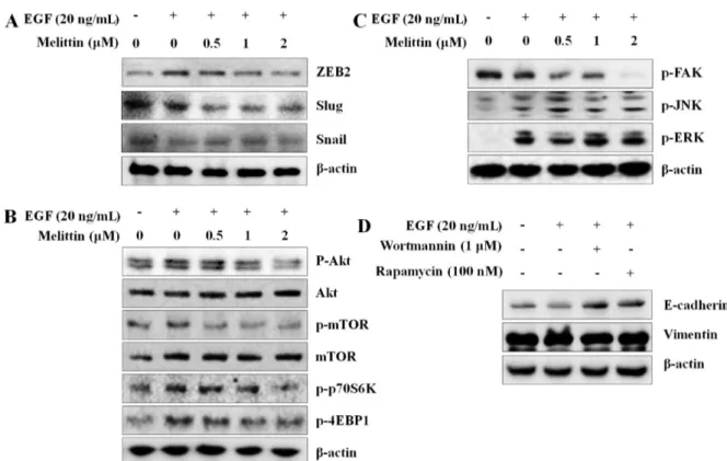
EMT 유도 시 발현이 증가되는 전사인자로 ZEB2, Slug, Snail 이 알려져 있으며, 이들 전사인자들의 활성은 이카드헤린을 감소 시키고 바이멘틴의 발현을 유도한다고 보고되었다(17-19). 멜리틴 의 EMT를 조절하는 전사인자에 대한 영향을 확인하기 위해 ZEB2, Slug, Snail의 발현을 웨스턴 블롯을 통해 분석하였다. 실 험결과 EGF 처리에 의해 증가된 이들 유전자의 발현이 멜리틴 처리에 의해 농도의존적으로 감소하였다(Fig. 4A). 이와 같은 결 과로 멜리틴이 EGF로 유도된 ZEB2와 Slug, Snail 발현 억제를 통해 EMT의 표적 인자인 이카드헤린과 바이멘틴의 발현을 조절 하여 EMT를 억제하는 것으로 예상된다.
멜리틴에 의한 AKT/mTOR와 FAK 활성 조절 효과
EMT는 mTOR (mammalian target of rapamycin) 신호 메커니 즘이나 FAK (focal adhesion kinase) 신호 메커니즘에 의해 조절 되는 것으로 알려져 있다(20-22). 최근 발표된 연구에서도 메트포 민(metformin)이 mTOR 메커니즘 억제를 통해 갑상샘암의 EMT 와 세포 이동과 침투를 억제한다고 확인되었다(23). 따라서 EGF 로 유도된 EMT 조건에서 멜리틴이 mTOR 신호 메커니즘에 미 치는 영향을 확인한 결과 EGF 처리에 의해 증가된 AKT/mTOR 의 인산화가 증가하였으며, 멜리틴에 의해 감소하였다(Fig. 4B). AKT/mTOR의 단백질 발현에는 멜리틴이 영향이 없었으므로 이 들의 인산화 조절을 통해 활성을 억제함을 알 수 있었다. 뿐만 아니라 AKT/mTOR의 하위 메커니즘인 p70S6K, 4EBP1의 인산 화가 멜리틴 처리에 의해 농도의존적으로 억제하는 것으로 나타 났다. 또한 FAK의 활성 억제는 EMT를 억제하며, 이에 따라 암 세포의 항암활성이 증가된다는 연구결과가 발표되었다(24,25). 따 라서 FAK의 활성을 확인한 결과 멜리틴 처리에 의해 농도의존 적으로 억제되었다. 그러나 A549세포에 EGF를 처리하였을 때 FAK가 증가하지 않았으며, FAK 하위 메커니즘으로 알려진 JNK 와 ERK는 EGF 처리에 의해 인산화는 증가하였으나 멜리틴 처 리에 의해 억제되지 않았다(Fig. 4C). 이와 같은 결과를 통해 FAK 의 활성이 억제 효능이 멜리틴 조절에 의한 EMT와는 관련이 없 을 것으로 예상되었다. 따라서 멜리틴이 AKT/mTOR 신호 메커니즘 억제를 통해 EMT 현상을 조절하는 것으로 예상되므로, AKT/mTOR의 억제제를 이 용하여 EMT의 변화를 관찰하였다. AKT억제제인 워트마닌(wort-mannin)과 mTOR의 라파마이신(rapamycin)을 처리한 결과 EGF에 의해 감소된 이카드헤린이 증가되었다(Fig. 4D). EGF에 의해 유 도된 바이멘틴의 발현은 워트마닌과 라파마이신에 의해 증가되 었으므로, AKT/mTOR의 경로를 통해 EMT가 조절됨을 확인하였 다. 본 연구 결과를 통해 멜리틴은 EGF로 유도된 AKT, mTOR, p70S6K, 4EBP1의 인산화 억제와 EMT 억제 전사인자인 Slug, Snail, ZEB2의 발현 억제를 통해 EMT를 억제하며, 최종적으로 폐암세포의 이동과 침투를 억제함을 도출하였다.
요
약
멜리틴은 봉독의 주요 성분 중 하나로 항염증과 항암활성 효 과를 가지고 있다. 우리는 폐암세포에서 멜리틴이 EMT 억제를 통해 암세포 이동과 침투를 억제하는 사실을 확인하였다. 멜리틴 은 EGF로 유도된 폐암 세포 이동과 침투를 억제하였을 뿐만 아 니라 EMT와 관련된 단백질인 이카드헤린의 발현을 증가시켰으 며, 바이멘틴과 피브로넥틴 발현은 감소시켰다. 또한 멜리틴에 의 Fig. 3. Inhibitory effect of melittin on EGF-induced EMT in lung cancer cells. (A) Lung cancer cells were pretreated with melittin for 2 h, and then treated with 20 ng/mL EGF for 1 h. F-actin was stained as described in methods and photographed. (B) Cells were pretreated with melittin for 1 h, and then treated with 20 ng/mL EGF for 24 h. Cells lysates were subjected to western blotting with E-cadherin and vimentin. β-Actin was used as a loading control. (C) mRNA was subjected to RT-PCR with primer and fibronectin. β-Actin was used as a control.한 EMT조절 전사인자인 ZEB2, Slug, Snail의 발현을 확인한 결 과 멜리틴 처리에 의해 농도의존적으로 발현이 감소하였다. 또한 작용 메커니즘을 확인하기 위해 mTOR와 FAK 메커니즘을 확인 한 실험에서 EGF 처리에 의해 증가한 AKT, mTOR, p70S6K, 4EBP1의 인산화가 멜리틴 농도의존적으로 감소하였다. 그러나 FAK는 EGF에 의해 변화가 없었으며, EKR, JNK 메커니즘은 EGF 처리에 의해 인산화가 증가하였으나 멜리틴 처리에 의해 아 무런 영향을 받지 않았다. 그러므로, 폐암세포의 세포 이동과 침 투에 대한 멜리틴의 억제효과는 AKT/mTOR/P70S6K/4EBP1 기전 억제를 통해 EMT를 억제하여 세포 이동과 침투를 억제하는 것 으로 보인다.
감사의 글
이 연구는 2015년도 대구가톨릭대학교 의과학연구소 연구비의 지원으로 이루어졌음.References
1. Jeong YJ, Choi Y, Shin JM, Cho HJ, Kang JH, Park KK, Choe JY, Bae YS, Han SM, Kim CH, Chang HW, Chang YC. Melittin suppresses EGF-induced cell motility and invasion by inhibiting PI3K/Akt/mTOR signaling pathway in breast cancer cells. Food Chem. Toxicol. 68: 218-225 (2014)
2. Yun Y, Gao R, Yue H, Guo L, Li G, Sang N. Sulfate aerosols
promote lung cancer metastasis by epigenetically regulating the epithelial-to-mesenchymal transition (EMT). Environ. Sci. Tech-nol. 51: 11401-11411 (2017)
3. Lamouille S, Xu J, Derynck R. Molecular mechanisms of epithe-lial-mesenchymal transition. Nat. Rev. Mol. Cell Biol. 15: 178-196 (2014)
4. Heerboth S, Housman G, Leary M, Longacre M, Byler S, Lapin-ska K, Willbanks A, Sarkar S. EMT and tumor metastasis. Clin. Transl. Med. 4: 6 (2015)
5. Kinugawa K, Minobe WA, Wood WM, Ridgway EC, Baxter JD, Ribeiro RC, Tawadrous MF, Lowes BA, Long CS, Bristow MR. Signaling pathways responsible for fetal gene induction in the failing human heart: evidence for altered thyroid hormone recep-tor gene expression. Circulation 103: 1089-1094 (2001)
6. Derksen PW, Liu X, Saridin F, van der Gulden H, Zevenhoven J, Evers B, van Beijnum JR, Griffioen AW, Vink J, Krimpenfort P, Peterse JL, Cardiff RD, Berns A, Jonkers J. Somatic inactivation of E-cadherin and p53 in mice leads to metastatic lobular mam-mary carcinoma through induction of anoikis resistance and angiogenesis. Cancer Cell 10: 437-449 (2006)
7. Zhang C, Liu T, Wang G, Wang H, Che X, Gao X, Liu H. Rac3 regulates cell invasion, migration and EMT in lung adenocarci-noma through p38 MAPK pathway. J. Cancer 8: 2511-2522 (2017)
8. Liu XL, Zhang XT, Meng J, Zhang HF, Zhao Y, Li C, Sun Y, Mei QB, Zhang F, Zhang T. ING5 knockdown enhances migra-tion and invasion of lung cancer cells by inducing EMT via EGFR/PI3K/Akt and IL-6/STAT3 signaling pathways. Oncotarget 8: 54265-54276 (2017)
9. Billingham ME, Morley J, Hanson JM, Shipolini RA, Vernon CA. Letter: An anti-inflammatory peptide from bee venom.
Fig. 4. Inhibitory effect of melittin on EMT related transcription factors and mTOR signaling pathway. (A) Cells were pretreated with melittin for 1 h, and then treated with 20 ng/mL EGF for 24 h. Cells lysates were subjected to western blotting with Slug, Snail, and ZEB2. β-Actin was used as a loading control. (B) Cells were pretreated with melittin for 1 h, and then treated with 20 ng/mL EGF for 30 min. The phosphorylation levels of AKT, mTOR, p70S6K, 4EBP1 and the total protein expression of AKT and mTOR were subjected to western blotting. (C) The phosphorylation levels of FAK, JNK, and ERK were subjected to western blotting. β-Actin was used as loading control. (D) Cells were pretreated with wortmannin (1μM) and rapamycin (100 nM) for 1 h, and then treated with 20 ng/mL EGF for 24 h. Cells lysates were subjected to western blotting with E-cadherin and vimentin. β-Actin was used as a loading control.
13. Park JH, Lee WR, Kim HS, Han SM, Chang YC, Park KK. Pro-tective effects of melittin on tumor necrosis factor-alpha induced hepatic damage through suppression of apoptotic pathway and nuclear factor-kappa B activation. Exp. Biol. Med. (Maywood) 239: 1705-1714 (2014)
14. Jeong YJ, Shin JM, Bae YS, Cho HJ, Park KK, Choe JY, Han SM, Moon SK, Kim WJ, Choi YH, Kim CH, Chang HW, Chang YC. Melittin has a chondroprotective effect by inhibiting MMP-1 and MMP-8 expressions via blocking NF-kappaB and AP-1 sig-naling pathway in chondrocytes. Int. Immunopharmacol. 25: 400-405 (2015)
15. Park JH, Jeong YJ, Park KK, Cho HJ, Chung IK, Min KS, Kim M, Lee KG, Yeo JH, Park KK, Chang YC. Melittin suppresses PMA-induced tumor cell invasion by inhibiting NF-kappaB and AP-1-dependent MMP-9 expression. Mol. Cells 29: 209-215 (2010)
16. Manupati K, Dhoke NR, Debnath T, Yeeravalli R, Guguloth K, Saeidpour S, De UC, Debnath S, Das A. Inhibiting epidermal growth factor receptor signalling potentiates mesenchymal-epithe-lial transition of breast cancer stem cells and their responsiveness
thelial-mesenchymal transition and tumor growth in pancreatic cancer via deactivating Akt/mTOR signaling. Biochem. Biophys. Res. Commun. 493: 455-460 (2017)
21. Xu M, Qin S, Cao F, Ding S, Li M. MicroRNA-379 inhibits metastasis and epithelial-mesenchymal transition via targeting FAK/AKT signaling in gastric cancer. Int. J. Oncol. 51: 867-876 (2017)
22. Cicchini C, Laudadio I, Citarella F, Corazzari M, Steindler C, Conigliaro A, Fantoni A, Amicone L, Tripodi M. TGFbeta-induced EMT requires focal adhesion kinase (FAK) signaling. Exp. Cell Res. 314: 143-152 (2008)
23. Han B, Cui H, Kang L, Zhang X, Jin Z, Lu L, Fan Z. Metformin inhibits thyroid cancer cell growth, migration, and EMT through the mTOR pathway. Tumour. Biol. 36: 6295-6304 (2015)
24. Bailey KM, Liu J. Caveolin-1 up-regulation during epithelial to mesenchymal transition is mediated by focal adhesion kinase. J. Biol. Chem. 283: 13714-13724 (2008)
25. Gonzalez DM, Medici D. Signaling mechanisms of the epithelial-mesenchymal transition. Sci. Signal. 7: re8 (2014)