Original Article Micro Deluxe Phantom을 통한 핀홀 콜리메이터 초점의 직경별 분해능 평가
삼성서울병원 핵의학과
안병호·연준호·김수영·최성욱
Resolution Evaluation of a Pinhole Collimator according to the Aperture Diameter using Micro Deluxe Phantom
Byung Ho An, Joon Ho Yeon, Soo Young Kim and Sung Wook Choi Dept. of Nuclear Medicine, Samsung Medical Center, Seoul, Korea
Purpose It is hard to obtain high quality images of knee and T.M joint because of a lot of soft tissues in the knee and T.M joint area. Most conventional system for high resolution scintigraphy was used by 4 mm aperture pinhole collimator. Performance comparison of high-resolution pinhole SPECT for Micro deluxe phantom using conventional system. the aim of this study is to evaluate performance of each aperture according to the diameter size and the usefulness of 24-hour delayed bone scintigraphy.
Materials and Methods In this study 6 mm, 8 mm diameter pinhole collimators were mounted on Siemens E.CAM systems. In order to evaluate performance evaluation of each aperture and Micro Deluxe phantom was used for performance comparison of conventional SPECT system, Projection data were obtained with 9 degree increment per 30 second. Transverse images were reconstructed using dedicated OSEM algorithm with recovery of detector blurring. 99mTc-HDP source was used for 24-hour delayed bone scintigraphy.
Results The knee joint images obtained with 24-hour delay were improved more than those obtained with 3-hour delay in our study. The 6 mm and 8 mm pinhole collimators FWHM have improved by 28% SNR and Uniformity have improved by 35%, Contrast has improved by 7% in 24-hour delayed knee joint image. While in 24-hour delayed T.M joint image of the 6 mm and 8 mm pinhole collimators FWHM have decreased by 60% SNR has decreased by 20% and Uniformity has decreased by 25%, Contrast has decreased significantly.
Conclusion Pinhole collimators with 6 mm and 8 mm diameter could offer a superior performance for 24-hour delayed bone scintigraphy. The use of 24-hour delayed image provides additional benefits for pinhole scintigraphy of knee joint. Therefore, we expect that it is useful for precise diagnosis of knee joint and it is applicable to others joint imaging.
Key Words Micro Deluxe phantom, Pinhole collimator, Resolution, Contrast, OSEM
1)
서 론
핀홀 콜리메이터를 이용한 검사는 뼈검사(wholebody bone scan)를 통한 정적검사(static scan)와 갑상선검사 (thyroid scan)가 있다. 2 mm 초점(aperture)은 일반적으로 소
∙ Received: 2015. 4. 3 Accepted: 2015. 4. 30
∙ Corresponding Author: Byung Ho An
Department of Nuclear Medicine, Samsung Medical Center, 50 Il won-dong, Kang Nam-gu, Seoul, 135-701, Korea Tel: +82-2-3410-2637, Fax: +82-2-3410-2639 E-mail: [email protected]
동물 검사에 주로 이용되고 있으며 2 mm 이하의 다중 초점 을 이용한 핀홀 콜리메이터는 고분해능으로 영상 질을 향상 시키고 있다1). 기존 감마카메라에 장착된 핀홀 콜리메이터 는 4 mm 초점만을 사용하고 있으며 현재, Gamma camera E.CAM (Siemens Germany) 핀홀 콜리메이터(Pinhole collimator) 초점은 4 mm, 6 mm, 8 mm, Gamma camera Infinia (GE USA) 핀홀 콜리메이터 초점은 2 mm, 4 mm, 8 mm가 제 작 및 판매되고 있다. 본원에서는 2 mm 초점을 자체 제작하 여 소동물 정적검사 및 SPECT 검사에 이용하고 있다. 핵의
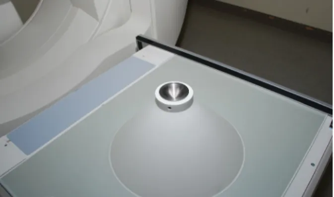
Fig. 1. Pinhole collimator was used conventional system.
학 체내감사에서 기존 감마카메라에 장착된 핀홀 콜리메이 터의 4 mm 초점만을 이용하여 갑상선 검사와 뼈검사 정적검 사에서 고관절(Hip joint), 악관절(T.M jpint), 슬관절(Knee joint), 천장관절(S.I joint)등을 검사를 한다2). 당일 검사를 하 는 이유는 Matrix size와 Zoom factor에 따른 시간에 따른 계 수치 한계로 인해 24시간 지연검사를 시행하기에는 어려운 부분이었다. 본원에서는 이러한 시간에 따른 계수치 한계성 을 극복하기 위하여 핀홀 콜리메이터의 6 mm, 8 mm 초점을 이용하여 고선량 99mTcO4-
222 M㏃ (6 mCi)와 저선량 11.1 M㏃
(300 μCi) Micro Deluxe Phantom을 이용하여 획득한 SPECT (Single Photon Computed Tomography) 재구성 영상을 평가 하고 핀홀 콜리메이터의 6 mm, 8mm 초점을 이용하여 획득 한 임상영상의 관심영역(Region of Interest, ROI)을 대상으 로 공간분해능(Spatial resolution), 신호대 잡음비(Signal to Noise Ratio, SNR), 대조도(Contrast), 균일도(Uniformity)를 비교 및 분석하여 본원에서는 핀홀 콜리메이터 각 초점 크기 별 성능평가 및 특성을 알아보고 24시간 지연검사에 유용한 초점을 찾아 효율성을 알아 보았다.
실험재료 및 방법
1. 장비 및 대상이 실험에 사용한 장비는 Gamma camera E.CAM (Siemens, Germany)이고 디텍터에 핀홀 콜리메이터를(Fig. 1) 장착 하
2. 연구 방법
1) 분해능 실험
핀홀 콜리메이터의 각 초점별 FWHM (Full Width at Half maximum) 평가를 위해 지름 0.58 mm의 capillary tube 안에 선원당 5.5 M㏃을 주입한 두 개의 선원을 1 cm 간격을 두고 평행하게 위치시킨 후 테이프로 고정하여 FWHM 평가용 선 선원(line source)를 제작하였고 기존 감마카메라에 장착한 2 mm, 4 mm, 6 mm, 8 mm 초점을 이용한 핀홀 콜리메이터로 SPECT 획득한 영상 프로토콜은 Matrix size 256×256, Zoom factor 2.0, frame당 30 sec씩 9° 간격으로 얻었으며 자체 제작 한 분석 프로그램을 이용하여 공간분해능을 평가 하였다 (Fig. 4).
2) 팬텀 실험
핀홀 콜리메이터 초점 중심과 테이블의 균형을 맞추고 점 선원을 이용하여 회전중심(Center of Rotation, COR) 보정을 한다. Micro Deluxe Phantom 내부는 1.2 mm, 1.6 mm, 2.4 mm, 3.2 mm, 4.0 mm, 4.8 mm 총 6종류의 hole로 구성되어 있으며 고선량 99mTcO4-
222 M㏃ (6 mCi)와 저선량 11.1 M㏃(300 μCi) 를 volume 15 ㎖ 증류수와 혼합한 Micro Deluxe Phantom을 기존 감마카메라에 장착한 6 mm, 8 mm 초점을 이용한 핀홀 콜리메이터로 SPECT를 시행한다. 검출기를 회전시켜 투사 상(projection image)를 얻은 후 OSEM(Odered Subset Expectation Maximization)을 사용하여 영상을 재구성 하였다(Fig. 5).
matrix size는 256x256, Zoom은 2.0으로 설정하고 검출기는 9°씩 회전시켜 frame당 30sec 씩 총 40개의 투사상을 획득하 였으며(Fig. 6) OSEM의 반복횟수는 3회 부분집합은 5로 설 정하여 재구성 하였다(Fig. 7)3).
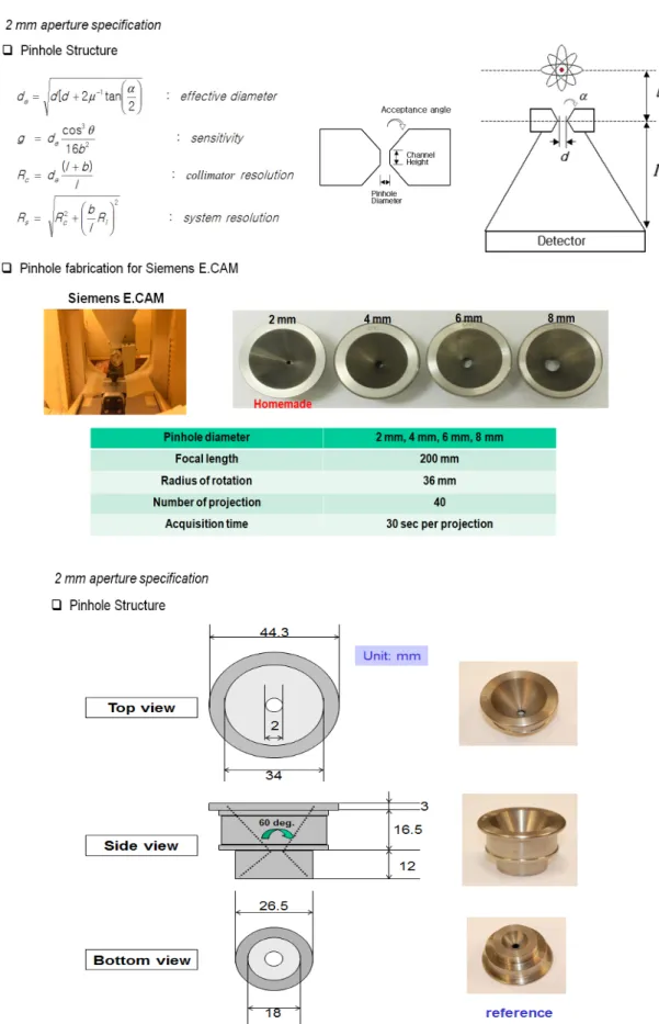
Fig. 2. Pinhole collimator design and 2 mm aperture specification
Fig. 3. Photographs of the Micro deluxe phantom with cold-rod (left) and hot-rod insert (right). The cylinder has a 45 mm inner diameter and 26 mm long rods in six sectors.
Fig. 4. We used X-Profile a homemade analysis program using IDL 6.4version.
Fig. 5. SPECT data acquisition procedure.
Fig. 6. Image reconstruction procedure.
+
=
t t j
i
t t j t j
i j j
i
a
S x a
x
, , 1
/ μ
System matrix (at,j) was obtained by the numerical calculation of point spread function (PSF) Fig. 7. Reconstructed equation using dedicated OSEM algorithm with recovery of detector blurring.
3)임상 영상
99mTc-HDP 방사성의약품을 740 M㏃ (20 mCi)를 정맥주 사하고 3시간후 전신영상을 획득하고(Fig. 8) 핀홀 콜리메이 터의 6 mm, 8 mm 초점을 이용하여 3시간 및 24시간 정적영
상을 얻는다(Fig. 9). 전신영상은 Matrix size 256x1024, Zoom factor 1.0, Scan speed 20이고 3시간 및 24시간 정적영 상은 Matrix size 256x1024, Zoom factor 1.45, 100kcount이다.
획득한 영상은 자체 제작한 분석 프로그램을 이용하여
Fig. 8. hour wholebody bone scintigraphy after 99mTc-HDP 740 M
㏃ (20 mCi) injection.
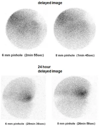
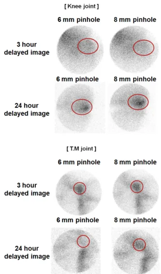
Fig. 9. 3 hour and 24 hour delayed image of knee joint using 6 mm, 8 mm aperture.
Fig. 10. 3 hour and 24 hour delayed image of T.M joint using 6 mm, 8 mm aperture.
FWHM을 분석하고 임상영상 관심영역(Region of Interest, ROI)을 대상으로 공간분해능(Spatial resolution), 신호대 잡 음비, 균일도, 대조도를 분석한다.
결 과
1. 분해능 평가핀홀 콜리메이터 cone길이, 초점크기, 입사각도 및 선 선 원과 초점간 거리를 고려하고 각 capillary tube간 거리와 x,y 프로파일에 나타난 2 점선원의 pixel값을 산정한다4-5). 선 선 원을 이용한 SPECT영상은 X축 프로파일 분석 프로그램을 이용하여 재구성된 FWHM 평가에서 2 mm 초점에서 2.2 mm FWHM, 4 mm에서 3.2 mm FWHM, 6 mm에서 5.4 mm FWHM, 8 mm에서 7.5 mm FWHM의 각 초점별 FWHM이 나타났다 (Fig. 11).
2. 팬텀 영상 평가
각 초점별 계수 설정법에 대한 영상은 다음과 같이 나타 났으며(Fig. 12) 고선량을 혼합한 Micro deluxe phantom을 통 한 직경이 2 mm일 때 2.4 mm, 4 mm일 때 3.2 mm, 6 mm일 때 4.8 mm 까지 육안 적 구분이 되었지만 8 mm의 경우 넓은 반
Fig. 11. Planar resolution using line source.
Fig. 12. SPECT imaging perfomance using 40 projection with micro-deluxe phantom.
치 폭 으로 인해 육안 적 식별이 불가능 하였다6). 저선량을 혼 합한 Micro deluxe phantom을 통한 직경이 2 mm, 4 mm, 6 mm 일 때는 4.8 mm 까지 육안적 구분이 되었고 8 mm의 경우 넓 은 반치폭 으로 인해 육안적 식별이 불가능 하였다(Fig. 12).
3. 임상 영상 평가
핀홀 콜리메이터의 6 mm, 8 mm 초점을 이용하여 슬관절 3시간 및 24시간 정적영상에서 염증질환 부위와 주변부의 ROI를 통한 신호대 잡음비, 균일도, 대조도 평가에서는 3시 간 정적검사 결과에서 신호대 잡음비는 6 mm, 8 mm일때 각
각 2.869, 2.853으로 24시간 지연 정적영상에서는 6 mm, 8 mm에서 3.804, 3.838이고 균일도는 6 mm, 8 mm일 때 각각 0.349, 0.351이고 24시간 지연 정적영상에서는 6 mm, 8 mm 에서 0.263, 0.261로 나타났으며 대조도는 6 mm, 8 mm일때 각각 2.291, 2.263으로 24시간 지연 정적영상에서는 6 mm, 8 mm에서 2.367, 2.364 결과값을 얻었다. 슬관절 24시간 지연 정적검사에서는 모두 향상된 값을 얻을수 있었지만 악관절 24시간 지연 정적검사에서는 신호대 잡음비, 균일도, 대조 도값 모두 현저히 감소됨을 알수 있었다(Table 1). 또한, FWHM평가는 슬관절 3시간 정적검사에서는 6 mm, 8 mm에 서 17.2 mm FWHM, 18.2 mm FWHM, 24시간 지연 정적영상
Table 1. SNR, Uniformity, Contrast Value of Knee and T.M joint
Knee joint R1 총계수
R1 표준분포
R1 평균계수
R2 총계수
R2 표준분포
R2
평균계수 SNR Uniformity Contrast
6 mm 3 hrs 2872 3.35 9.61 867 1.83 2.92 2.869 0.349 2.291
8 mm 3 hrs 2800 3.26 9.30 850 1.66 2.85 2.853 0.351 2.263
6 mm 24 hrs 5707 5.00 19.0 1694 2.50 5.65 3.804 0.263 2.367
8mm 24 hrs 5703 5.02 19.3 1745 2.41 5.78 3.838 0.261 2.334
T.M joint R1 총계수
R1 표준분포
R1 평균계수
R2 총계수
R2 표준분포
R2
평균계수 SNR Uniformity Contrast
6 mm 3 hrs 2853 3.96 19.4 110 1.83 2.97 4.902 0.204 5.535
8 mm 3 hrs 2717 4.71 18.9 112 1.76 3.03 4.006 0.249 5.228
6 mm 24 hrs 1833 3.45 12.2 161 2.26 4.35 3.542 0.282 1.809
8 mm 24 hrs 1559 2.86 10.4 117 1.52 2.54 3.633 0.275 3.091
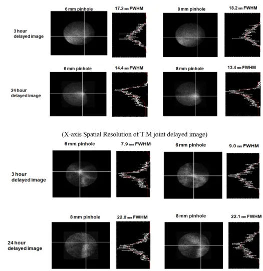
(X-axis Spatial Resolution of Knee joint delayed image)
(X-axis Spatial Resolution of T.M joint delayed image)
Fig. 13. Knee and T.M joint imaging perfomance using X-Profile a homemade analysis program.
Fig. 14. Region of interest of knee and T.M joint delayed image using 6 mm, 8 mm aperture.
에서는 6 mm, 8 mm에서는 14.4 mm FWHM, 13.4 mm FWHM값을 나타냈었고 악관절 3시간 정적검사에서는 6 mm, 8 mm에서 7.9 mm FWHM, 22.0 mm FWHM, 24시간 지 연 정적영상에서는 6 mm, 8 mm에서는 9.0 mm FWHM, 22.1 mm FWHM값을 산출 하였다(Fig. 13,14).
고 찰
본원에서 시행한 2 mm, 4 mm, 6 mm, 8mm 초점을 이용한 핀홀 콜리메이터 SPECT 검사시 첫 번째로 가장 고려해야 할 점은 핀홀 콜리메이터와 테이블간 회전중심 보정이었다. 기 존 테이블에 소동물 미니 테이블을 올려놓고 실험을 해야 하 므로 선 선원과 Micro deluxe phantom을 이용한 핀홀 콜리메 이터 SPECT 검사전 정확한 회전중심 보정이 요구 되어 장시
값을 입력하여 자체 분석 프로그램을 사용해야 하는점이다.
마지막으로 임상의 많은 환자를 대상으로 다양한 부위의 24 시간 지연검사를 시행하지 못하는 아쉬움이 남는다. 고분해 능을 얻을 수 있는 소동물 SPECT 장비가 없어도 기존 감마카 메라에 핀홀 콜리메이터를 장착하여 소동물 실험 및 24시간 지 연검사를 통해 고분해능 및 대조도가 높은 영상 획득이 가능 하 였다7-8).
결 론
일반적으로 24시간 지연검사시 방사성동위원소 감쇄로 검사시간 소요시간이 길어 원활한 검사 진행을 못하는 경우 가 많았지만 본 연구를 통해 임상에서 지연검사시 핀홀 콜리 메이터의 6 mm, 8 mm 초점을 이용하여 염증질환 환자 연조 직이 많이 포함된 무릎관절, 고관절, 천장관절에서 충분히 질적인 영상을 얻을 수 있다. 핀홀 콜리메이터 초점 직경 크 기에 따라 저선량 Micro deluxe phantom을 통한 분해능 변화 는 큰 차이가 없음을 알 수 있었다. 24시간 이후 지연 정적영 상 획득 시 고선량 Micro deluxe phantom의 경우 2 mm, 4 mm 초점에서 분해능이 가장 우수했지만 1시간 이상 소요되는 단점이 있으며, 상대적으로 저 선량일 경우 분해능이 다소 떨어지지만 감도가 우수한 6 mm, 8 mm 초점을 이용한다면 30분 이내 소요시간 영상을 재현이 가능하다는 장점이 있다.
팬텀영상의 분해능이 2 mm, 4 mm 초점보다 떨어지는 것은 선 선원에서 6 mm에서 5.4 mm FWHM, 8 mm에서 7.5 mm값 을 나타내므로 이번 실험에서 사용한 팬텀은 소동물 전용 팬 텀으로 최대로 구별이 가능한 크기가 4.8 mm FWHM이므로 6 mm, 8 mm 초점의 분해능을 나타내기에는 다소 어려웠다.
6 mm, 8 mm 초점에 맞는 팬텀을 사용한다면 충분히 질적인 영상 재현이 가능하다고 사료된다. 임상 지연영상에서 슬관 절 지연영상에서 8 mm 초점의 FWHM값이 좋은 것은 작은 계수욜(Low count rate)에서 핀홀직경이 큰 것이 검출효율이 높아서 핀홀 중심부보다 계수치가 많아져서 curve fiting이 잘되었기 때문이다9-10). 그러나, 잡음이 워낙 많아 두 결과 차 이는 미비하므로 지연 24시간 영상에서 6 mm, 8 mm 모두 좋
은 것을 알수 있었다. 악관절(T.M joint)은 상대적으로 FWHM값이 높게 나타난 것은 연조직이 뼈조직에 비해 상대 적으로 작기 때문이다. 핀홀 콜리메이터 초점 직경 크기에 따라 저선량 Micro deluxe phantom을 통한 분해능 변화는 큰 차이가 없음을 알 수 있었다. 24시간 이후 지연 정적영상 획 득 시 고선량 Micro deluxe phantom의 경우 2 mm, 4 mm 초점 에서 분해능이 가장 우수했지만 1시간 이상 소요되는 단점 이 있으며, 상대적으로 저 선량일 경우 분해능이 다소 떨어 지지만 감도가 우수한 6 mm, 8 mm 초점을 이용한다면 30분 이내 소요시간 영상을 재현이 가능하다는 장점이 있다. 임상 에서 염증질환 환자 연조직이 많이 포함된 무릎관절, 고관 절, 천장관절에서 충분히 질적인 영상을 얻을 수 있다고 사 료된다.
요 약
기존 감마카메라에 장착된 핀홀 콜리메이터 4 mm 초점을 이용하여 24시간 지연검사에는 슬관절, 악관절 연조직 계수 치가 작아 높은 질영상을 얻기에는 매우 어려운 부분이 있 다. 대부분 고분해능 영상 획득시 4 mm 직경의 초점만을 이 용한 검사를 시행해 왔다. 기존 감마카메라를 이용하여 Micro deluxe phantom의 고분해능 핀홀 콜리메이터 SPECT 을 비교 평가해 보았다. 본 연구에서는 각 초점 직경의 비교 평가와 24시간 지연검사 유용성을 평가해 보았다. 선 선원을 이용하여 6 mm, 8 mm 직경 초점을 핀홀 콜리메이터에 장착 하고 각 초점의 분해능을 평가해 보았으며 Micro deluxe phantom을 고선량 및 저선량으로 혼합하여 SPECT 영상을 획득하여 OSEM 알고리즘을 이용한 프로그램으로 영상을 재구성 및 분해능 평가를 하였다. 임상영상은 염증질환이 있 는 슬관절과 악관절의 3시간, 24시간 지연영상을 획득하여 관심영역과 주변부를 150 mm로 설정하여 신호대 잡음비, 대조도, 균일도를 비교 및 분석하였다. 슬관절 24시간 지연 영상에서 신호대 잡음비, 대조도, 균일도가 향상되었으나 악관절에서는 신호대 잡음비, 균일도는 저하 되었고 대조도 는 현저히 감소됨을 알수 있었다.
6 mm, 8 mm 초점을 이용한 핀홀 콜리메이터는 24시간 지 연영상에서 좀더 나은 정보를 얻을수 있으며 슬관절뿐만 아 니라 연조직이 많이 포함된 고관절, 천장관절에서 충분히 질적인 임상영상을 얻을 수 있다고 사료된다.
REFERENCES
1. 김중현, 이재성, 김진수, 이병일, 김수미, 정인순 등. Philips ARUS 감마카메라와 바늘구멍조준기를 이용한 소동물 SPECT 시스템의 개발 및 성능평가. 핵의학 분자영상 2005;39:6445-455.
2. 고창순, 핵의학, 제3판, 고려의학, 2008;101-103.
3. 안병호, 차은선, 석재동, 진광호, 핀홀콜리메이터를 이용하 여 SPECT 방법에 대한 고찰. 대한핵의학기술학지. 2007;
11:203-208.
4. 이광훈, 안병호, 김수영, 최성욱, 핀홀콜리메이터 초점의 직 경 크기 별 성능비교 및 평가. 대한핵의학기술학지. 2014;
18(1):104-109.
5. Byungho An, Byungjun Min, Junsang Bae, Yong Choi, KyungHan Lee and Byung-Tae Kim "Performance comparison of high-resolution pinhole SPECT for small animal imaging using two conventional SPECT systems" J Nucl Med. 2008; 49 (Supplement 1):417P.
6. J. H. Kim, Y. Choi, K. S. Joo, B. S. Sihn, J. W. Chong, S. E.
Kim, K-H. Lee, Y. S. Choe, B-T. Kim. "Development of a miniature scintillation camera using NaI(Tl) scintillator and PSPMT for scintimammography," Phys. Med. Biol., vol. 45, pp. 3481-3488, 2000.
7. D. P. McElroy, L. R. MacDonald, F. J. Beekman, Y. Wang, B.
E. Patt, J. S. Iwanczyk, et al., "Performance evaluation of A-SPECT: A high resolution desktop pinhole SPECT system for imaging small animals," IEEE Trans. Nucl. Sci., vol. 49, pp.
2139-2147, 2002.
8. Baek CH, An SJ, Kim HI, Kwak SW, Chung YH.
Development of a pinhole gamma camera for environmental monitoring. Radiation measurements. 2013;59:114-118.
9. Jacobowitz, Metzler H, Scottd. Geometric sensitivity of a pinhole collimator. International journal of mathematics and mathematical sciences. 2010:915.
10. Gauthe M, Mesras D, Canavese F, Samba A, Cachin F.
Differential diagnosis of trampoline fracture from osteomyelitis by bone scan with pinhole collimator. Annals of nuclear medicine. 2014;28:163-166.