Kawasaki disease (KD) is a systemic vasculitis characterized by fever, cervical lymphadenopathy, conjunctival injection, oral lesions, indurative edema, late desquamation of extremities, and polymorphous skin rashes. Complications of KD include coronary artery aneurysms and thrombosis, making it an important cause of acquired heart disease in children.1Clinical and epidemiological features strongly suggest that the etiology of KD is an infection.2These include young age of affected individuals range, (6 months to 5 years old), clinical features of the illness such as fever and self-limited course, the occurrence of epidemics with periodicity, and the geographic wave-like spread of illness during epidemics.2
Immunoglobulin V H Chain Gene Analysis of Peripheral Blood IgM-Producing B Cells in Patients
with Kawasaki Disease
Hyun Hee Lee,
1Jun Soo Shin,
2and Dong Soo Kim
31Department of Pediatrics, Kwandong University College of Medicine, Goyang;
Departments of 2Microbiology, 3Pediatrics, Yonsei University College of Medicine, Seoul, Korea.
Purpose:Kawasaki disease is a systemic vasculitis, and its etiology and pathogenesis are still not clear. Our study was undertaken to investigate the characteristics of the activation of B cells in the peripheral blood of Kawasaki disease (KD) patients and evidence of stimulation by superantigens. Materials and Methods:Blood samples were obtained from three patients (2 males, 1 female) with KD, who were admitted to our Hospital, Seoul, Korea. The mean age was 1.2 years. Distribution of B cells was studied in the acute and subacute phases of KD patients. From the RNA of B cells, we obtained complementary DNA (cDNA) and performed polymerase chain reaction (PCR).
To determine the oligoclonal expansion of immunoglobulin M (IgM) VHfamily, we cloned and sequenced the PCR products from each group and analyzed DNA. Results:In the peripheral blood of acute phase patients, T cells were significantly decreased (p < 0.05), whereas B cells were significantly increased (p < 0.05). When the first PCR was done on the B cell chains, VH1 to VH6 were all found to be expressed. The number of µ gene clones obtained from 3 patients was 312, and they belonged to VH3, VH4 and VH5 family. M99686 germ line was most frequently used and the next most frequently used, were X92224/J, L21967 and L21964. A similar order was seen in patients. Among the clones, 20 sets of clones showed the same base sequence and this was frequent between VH2 and VH5. There was one set, which showed almost the same base sequence between different patients, and the homology was 99.5%. Twenty sets of clones that had the same base sequence showed high similarity to the germ line (94 - 100%).
Among these, the clones that utilized the M99686 germ line were 4 sets which were most frequent. The 3- dimensional structure of one of these clones showed typical β‚ sheet structure of immunoglobulin chains.
Conclusion:The IgM transcripts expressed by the B cells in the peripheral blood of KD patients in the acute phase of the disease clearly showed an oligoclonal expansion, suggesting that KD is caused not by stimulation of a superantigen, but rather by a conventional antigen.
Key Words : Kawasaki disease, B cell, oligoclonal expansion, superantigen, conventional antigen
Received: October 15, 2008 Revised: January 19, 2009 Accepted: February 10, 2009
Corresponding author: Dr. Dong Soo Kim, Department of Pediatrics, Yonsei University College of Medicine, 250 Seongsan-ro, Seodaemun-gu, Seoul 120-752, Korea.
Tel: 82-2-2228-2057, Fax: 82-2-393-9118 E-mail: [email protected]
∙The authors have no financial conflicts of interest.
© Copyright:
Yonsei University College of Medicine 2009
INTRODUCTION
Immunologic mechanisms are thought to be important in disease activation, progression, and complications. The selective expansion of T cells expressing Vβ2 and Vβ8 families in the peripheral blood of acute KD patients has been observed, suggesting the involvement of superan- tigens.3Based upon a single study,4toxic shock syndrome toxin-1 or streptococcal superantigens were proposed to be related etiologically to KD, however, other investigators have not been able to confirm these findings. Over the past few years, it has been debated whether KD is developed as a response to a superantigen or to a conventional antigen.
Rowley et al.5reported that immunoglobulin (Ig) A produced in acute KD vascular tissue is oligoclonal, con- sistent with an antigen-driven immune response. In young infants, primary antigenic stimulation of peripheral blood induces B cells to predominantly secrete IgM.6,7In this study, we examined the clonality of the IgM response in peripheral blood during acute phase of the disease to determine actual immunologic response.
Patients
Blood samples were obtained from 3 patients (2 males, 1 female) with KD, who were admitted to our Hospital, Seoul, Korea. All patients satisfied at least 5 of the 6 diagnostic criteria for KD.8They exhibited typical clinical symptoms and signs of KD, and did not have any complications of coronary artery aneurysm during 6 months after diagnosis.
The mean age was 1.2 years (11, 15, and 18 months). All patients were treated with intravenous gammaglobulin in addition to high-dose aspirin. Whole blood was collected during the acute diagnostic phase before the initiation of any therapy and convalescent phase after therapy. Blood samples from the patients were obtained on the first day of diagnosis for each child and again on the 7th day of diag- nosis during the convalescent phase. Informed consent was obtained from the parents of the patients included in the study.
Cell preparation, RNA isolation and cDNA synthesis Mononuclear cells were isolated by Ficoll-Hypaque density gradient centrifugation, followed by B cell separation using anti-CD19-coated magnetic beads (Dynabeads M450 Pan B; Dynal, Oslo, Norway). Messenger RNA (mRNA) was isolated from 1×10⁶B cells by using the Oligotex Direct mRNA-Kit (Quiagen Inc., Chatsworth, CA, USA), and com- plementary DNA (cDNA) was prepared with the Supers- cript Preamplification System (GIBCO BRL, Gaithers- burg, MD, USA) and a random hexamer nucleotide mix.
Amplification of VHgenes
For analysis of VH genes, a set of 5’ primers specific for human VH1, VH2, VH3, VH4, VH5, and VH6 gene families in combination with a 3’ primer specific for the first exon of the µ constant region were employed (Table 1).9
All polymerase chain reaction (PCR) reactions were performed in a final volume of 100 µL with 20 pmol each of primer, 200 µM dNTP, 2.5 U of Taq DNA Polymerase (Promega, Madison, WI, USA) and 10 µL of 10 ×reaction buffer. Amplification was performed in a thermal cycler and consisted of 35 cycles of 1 min denaturation at 95˚C, 1 min primer annealing at 58˚C, and 1.5 min extension at 72˚C, with a final extension of 5 min at 72˚C PCR pro- ducts were analyzed in a 2% agarose gel, slices containing the specific band of 500 bp were excised, and DNA was purified using QIAquick Gel Extraction Kit (Quiagen).
Cloning of PCR products
Cloning of the PCR product was performed using pCR® 2.1-TOPO®kit and One Shot®competent E. coli (Invitro- gen, Carlsbad, CA, USA) according to the manufacturer’s protocol. After mixing PCR products and using pCR®2.1- TOPO®vector, ligation was performed for 5 min at room temperature. One Shot®competent E. coli was mixed and incubated for 5 min on ice. Then, SOC culture media were added after heating for 30 sec in a 42˚C water bath. After incubation in selective media containing X-gal (Sigma Co., St. Louis, MO, USA) and ampicillin (100 µg/mL), trans- formed positive clones were selected. After these clones
MATERIALS AND METHODS
Table 1. Sequences of Primers Used for Specific Amplification of IgM VHTranscripts Sense
VH1 5’-CCATCGACTGGACCTGG-3’
VH2 5’-ATGGACATACTTTGTTCCAC-3’
VH3 5’-ATGGAGTCTGGGCTGAGCTGGCTTT-3’
VH4 5’-ATGAAACACCTGTGGTTCTT-3’
VH5 5’-ATGGGGTCAACCGCCATCCT-3’
VH6 5’-ATGTCTGTCTCCTTCCTCAT-3’
Antisense
Cµ 5’-GCTCTAGAAGACGAGGGGGAAAAGGGTT-3’
IgM, immunoglobulin M.
were cultured in Luria-Bertani media containing ampicillin (100 µg/mL), plasmids were obtained and DNA was quantified using QIAprep®Miniprep kit (Qiagen, Valencia, CA, USA).
Sequencing of DNA
After plasmid DNA was mixed with BigDye®terminator mix (PE Applied Biosystems, Foster City, CA, USA) and sequencing primer, DNA was amplified for 10 sec at 96˚C, 5 sec at 50˚C, and 4 min at 60˚C for 25 cycles. Amplified DNA was washed with ethanol, and sequences were deter- mined with an ABI PRISM® 310. Each sequence was analyzed directionally using M13 forward primer (5’- GTAAAACGAGGCCAG-3’) and M13 reverse primer (5’-CAGGAAACAGCTATGAC-3’) by Macrogen Co.
Ltd (Seoul, Korea). Sequences were compared using DNA sequencing analysis software (PE Applied Biosystems, Wellesley, MA, USA) and DNAssist v 2.0 (Bellville, South Africa). These sequences were also compared with VH complementarity determining regions (CDRs) and human immunoglobulin germ line genes using http://imgt.
cines.fr and advanced Blast search program from the Genbank data base (National Center for Biotechnology Information, NIH, Bethesda, MD, USA).
Distribution of T and B cells in Kawasaki disease We observed significantly decreased T cells (p < 0.05) and increased B cells (p < 0.05) during the acute phase, when blood samples during the acute and convalescent phase were separately analyzed (Table 2). Serum IgM showed no significant difference during the acute phase (988
±
218 mg/dL) compared to control group (1046±
264 mg/dL).Serum IgM level was increased to 2723
±
259 mg/dL 24 hours after administration of intravenous gammaglobulin, and the increased level was maintained at 2470±
311 mg/dL 7 days thereafter.Identification of immunoglobulin V chain gene by RT- PCR
Reverse transcription polymerase chain reaction (RT-PCR) was performed to identify oligoclonal expansion of heavy chains in B-cells of peripheral blood during acute phase.
As shown in Fig. 1, all heavy chains from VH1 to VH6 were evenly distributed.
Table 2. Percentage of T and B Cells in Kawasaki Disease
Cell Stage
Controls
Acute Convalescent
T cell 62.5
±
9.5* 69.2±
7.9 70.0±
7.2B cell 14.8
±
6.5* 13.1±
6.0 8.4±
3.5Peripheral blood mononuclear cells were stained with anti-CD19 or anti-CD3 monoclonal antobodies and total 5,000 to 20,000 cells were analyzed.
*p < 0.05 compared with normal controls.
RESULTS
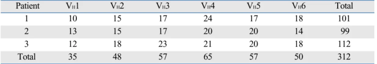
Table 3. Distribution of µ Gene Clones from cDNA of 3 Korean Patients with Kawasaki Disease
Patient VH1 VH2 VH3 VH4 VH5 VH6 Total
1 10 15 17 24 17 18 101
2 13 15 17 20 20 14 99
3 12 18 23 21 20 18 112
Total 35 48 57 65 57 50 312
cDNA, complementary DNA.
Fig. 1. Amplification of V regions of Ig µ heavy chain genes (VH1 - VH6) by RT-PCR.
M, marker; RT-PCR, reverse transcription polymerase chain reaction.
M VH1 VH2 VH3 VH4 VH5 VH6
IgM
VHfamily utilization in µ transcripts
The nucleotide and amino acid sequences of a total of 312 clones were analyzed (Table 3). A total of 101 clones from patient #1, 99 clones from patient #2 and 112 clones from patient #3 were analyzed. In all three patients, a preferential usage or deletion of a certain VHfamily seemed to be unlikely.
VHgene utilization
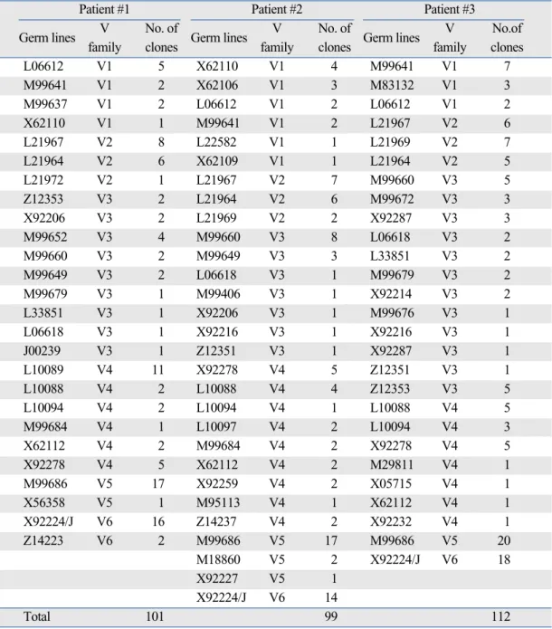
Fifty-four out of 312 clones (17.3%) used M99686 germ line gene of the VH5 family which was the most frequently used clone for the 3 patients. The X92224/J of the VH6 family was the second most frequently used (48 out of 312 clones: 15.4%) (Table 4). These findings suggest that there is a clonal relation among the respective B cells.
Somatic mutations in the µVHsegments
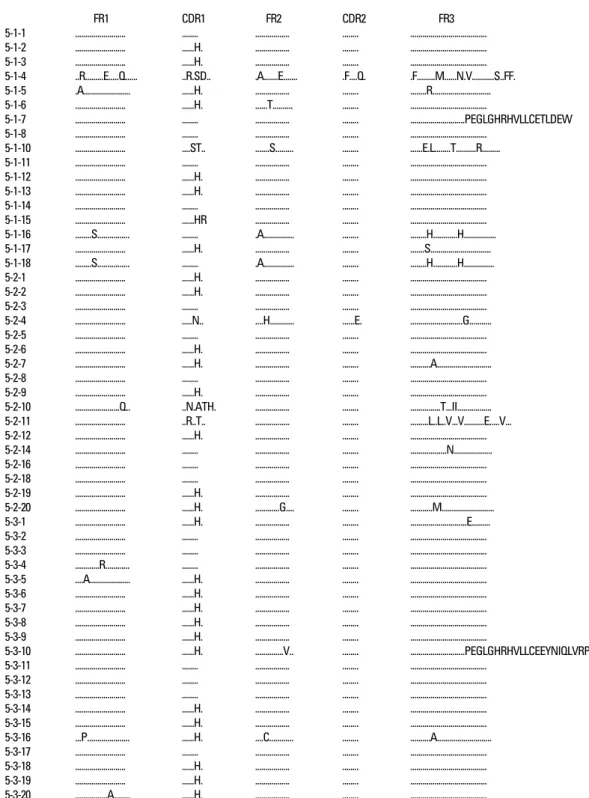
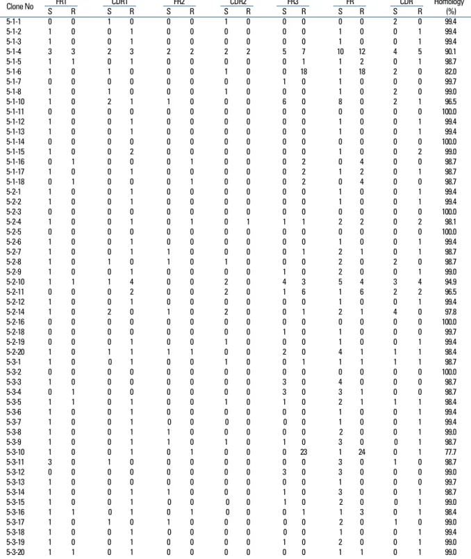
The VHgene segments were aligned to the germ-line sequence sharing the highest nucleotide identity. In general, both mutated and unmutated VH genes were found to be expressed in IgM-producing B cells of patients with KD.
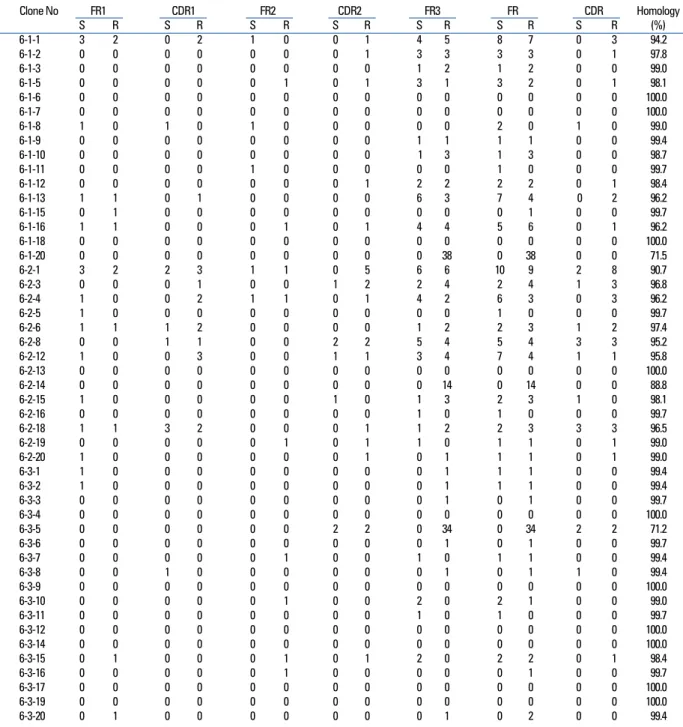
Eighty seven percent (47/54) of M99686 derived clones were more than 98% identical to the germ-line gene at the nucleotide level, and 70.8% (34/48) of X92224/J derived clones displayed greater than 98% homology. Two M99686 derived clones (clone #5-1-6 and clone #5-3-10) and 3 X92224/J derived clones (clone #6-1-20, clone #6-2-14 and clone #6-3-5) displayed extensive somatic mutations, especially in the FR3 region, whereas others showed only a few somatic mutations and some showed nearly identical
Table 4. Frequently Used Germ Lines in Each Patient
Patient #1 Patient #2 Patient #3
Germ lines V No. of
Germ lines V No. of
Germ lines V No.of
family clones family clones family clones
L06612 V1 5 X62110 V1 4 M99641 V1 7
M99641 V1 2 X62106 V1 3 M83132 V1 3
M99637 V1 2 L06612 V1 2 L06612 V1 2
X62110 V1 1 M99641 V1 2 L21967 V2 6
L21967 V2 8 L22582 V1 1 L21969 V2 7
L21964 V2 6 X62109 V1 1 L21964 V2 5
L21972 V2 1 L21967 V2 7 M99660 V3 5
Z12353 V3 2 L21964 V2 6 M99672 V3 3
X92206 V3 2 L21969 V2 2 X92287 V3 3
M99652 V3 4 M99660 V3 8 L06618 V3 2
M99660 V3 2 M99649 V3 3 L33851 V3 2
M99649 V3 2 L06618 V3 1 M99679 V3 2
M99679 V3 1 M99406 V3 1 X92214 V3 2
L33851 V3 1 X92206 V3 1 M99676 V3 1
L06618 V3 1 X92216 V3 1 X92216 V3 1
J00239 V3 1 Z12351 V3 1 X92287 V3 1
L10089 V4 11 X92278 V4 5 Z12351 V3 1
L10088 V4 2 L10088 V4 4 Z12353 V3 5
L10094 V4 2 L10094 V4 1 L10088 V4 5
M99684 V4 1 L10097 V4 2 L10094 V4 3
X62112 V4 2 M99684 V4 2 X92278 V4 5
X92278 V4 5 X62112 V4 2 M29811 V4 1
M99686 V5 17 X92259 V4 2 X05715 V4 1
X56358 V5 1 M95113 V4 1 X62112 V4 1
X92224/J V6 16 Z14237 V4 2 X92232 V4 1
Z14223 V6 2 M99686 V5 17 M99686 V5 20
M18860 V5 2 X92224/J V6 18
X92227 V5 1
X92224/J V6 14
Total 101 99 112
germ-line. The M99686 derived clones differed in a number of replacement mutations, mainly in CDR2, however, mutations in X92224/J derived clones were distributed more or less randomly over both CDR and FR segments (Figs. 2-7). Interestingly, two clones (clone #5-1-8 and clone #5-3-4) from 2 different patients were almost identical (Fig. 8). These were M99686 derived clones.
D and JHgene usage in µ transcripts
The analyzed CDR3 gene segments of M99686 derived clones appeared to differ markedly in length, ranging from 20 to 29 amino acids of 60 - 87 nucleotides (Fig. 3), while the analyzed CDR3 gene segments of X92224/J derived clones appeared to differ markedly in length, ranging from 15 to 34 amino acids of 45 - 102 nucleotides (Fig. 5).
FR1 CDR1 FR2 CDR2 FR3
5-1-1 ... ... ... ... ...
5-1-2 ... ...H. ... ... ...
5-1-3 ... ...H. ... ... ...
5-1-4 ..R...E...Q... ..R.SD.. .A...E... .F....Q. .F...M...N.V...S..FF.
5-1-5 .A... ...H. ... ... ...R...
5-1-6 ... ...H. ...T... ... ...
5-1-7 ... ... ... ... ...PEGLGHRHVLLCETLDEW 5-1-8 ... ... ... ... ...
5-1-10 ... ....ST.. ...S... ... ...E.L...T...R...
5-1-11 ... ... ... ... ...
5-1-12 ... ...H. ... ... ...
5-1-13 ... ...H. ... ... ...
5-1-14 ... ... ... ... ...
5-1-15 ... ...HR ... ... ...
5-1-16 ...S... ... .A... ... ...H...H...
5-1-17 ... ...H. ... ... ...S...
5-1-18 ...S... ... .A... ... ...H...H...
5-2-1 ... ...H. ... ... ...
5-2-2 ... ...H. ... ... ...
5-2-3 ... ... ... ... ...
5-2-4 ... ...N.. ....H... ...E. ...G...
5-2-5 ... ... ... ... ...
5-2-6 ... ...H. ... ... ...
5-2-7 ... ...H. ... ... ...A...
5-2-8 ... ... ... ... ...
5-2-9 ... ...H. ... ... ...
5-2-10 ...Q.. ..N.ATH. ... ... ...T...II...
5-2-11 ... ..R..T.. ... ... ...L..L..V...V...E...V...
5-2-12 ... ...H. ... ... ...
5-2-14 ... ... ... ... ...N...
5-2-16 ... ... ... ... ...
5-2-18 ... ... ... ... ...
5-2-19 ... ...H. ... ... ...
5-2-20 ... ...H. ...G.... ... ...M...
5-3-1 ... ...H. ... ... ...E...
5-3-2 ... ... ... ... ...
5-3-3 ... ... ... ... ...
5-3-4 ...R... ... ... ... ...
5-3-5 ....A... ...H. ... ... ...
5-3-6 ... ...H. ... ... ...
5-3-7 ... ...H. ... ... ...
5-3-8 ... ...H. ... ... ...
5-3-9 ... ...H. ... ... ...
5-3-10 ... ...H. ...V.. ... ...PEGLGHRHVLLCEEYNIQLVRPL 5-3-11 ... ... ... ... ...
5-3-12 ... ... ... ... ...
5-3-13 ... ... ... ... ...
5-3-14 ... ...H. ... ... ...
5-3-15 ... ...H. ... ... ...
5-3-16 ...P... ...H. ....C... ... ...A...
5-3-17 ... ... ... ... ...
5-3-18 ... ...H. ... ... ...
5-3-19 ... ...H. ... ... ...
5-3-20 ...A... ...H. ... ... ...
Fig. 2. The deduced amino acid sequences of VHgerm line gene segments of 54 clones isolated from three different patients are shown in comparison with germ line M99686.
There has been much controversy as to whether a superan- tigen or conventional antigen plays a role in KD.10,11Evi- dence in support of the superantigen theory is that KD is
clinically similar to toxin-mediated diseases such as toxic shock syndrome, supported by an increase in peripheral T cells with Vβ2 family T cell receptors (TCR). Furthermore, the isolation of toxin-producing staphylococcus from KD patients gives credence to this theory.4However, others failed to isolate staphylococcus from KD patients.
VH D/J CµM
5-1-1 TGTGCGAGG AGTACAACACACAACTGGTTCGACCCCTGGGGCCAGGGAACCCTGGTCACCGTCTCCTCA GGGAGTGCATCC
5-1-2 TGTGCGAGA CGCACTTTTACTATGATAGTCGACTACTGGGGCCAGGGAACCCTGGTCACCGTCTCCTCA GGGAGTGCATCC
5-1-3 TGTGCGAGG AGTACAACATACAACTGGTTCGACCCCTGGGGCCAGGGAACCCTGGTCACCGTCTCCTCA GGGAGTGCATCC
5-1-4 TGTGCGAGA TTTGGGGCTCCGACCTTCTCGGGAAATTATTATGACTTTTGGGGCCAGGGAACCCAGGTTACCGTCTCCTCG GGGAGTGCACCC
5-1-5 TGTGCGAGG AGTACAACATACAACTGGTTCGACCCCTGGGGCCAGGGAACCCTGGTCACCGTCTCCTCA GGGAGTGCATCC
5-1-6 TGTGCGAGG AGTACAACATACAACTGGTTCGACCCCTGGGGCCAGGGAACCCTGGTCACCGTCTCCTCA GGGAGTGCATCC
5-1-7 TGTGCGAGA CATTGGATGAGTGGTAGCTGCTACCCCGACTACTGGGGCCAGGGAACCCTGGTCACCGTCTCCTCA GGGAGTGCATCC
5-1-8 TGTGCGAGA CGGCCTTATTACGATATCTTGACTGGCTCTGATGCTTTTGACATCTGGGGCCAAGGGACAATGGTCACCGTCTCTTCA GGGAGTGCATCC 5-1-10 TGTGCGAGG GCCCAGTATAACTGGGTAACAAACGCACACCGCTACTTTGACTACTGGGGCCAGGGAAGCCGGGTCACCGTCTCCTCA GGGAGTGCATCC
5-1-11 TGTGCGAGA CTAGGCGTAGAGATGGCTACAACGCCGGGCTACTGGGGCCAGGGAACCCTGGTCACCGTCTCCTCA GGGAGTGCATCC
5-1-12 TGTGCGAGG AGTACAACATACAACTGGTTCGACCCCTGGGGCCAGGGAACCCTGGTCACCGTCTCCTCA GGGAGTGCATCC
5-1-13 TGTGCGAGG AGTACAACATACAACTGGTTTGACCCCTGGGGCCAGGGAACCCTGGTCACCGTCTCCTCA GGGAGTGCATCC
5-1-14 TGTGCGAGA CTAGGCGTAGAGATGGCTACAACGCCGGGCTACTGGGGCCAGGGAACCCTGGTCACCGTCTCCTCA GGGAGTGCATCC
5-1-15 TGTGCGAGG AGTACAACATACAACTGGTTCGACCCCTGGGGCCAGGGAACCCTGGTCACCGTCTCCTCA GGGAGTGCATCC
5-1-16 TGTGCGAGA CATCCGGGGCAGCAGTGGCCGTCTGACTACTGGGGCCAGGGAACCCTGGTCACCGTCTCCTCA GGGAGTGCATCC
5-1-17 TGTGCGAGG AGTACAACATACAACTGGTTCGACCCCTGGGGCCAGGGAACCCTGGTCACCGTCTCCTCA GGGAGTGCATCC
5-1-18 TGTGCGAGA CATCCGGGGCAGCAGTGGCCGTCTGACTACTGGGGCCAGGGAACCCTGGTCACCGTCTCCTCA GGGAGTGCATCC
5-2-1 TGTGCGAGG AGTACAACATACAACTGGTTCGACCCCTGGGGCCAGGGAACCCTGGTCACCGTCTCCTCA GGGAGTGCATCC
5-2-2 TGTGCGAGA CAGGGGTACCAGCTGCTCACCGCGAATGCTTTTGATATCTGGGGCCAAGGGACAATGGTCACCGTCTCTTCA GGGAGTGCATCC 5-2-3 TGTGCGAGA CAATCGGTCGGATATTGTAGTAGTACCAGCTGCATAGGGCATGCTTTTGATATCTGGGGCCAAGGGACAATGGTCACC GGGAGTGCATCC
GTCTCTTCA
5-2-4 TGTGCGAGA CTGGGGCCAGGGGGGTATAGCAGTGGCATTGACTCCTGGGGCCAGGGAACCCTGGTCACCGTCTCCTCA GGGAGTGCATCC
5-2-5 TGTGCGAGA CTGGATTACTATGGTTCGGGGTACTACTTTGACTACTGGGGCCAGGGAACCCTGGTCACCGTCTCCTCA GGGAGTGCATCC
5-2-6 TGTGCGAGG AGTACAACATACAACTGGTTCGACCCCTGGGGCCAGGGAACCCTGGTCACCGTCTCCTCA GGGAGTGCATCC
5-2-7 TGTGCGAGG AGTACAACATACAACTGGTTCGACCCCTGGGGCCAGGGAACCCTGGTCACCGTCTCCTCA GGGAGTGCATCC
5-2-8 TGTGCGAGG AGTACAACATACAACTGGTTCGACCCCTGGGGCCAGGGAACCCTGGTCACCGTCTCCTCA GGGAGTGCATCC
5-2-9 TGTGCGAGG AGTACAACATACAACTGGTTCGACCCCTGGGGCCAGGGAACCCTGGTCACCGTCTCCTCA GGGAGTGCATCC
5-2-10 TGTGCGAGG ATGGACTACGGTGGTAGTGAATTGGTTGACTCCTGGGGCCAGGGAACCCTGGTCAGCGTCTCCTCA GGGAGTGCATCC
5-2-11 TGTGCGAGA CAGGGTGGTAACTCCGTCTCGATTGACTACTGGGGCCAGGGAACCCTGGTCACCGTCTCCTCC GGGAGTGCATCC
5-2-12 TGTGCGAGG AGTACAACATACAACTGGTTCGACCCCTGGGGCCAGGGAACCCTGGTCACCGTCTCCTCA GGGAGTGCATCC
5-2-14 TGTGCGAGA TGGGGCTCGCCTAGTTTGACGGTGGTGACCTTTGACTTCTGGGGCCAGGGAACCCTGGTCACCGTCTCCTCA GGGAGTGCATCC 5-2-16 TGTGCGAGA ACGGGGGCGAATAGTGGGAGCCGCCCCTACGGTATGGACGTCTGGGGCCAAGGGACCACGGTCACCGTCTCCTCA GGGAGTGCATCC 5-2-18 TGTGCGAGA CTGTTATGGCCGAACTATTGGAGCAGCTGGTTCTTTGACTACTGGGGCCAGGGAACCCTGGTCACCGTCTCCTCA GGGAGTGCATCC
5-2-19 TGTGCGAGG AGTACAACATACAACTGGTTCGACCCCTGGGGCCAGGGAACCCTGGTCACCGTCTCCTCA GGGAGTGCATCC
5-2-20 TGTGTGAGA CGGGCGGGTTACTATGGTTCGTCACAAAACTACGGTATGGACGTCTGGGGCCAAGGGACCGCGGTCACCGTTTCCTCA GGGAGTGCATCC
5-3-1 TGTGCGAGG AGTACAACATACAACTGGTTCGACCCCTGGGGCCAGGGAACCCTGGTCACCGTCTCCTCA GGGAGTGCATCC
5-3-2 TGTGCGAGA CTAAGGAGTCATTACTATGGTTCGGGGAGTTATCAGAACTGGTTCGACCCCTGGGGCCAGGGAACCCTGGTCACCGTC GGGAGTGCATCC TCCTCA
5-3-3 TGTGCGAGA CGGCCTTATTATGATATCTTGACTGGCTCTGATGCTTTTGACATCTGGGGCCAAGGGACAATGGTCACCGTCTCTTCA GGGAGTGCATCC 5-3-4 TGTGCGAGA CGGCCTTATTACGATATCTTGACTGGCTCTGATGCTTTTGACATCTGGGGCCAAGGGACAATGGTCACCGTCTCTTCA GGGAGTGCATCC
5-3-5 TGTGCGAGG AGTACAACATACAACTGGTTCGACCCCTGGGGCCAGGGAACCCTGGTCACCGTCTCCTCA GGGAGTGCATCC
5-3-6 TGTGCGAGG AGTACAACATACAACTGGTTCGACCCCTGGGGCCAGGGAACCCTGGTCACCGTCTCCTCA GGGAGTGCATCC
5-3-7 TGTGCGAGG AGTACAACATACAACTGGTTCGACCCCTGGGGCCAGGGAACCCTGGTCACCGTCTCCTCA GGGAGTGCATCC
5-3-8 TGTGCGAGG AGTACAACATACAACTGGTTCGACCCCTGGGGCCAGGGAACCCTGGTCACCGTCTCCTCA GGGAGTGCATCC
5-3-9 TGTGCGAGG AGTACAACATACAACTGGTTCGACCCCTGGGGCCAGGGAACCCTGGTCACCGTCTCCTCA GGGAGTGCATCC
5-3-10 TGTGCGAGG AGTACAACATACAACTGGTTCGACCCCTGGGGCCAGGGAACCCTGGTCACCGTCTCCTCA GGGAGTGCATCC
5-3-11 TGTGCGAGG GGCTACTATGATAGTAGTGGTTATTTTGACTACTGGGGCCAGGGAACCCTGGTCACCGTCTCCTCA GGGAGTGCATCC
5-3-12 TGTGCGAGA CGGCCTTATTACGATATCTTGACTGGCTCTGATGCTTTTGACATCTGGGGCCAAGGGACAATGGTCACCGTCTCTTCA GGGAGTGCATCC
5-3-13 TGTGCGAGA CATTCCTCGGGTATAGCAGCAGCTGGTACTGACTACTGGGGCCAGGGAACCCTGGTCACCGTCTCCTCA GGGAGTGCATCC
5-3-14 TGTGCGAGG AGTACAACGTACAACTGGTTCGACCCCTGGGGCCAGGGAACCCTGGTCACCGTCTCCTCA GGGAGTGCATCC
5-3-15 TGTGCGAGA CGGCCTTATTACGATATCTTGACTGGCTCTGATGCTTTTGACATCTGGGGCCAAGGGACAATGGTCACCGTCTCTTCA GGGAGTGCATCC
5-3-16 TGTGCGAGG AGTACAACATACAACTGGTTCGACCCCTGGGGCCAGGGAACCCTGGTCACCGTCTCCTCA GGGAGTGCATCC
5-3-17 TGTGCGAGA CGGCCTTATTGCGATATCTTGACTGGCTCTGATGCTTTTGACATCTGGGGCCAAGGGACAATGGTCACCGTCTCTTCA GGGAGTGCATCC
5-3-18 TGTGCGAGG AGTACAACATACAACTGGCTCGACCCCTGGGGCCAGGGAACCCTGGTCACCGTCTCCTCA GGGAGTGCATCC
5-3-19 TGAGCGAGG AGTACAACATACAACTGGTTCGACCCCTGGGGCCAGGGAACCCTGGTCACCGTCTCCTCA GGGAGTGCATCC
5-3-20 TGTGCGAGG AGTACAACATACAACTGGTTCGACCCCTGGGGCCAGGGAACCCTGGTCGCCGTCTCCTCA GGGAGTGCATCC
Fig. 3. The nucleotide sequences of CDR3 / FR4 and adjacent regions of VHgerm line gene segments of 54 clones homologous to M99686 isolated from 3 different patients are shown in comparison with germ line M99686.
DISCUSSION
In the study on the mechanism of immunoglobulin in the treatment of KD patients, Leucht et al.12found that B-cells were produced as a response to superantigens, and Duong et al.13 reported that superantigens were the cause of the damage to coronary arteries in mice model of KD. On the other hand, however, many other studies have shown that KD arises from a conventional antigen stimulus rather than a superantigen stimulus. Choi et al.14studied T cell response
to superantigens, and reported clonal expansion of CD8+T cells to a conventional antigen, however, they could not demonstrate an increase of T cells with Vβ family TCR to a superantigen stimulus in peripheral blood of KD patients.
Post-mortem examination of patients who died of com- plications from Kawasaki disease showed infiltration of coronary arteries by IgA-producing plasma cells, providing further evidence that KD is due to a conventional antigen
Clone No FR1 CDR1 FR2 CDR2 FR3 FR CDR Homology
S R S R S R S R S R S R S R (%)
5-1-1 0 0 1 0 0 0 1 0 0 0 0 0 2 0 99.4
5-1-2 1 0 0 1 0 0 0 0 0 0 1 0 0 1 99.4
5-1-3 1 0 0 1 0 0 0 0 0 0 1 0 0 1 99.4
5-1-4 3 3 2 3 2 2 2 2 5 7 10 12 4 5 90.1
5-1-5 1 1 0 1 0 0 0 0 0 1 1 2 0 1 98.7
5-1-6 1 0 1 0 0 0 1 0 0 18 1 18 2 0 82.0
5-1-7 0 0 0 0 0 0 0 0 1 0 1 0 0 0 99.7
5-1-8 1 0 1 0 0 0 1 0 0 0 1 0 2 0 99.0
5-1-10 1 0 2 1 1 0 0 0 6 0 8 0 2 1 96.5
5-1-11 0 0 0 0 0 0 0 0 0 0 0 0 0 0 100.0
5-1-12 1 0 0 1 0 0 0 0 0 0 1 0 0 1 99.4
5-1-13 1 0 0 1 0 0 0 0 0 0 1 0 0 1 99.4
5-1-14 0 0 0 0 0 0 0 0 0 0 0 0 0 0 100.0
5-1-15 1 0 0 2 0 0 0 0 0 0 1 0 0 2 99.0
5-1-16 0 1 0 0 0 1 0 0 0 2 0 4 0 0 98.7
5-1-17 1 0 0 1 0 0 0 0 0 2 1 2 0 1 98.7
5-1-18 0 1 0 0 0 1 0 0 0 2 0 4 0 0 98.7
5-2-1 1 0 0 1 0 0 0 0 0 0 1 0 0 1 99.4
5-2-2 1 0 0 1 0 0 0 0 0 0 1 0 0 1 99.4
5-2-3 0 0 0 0 0 0 0 0 0 0 0 0 0 0 100.0
5-2-4 1 0 0 1 0 1 0 1 1 1 2 2 0 2 98.1
5-2-5 0 0 0 0 0 0 0 0 0 0 0 0 0 0 100.0
5-2-6 1 0 0 1 0 0 0 0 0 0 1 0 0 1 99.4
5-2-7 1 0 0 1 1 0 0 0 0 1 2 1 0 1 98.7
5-2-8 1 0 1 0 1 0 1 0 0 0 2 0 2 0 98.7
5-2-9 1 0 0 1 0 0 0 0 1 0 2 0 0 1 99.0
5-2-10 1 1 1 4 0 0 2 0 4 3 5 4 3 4 94.9
5-2-11 0 0 0 2 0 0 2 0 1 6 1 6 2 2 96.5
5-2-12 1 0 0 1 0 0 0 0 0 0 1 0 0 1 99.4
5-2-14 1 0 2 0 1 0 2 0 0 1 2 1 4 0 97.8
5-2-16 0 0 0 0 0 0 0 0 0 0 0 0 0 0 100.0
5-2-18 0 0 0 0 0 0 0 0 1 0 1 0 0 0 99.7
5-2-19 0 0 0 1 0 0 1 0 0 0 1 0 0 1 99.4
5-2-20 1 0 1 1 1 1 0 0 2 0 4 1 1 1 98.4
5-3-1 1 0 0 1 0 0 1 0 0 1 1 1 1 1 98.7
5-3-2 0 0 0 0 0 0 0 0 0 0 0 0 0 0 100.0
5-3-3 1 0 0 0 0 0 0 0 3 0 4 0 0 0 98.7
5-3-4 0 1 0 0 0 0 0 0 3 0 3 1 0 0 98.7
5-3-5 1 1 0 1 0 0 1 0 1 0 2 1 1 1 98.4
5-3-6 1 0 0 1 0 0 0 0 0 0 1 0 0 1 99.4
5-3-7 1 0 0 1 0 0 0 0 0 0 1 0 0 1 99.4
5-3-8 1 0 0 1 1 0 0 0 0 0 2 0 0 1 99.0
5-3-9 1 0 0 1 1 0 1 0 1 0 3 0 0 1 98.7
5-3-10 1 0 0 1 0 1 0 0 0 23 1 24 0 1 77.7
5-3-11 3 0 1 0 0 0 0 0 0 0 3 0 1 0 98.7
5-3-12 0 0 0 0 0 0 0 0 3 0 3 0 0 0 99.0
5-3-13 1 0 0 0 0 0 0 0 0 0 1 0 0 0 99.7
5-3-14 1 0 0 1 1 0 0 0 1 0 3 0 0 1 98.7
5-3-15 1 0 0 1 0 0 0 0 1 0 2 0 0 1 99.0
5-3-16 1 1 0 1 0 1 0 0 0 1 1 3 0 1 98.4
5-3-17 1 0 1 0 1 0 0 0 0 0 2 0 1 0 99.0
5-3-18 1 0 0 1 0 0 0 0 0 0 1 0 0 1 99.4
5-3-19 1 0 0 1 0 0 0 0 1 0 2 0 0 1 99.0
5-3-20 1 1 0 1 0 0 0 0 0 0 1 1 0 1 99.0
Fig. 4. Mutation analysis of 54 M99686 derived clones from the peripheral B cells of 3 different patients.
stimulus.15IgA-mediated immune mechanism is thought to be due to an antigen stimulus in which the antigen invades the mucosal barrier of the host. Recently, Rowley et al.16 reported that KD-associated antigen may be present in cytoplasmic inclusion bodies of ciliated bronchial epithelial cells in acute fatal cases. These inclusions would be aggre- gates of viral proteins and nucleic acids. These findings suggest a common, persistent RNA virus of conventional antigen as the etiological agent of KD.
Though there has been active research going on whether KD is due to a conventional antigen or superantigen through T-cell activation, there are only a few reports on B-cell
activation. Barron et al.17were the first to study the B-cell activation and reported that polyclonal IgM and IgG antibodies were increased. The presence of cytotoxic anti- bodies against endothelial cells in the sera of KD patients was reported, and these antibodies which react against endothelial antigens are thought to be activated by cyto- kines.18,19Kim et al.20reported that the B-cell clones which contained unusually long (11 amino acid codons) VkIII derived light chain CDR3 regions proliferated significantly in the acute phase of KD and decreased in the convalescent phase. Although they reported the expansion of immuno- globulin light chain clones, however, the study did not
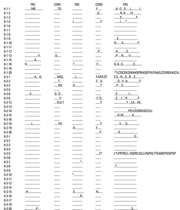
FR1 CDR1 FR2 CDR2 FR3
6-1-1 ...ME... ...TG.. ... .F... ...A...G....D...L...I...
6-1-2 ... ... ... ...G... ...N..A...H...
6-1-3 ... ... ... ... ...S...F..
6-1-5 ... ... .I... ...Y ...I...*...
6-1-6 ... ... ... ... ...
6-1-7 ... ... ... ... ...
6-1-8 ... ... ... ... ...
6-1-9 ... ... ... ... ...E...
6-1-10 ... ... ... ... N...S...F..
6-1-11 ... ... ... ... ...
6-1-12 ... ... ... ....P.... ...A...S...
6-1-13 ...V.. ..G... ... ... ...P...N...V...
6-1-15 ...A... ... ... ... ...
6-1-16 H... ... .T... ..F... G..A...G...S...
6-1-18 ... ... ... ... ...
6-1-20 ... ... ... ... **LCSICEKSNNHQPRHIQEPVLPAAELCDSRGHGCVL 6-2-1 ...A...G.. ...NSG.. ...L... .F.M.R.ST E.S....N....S....R...E...
6-2-3 ... ...T.. ... .F...K. ...Q....V...A...F.
6-2-4 ... ...NV. .H... ...T ....P...S...
6-2-5 ... ... ... ... ...
6-2-6 ...S... .G...D.... ... ... ...E...L...
6-2-8 ... ...V ... ..F..Q... ...E...I...R...F.
6-2-12 ... ...N.G.T ... ...Y ...Y...LA...M...
6-2-13 ... ... ... ... ...
6-2-14 ... ... ... ... ...PELCDSRGHGCVLL 6-2-15 ... ... ... ... ...R..M...K...
6-2-16 ... ... ... ... ...
6-2-18 ...L... ...VS. ... ...Y ...V...Q...
6-2-19 ... ... .H... .F... ...
6-2-20 ... ... ... ...F. ...G...
6-3-1 ... ... ... ... ...G...
6-3-2 ... ... ... ... ...
6-3-3 ... ... ... ... ...
6-3-4 ... ... ... ... ...
6-3-5 ... ... ... ...C*. L*LPRTRLC..VQERV.EQ.LVNPHLTTGAREPWSPSP 6-3-6 ... ... ... ... ...
6-3-7 ... ... ...*... ... ...
6-3-8 ... ... ... ... ..T...
6-3-9 ... ... ... ... ...
6-3-10 ... ... *... ... ...
6-3-11 ... ... ... ... ...
6-3-12 ... ... ... ... ...
6-3-14 ... ... ... ... ...
6-3-15 ..H... ... .S... .N... ...
6-3-16 ... ... ...R... ... ...
6-3-17 ... ... ... ... ...
6-3-19 ... ... ... ... ...
6-3-20 ...P... ... ... ... ...S...
Fig. 5. The deduced amino acid sequences of VHgerm line gene segments of 54 clones isolated from 3 different patients are shown in comparison with germ line X92224/J.
demonstrate oligoclonality through sequencing. Interes- tingly, in a post-mortem study of KD patients, Rowley et al.5reported that the plasma cells infiltrating the coronary arteries were mostly IgA and IgM-secreting cells, and that oligoclonal IgA reactivity could be observed, suggesting that KD is caused by conventional antigens rather than superantigens.15
However, when the peripheral blood of infants is stimul-
ated with a polyclonal stimulus, IgM is secreted mostly by B-cells, while there are few IgG and IgA-secreting B- cells.6,7The increase in IgG and IgA-secreting B-cells is observed at 1-2 years of age. Moreover, the primary immune reaction takes place first when the antigen is introduced into the host, and it is well known that the characteristics of this reaction is that IgM is mostly produced. The affinity and specificity of the antibodies for this antigen is low, and
VH D/J CµM
6-1-1 TGTGCAAGA GAGGGCGGTGGGAACTACTTGTACTACTTTGACTACTGGGGCCAGGGAGCCCAGGTCACCGTCTCCTCA GGGAGTGCATCC
6-1-2 TGTGCAAGG TCGCATACCAACAACTTGGGCTACTGGGGCCAGGGAACCCTGGTCACCGTCTCCTCA GGGAGTGCATCC
6-1-3 TGTGCAAGA GGTCATAGCAGTGACTTGTCCGCTTTTGATATCTGGGGCCAAGGGACAATGGTCACCGTCTCTTCA GGGAGTGCATCC
6-1-5 TGTGCAAGA GGGCCCCACGCTATGGACGTCTGGGGCCAAGGGACCACGGTCACCGTCTCCGCA GGGAGTGCATCC
6-1-6 TGTGCAAGA GCTAGGGACCCGTGGACAAATGCTTTTGATATCTGGGGCCAAGGGACAATGGTCACCGTCTCTTCA GGGAGTGCATCC
6-1-7 TGTGCAAGA GAGGGCCCCCTAAGGAACGGCTATTGTAGTGGTGGTAGCTGCTACCTCCGGGACAACTGGTTCGACCCCTGGGGCCA GGGAGTGCATCC GGGAACCCTGGTCACCGTCTCCTCA
6-1-8 TGTGCAAGA GGAATTTATGGTTCGGGGAGTCAGATCCCATTTGACTACTGGGGCCAGGGAACCCTGGTCACCGTCTCCTCA GGGAGTGCATCC
6-1-9 TGTGCAAGA GGGGAGGTTGGAACGACACGAAAATTTGACTACTGGGGCCAGGGAACCCTGGTCACCGTCTCCTCA GGGAGTGCATCC
6-1-10 TGTGCAAGA GGTCATAGCAGTGACTTATCCGCTTTTGATATCTGGGGCCAAGGGACAATGGTCACCGTCTCTTCA GGGAGTGCATCC
6-1-11 TGTGCAAGA GCTAGGGACCCGTGGACAAATGCTTCTGATATCTGGGGCCAAGGGACAATGGTCACCGTCTCTTCA GGGAGTGCATCC
6-1-12 TGTGCAAGA AGTATAGCAGCAGCTGGTACAGGTAGTGTTAGGTACTTCGATCTCTGGGGCCGTGGCACCCTGGTCACTGTCTCCTCA GGGAGTGCATCC
6-1-13 TGTGCAAAG TGGGCGGACTACTGGGGCCAGGGAAGCCCGGTCACCGTCTCCTCA GGGAGTGCATCC
6-1-15 TGTGCAAGA GCTAGGGACCCGTGGACAAATGCTTTTGATATCTGGGGCCAAGGGACAATGGTCACCGTCTCTTCA GGGAGTGCATCC
6-1-16 TGTGCAGGC AGTTTAACTGGGAATCTGGACTACTGGGGCCAGGGAACCCTGGTCACCGTCTCCTCA GGGAGTGCATCC
6-1-18 TGTGCAAGA GAGGGCCCCCTAAGGAACGGCTATTGTAGTGGTGGTAGCTGCTACCTCCGGGACAACTGGTTCGACCCCTGGGGCCA GGGAGTGCATCC GGGAACCCTGGTCACCGTCTCCTCA
6-1-20 TGTGCAAGA GGGGAGGTTGGAACGACACGAAAATTTGACTACTGGGGCCAGGGAACCCTGGTCACCGTCTCCTCA GGGAGTGCATCC
6-2-1 TGTGCAAGG CAACGTTATCGAGCTTTTGACATCTGGGGCCAAGGGACATCGGTTACTGTCTCCTCA GGGAGTGCATCC
6-2-3 TGTGCAAGA GATGACGACGGTTATGCTAATGCCTTTGACTCCTGGGGCCAGGGAACCCTGGTCACCGTCTCCTCA GGGAGTGCATCC
6-2-4 TGTACAAGA TCAGCGACTGGATCTTTACAGAACTGGGGCCAGGGAACCCTGGTCACCGTCTCCTCA GGGAGTGCATCC
6-2-5 TGTGCAAGG TCGGAGCTCGTCGGGGGCGAAAGCCGGAACTGGTTCGACCCCTGGGGCCAGGGAACCCTGGTCACCGTCTCCTCA GGGAGTGCATCC
6-2-6 TGTGCAAGA CAGTATGGGCACTTTGACTACTGGGGCCAGGGAACCCTGGTCACCGTCTCCTCA GGGAGTGCATCC
6-2-8 TGTGCAAAA AGGGGATTAGGGGGTGATGCTTTTGATGTCTGGGGCCAAGGGACAATGGTCATCGTCTCCTCA GGGAGTGCATCC
6-2-12 TGTGCAAGG GATGGGCCAGCAGCAACTGGTCTCCTTGACTACTGGGGTCAGGGAACCCTGGTCACCGTCTCCTCA GGGAGTGCATCC
6-2-13 TGTGCAAGA GAATTTTACCCGGGTATACCAGTGGCTGGTACCTCGGTATACTTCCAGCACTGGGGCCAGGGCACCCTGGTCACCGTC GGGAGTGCATCC TCCTCA
6-2-14 TGTGCAAGA GATACCGGGTATAGCAGCAGCTGGTACTACTTTGACTACTGGGGCCAGGGAACCCTGGTCACCGTCTCCTCA GGGAGTGCATCC
6-2-15 TGTGCAAGA GATCTTAGTGGGAGGTATCCGGACTGGGGCCAGGGAACCCTGGTCACCGTCTCCTCA GGGAGTGCATCC
6-2-16 TGTGCAAGA GGTCTCGAGTATAGCAGCTCGAACTGGTTCGACCCCTGGGGCCAGGAACCCTGGTCACCGTCTCCTCA GGGAGTGCATCC
6-2-18 TGTGCAAGA GGCCCAGGTGGGAGCTACCTATTTGACTACTGGGGCCAGGGAACCCTGGTCACCGTCTCCTCT GGGAGTGCATCC
6-2-19 TGTGCAAGA GATGGTGAACTGGGGATTGGTGACTACTGGGGCCAGGGAACCCTGGTCAGCGTCTCCTCA GGGAGTGCATCC
6-2-20 TGTACAAGA GACCGTACCCACTGCTTTGACTGCTGGGGCCAGGGAACCCTGGTCACCGTCTCCTCA GGGAGTGCATCC
6-3-1 TGTGCAAGA GATCGAGGCGCTTTTGATATCTGGGGCCAAGGGACAATGGTCACCGTCTCTTCA GGGAGTGCATCC
6-3-2 TGTGCAAGA GGTTTGACTGGTTATTATATATTTCCCCCTTGGTTTGACTACTGGGGCCAGGGAACCCTGGTCACCGTCTCCTCA GGGAGTGCATCC
6-3-3 TGTGCAAGA GACTACTACTACGGTATGGACGTCTGGGGCCAAGGGACCACGGTCACCGTCTCCTCA GGGAGTGCATCC
6-3-4 TGTGCAAGG GGCCCCACGGGATTTGACTACTGGGGCCAGGGAACCCTGGTCACCGTCTCCTCA GGGAGTTCGTCC
6-3-5 TGTGCAAGA GAGGGTATCGGAGCAGCAGCTGGTAAACCCGCATTTGACTACTGGGGCCAGGGAACCCTGGTCACCGTCTCCTCA GGGAGTGCATCC
6-3-6 TGTGCAAGA GACTACTACTACGGTATGGACGTCTGGGGCCAAGGGACCACGGTCACCGTCTCCTCA GGGAGTGCATCC
6-3-7 TGTGCAAGA GGCCGTGCAGCAGCTGGTACAATTAGGAACTGGTTCGACCCCTGGGGCCAGGGAACCCTGGTCACCGTCTCCTCA GGGAGTGCATCC 6-3-8 TGTGCAAGC GAGGTTTATAGCAGCAGCTGGGAGGATGCTTTTGATATCTGGGGCCAAGGGACAATGGTCACCCGTCTCTTCA GGGAGTGCATCC 6-3-9 TGTGCAAGA GATCCTGGGAATAGTGGGAGCTACTACGTTTGACGTTGGACTACTGGGGCCAGGGAACCCTGGTCACCGTCTCCTCA GGGAGTGCATCC 6-3-10 TGTGCAAGA GATCTGGCTATAGCAGTGGCTGGGCCGATCTGGTACTTCGATCTCTGGGGCCGTGGCACCCTGGTCACTGTCTCCTCA GGGAGTGCATCC
6-3-11 TGTGCAAGA GACTACTACTACGGTATGGACGTCTGGGGCCAAGGGACCACGGTCACCGTCTCCTCA GGGAGTGCATCC
6-3-12 TGTGCAAGA GAGGGGACTGGGGATAGGGGGGGGCTCTTTGACTACTGGGGCCAGGGAACCCTGGTCACCGTCTCCTCA GGGAGTGCATCC
6-3-14 TGTGCAAGA GGGATCTTAACTGCTGCCCCTTCATACTTTGACTACTGGGGCCAGGGAACCCTGGTCACCGTCTCCTCA GGGAGTGCATCC
6-3-15 TGTGCAAGA GCCTATTCTGGATACAATGATGCTTTTCATATCTGGGGCCAAGGGACACTGGTCACCGTCTCTTCA GGGAGTGCATCC
6-3-16 TGTGCAAGA GTTTCAAGTCAATATAGCAGCAGCTGGTACGAGGCGCCGGCGGGTGACTACTGGGGCCAGGGAACCCTGGTCACCGT GGGAGTGCATCC CTCCTCA
6-3-17 TGTGCAAGA GTGCCACACCTGTATAGCAGCAGCTGGTAACACGTTTGACTACTGGGGCCAGGGAACCCTGGTCACCGTCTCCTCA GGGAGTGCATCC
6-3-19 TGTGCAAGA TCACCCAATTTAAGGGATTGGAACTACTTTGACTACTGGGGCCAGGGAACCCTGGTCACCGTCTCCTCA GGGAGTGCATCC
6-3-20 TGTGCAAGA GATCGCGACCCAGCAGCTCGGCACAAGCTTGGCACCGATGCTTTTGATATCTGGGGCCAAGGGACAATGGTCACCGT GGGAGTGCATCC CTCTTCA
Fig. 6. The nucleotide sequences of CDR3 / FR4 and adjacent regions of VH germ line gene segments of 54 clones homologous to X92224/J isolated from 3 different patients are shown.