• 검색 결과가 없습니다.

Wolff-Parkinson-White Syndrome in a Patient With Mitochondrial Encephalopathy, Lactic Acidosis and Stroke-Like Episodes Syndrome

N/A
N/A
Protected

Academic year: 2022

Share "Wolff-Parkinson-White Syndrome in a Patient With Mitochondrial Encephalopathy, Lactic Acidosis and Stroke-Like Episodes Syndrome"

Copied!
3
0
0

로드 중.... (전체 텍스트 보기)

전체 글

(1)

674

Print ISSN 1738-5520 / On-line ISSN 1738-5555 Copyright © 2011 The Korean Society of Cardiology CASE REPORT

http://dx.doi.org/10.4070/kcj.2011.41.11.674

Open Access

Wolff-Parkinson-White Syndrome in a Patient With Mitochondrial Encephalopathy, Lactic Acidosis and Stroke-Like Episodes Syndrome

Min-Ho Lee, MD, Young-Jun Sung, MD, Jung-Han Yoon, MD, Jiyeong Kim, MD, Il-Young Oh, MD, Eue-Keun Choi, MD, and Seil Oh, MD

Department of Internal Medicine, Seoul National University College of Medicine and the Cardiovascular Center, Seoul National University Hospital, Seoul, Korea

ABSTRACT

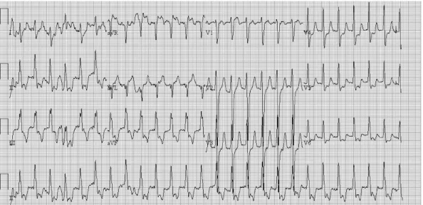
Mitochondrial encephalopathy, lactic acidosis and stroke-like episodes (MELAS) syndrome is a multisystem disorder, which is clinically characterized by encephalopathy, dementia, seizures and stroke-like episodes. Multiple organs can be affected and cardiac involvement often dominates the clinical picture because of its high energy requirement. We report a case of a 21-year-old woman with MELAS syndrome who had pre-excitation ECG and one episode of tachycardia attack. (Korean Circ J 2011;41:674-676)

KEY WORDS: MELAS syndrome; Wolff-Parkinson-White syndrome.

Received: October 4, 2010

Revision Received: December 21, 2010 Accepted: February 15, 2011

Correspondence: Seil Oh, MD, Department of Internal Medicine, Seoul National University College of Medicine, 101 Daehak-ro, Jongno-gu, Seoul 110-744, Korea

Tel: 82-2-2072-2088, Fax: 82-2-762-9662 E-mail: [email protected]

• The authors have no financial conflicts of interest.

cc

This is an Open Access article distributed under the terms of the Cre- ative Commons Attribution Non-Commercial License (http://creativecom- mons.org/licenses/by-nc/3.0) which permits unrestricted non-commer- cial use, distribution, and reproduction in any medium, provided the origi- nal work is properly cited.

Introduction

Mitochondrial encephalopathy, lactic acidosis and stroke- like episodes (MELAS) syndrome is a multisystem disorder, which is clinically characterized by encephalopathy, frequ- ently manifesting as dementia, seizures and stroke-like epi- sodes at a young age with evidence of mitochondrial dys- function in the form of lactic acidosis and ragged-red fibers.

Multiple organs can be affected, but tissues with high energy demand are the most vulnerable. Due to the high energy re- quirement of cardiac muscles, cardiac disease features promi- nently among the systemic manifestations of MELAS syn- drome. Although cardiomyopathy is the most common car- diac manifestation, Wolff-Parkinson-White (WPW) synd- rome has also been reported in association with MELAS sy- ndrome. We report a case of a WPW syndrome in a patient

with MELAS syndrome.

Case

A 21-year-old woman was admitted to the hospital for a sei- zure-like episode lasting for approximately five minutes and subsiding spontaneously. The patient had frequent and insi- dious onset of seizure-like episodes, dysarthria, gait disturb- ance and a right-sided visual field defect that had started four years ago without any history of essential hypertension, dia- betes mellitus and dyslipidemia.

The patient initially presented with blood pressure of 111/

59 mm Hg, pulse rate of 109 beats/min, respiratory rate of 20/

min, and body temperature of 36.0°C. Laboratory results were raised cerebrospinal fluid (CSF) lactate of 5.2 mmol/L.

Electroencephalography (EEG) showed diffuse cerebral dys- function. Electromyography revealed sensorimotor polyneu- ropathy and chronic myopathy. Brain MRI showed infarc- tion in the right temporal lobe, ischemia in the left posterior frontoparietal cortex and basal ganglia, and cystic lesion in the pineal gland with brainstem and cerebellar atrophy (Fig.

1). Biopsy of left vastus lateralis showed neurogenic atrophy and slightly increased lipid vacuoles without paracrystalline inclusion in the mitochondria (Fig. 2). Based on these clinical findings, the patient was diagnosed with MELAS syndrome.

Ten years later, she went to a dentist for treatment of dental

caries. Five minutes after local anesthesia with lidocaine, her

(2)

Min-Ho Lee, et al. 675

consciousness changed suddenly and she was urgently tr- ansferred to the emergency room of our institution.

On arrival, she was stuporous with blood pressure of 209/

147 mm Hg, pulse rate of 140 beats/min, respiratory rate of 25/

min, and body temperature of 36.5°C. At that time, her elec- trocardiography (ECG) showed supraventricular tachycar- dia. After intravenous injections of 300 mg amiodarone, she recovered consciousness, and follow-up ECG showed sinus rhythm with pre-excitation (Fig. 3).

Transthoracic echocardiography revealed decreased early diastolic mitral annulus velocity (E’ velocity) and abnormal myocardial texture which were possibly associated with the initial phase of restrictive cardiomyopathy. Considering her

bedridden status due to underlying MELAS syndrome, me- dical treatment with propafenone was started. The patient was discharged and has been followed up without tachycardia attack.

Discussion

To the best of our knowledge, this is the first case to report on WPW syndrome in a patient with MELAS syndrome in the Korean literature. MELAS syndrome is a maternally inhe- rited disorder caused by mutations of mitochondrial DNA that presents with a wide range of clinical expression. MELAS syndrome is clinically characterized by 1) encephalopathy, frequently manifesting as dementia, seizures, or both; 2) st- roke-like episodes at a young age (typically by age 40 years);

and 3) evidence of mitochondrial dysfunction in the form of lactic acidosis, ragged-red fibers or both.

1)

Age at onset was initially described to range from under 2 years to over 60 years.

2)

Multiple organs can be affected, but

Fig. 1. Axial T2 brain MRI scan revealed infarction in the right temporal lobe, ischemia in the left posterior frontoparietal cortex and basal ganglia.

Fig. 2. Hematoxylin-eosin staining showed small angulated mus- cle fibers with variations in size suggesting neurogenic atrophy and slightly increased lipid vacuoles in the subsarcolemmal area.

Fig. 3. Electrocardiography revealed a short PR interval, delta wave and widened QRS complex which were compatible with WPW syn-

drome.

(3)

676 MELAS Syndrome

tissues with high energy demand are the most vulnerable. Th- erefore, nervous system and muscle impairment often domi- nate the clinical picture.

3)

Cardiomyopathy is an early recogniz- ed cardiac manifestation and is now a widely accepted com- plication of MELAS syndrome. Kim et al.

4)

reported the first case of myocardial involvement in a patient with MELAS syn- drome in the Korean literature, and Son et al.

5)

also reported a case of myocardial involvement in a patient with mitochon- drial myopathy.

Conduction defects, including WPW syndrome, have also been reported in association with MELAS syndrome.

6)

Recent- ly, Sproule et al.

7)

reported that four (13%) of 30 patients with MELAS syndrome had a clinical history of, or ECG findings consistent with, WPW syndrome.

Laboratory evaluation is a logical initial step in the diag- nostic evaluation of suspected MELAS syndrome and an el- evated level of lactate in the CSF is helpful. MRI is a power- ful tool in the diagnostic evaluation of suspected cases of ME- LAS syndrome. The Brain MRI of patients with MELAS syn- drome typically demonstrates asymmetric lesions of the oc- cipital and parietal lobes that mimic ischemia.

1)3)

Muscle bio- psy can be vital in the evaluation of MELAS syndrome. The presence of ragged red fibers on muscle biopsy is a classical finding and scattered vacuolated muscle fibers with a clear surrounding rim as well as the presence of many basophilic inclusions on microscopic examination.

6)

The percentage of heteroplasmy for a mitochondrial RNA mutation may vary widely depending on the specific tissue sampled.

8)

Blood leukocyte samples have traditionally been the most common specimens, and urine sediment, cheek mu- cosa and skin fibroblasts are often used and easily accessible alternate specimens.

9)

Although the m.3243A>G common MELAS mutation is responsible for more than 80% of report- ed MELAS cases,

1)

most of these reports have come from Wes- tern countries. Abu-Amero et al.

10)

reported the case of a pa- tient with typical clinical features of MELAS syndrome, but mutations in recognized genes were not identified.

Despite considerable advances in genetics and molecular biology, therapeutic options for MELAS syndrome remain li- mited. Primary current treatment involves symptomatic ma- nagement, in addition to supplementation with a so-called

“mitochondrial cocktail”. Although recent studies of a poten- tial therapeutic role for L-arginine have been promising, these observations needs to be confirmed by controlled trials.

6)

Because of significant morbidity and mortality, patients with MELAS syndrome should be screened at the time of di- agnosis and followed regularly for the possibility of concurrent cardiac complications such as cardiomyopathy and WPW syndrome.

REFERENCES

1) Hirano M, Ricci E, Koenigsberger MR, et al. Melas: an original case and clinical criteria for diagnosis. Neuromuscul Disord 1992;2:125- 2) Hirano M, Pavlakis SG. Mitochondrial myopathy, encephalopathy, 35.

lactic acidosis, and strokelike episodes (MELAS): current concepts. J Child Neurol 1994;9:4-13.

3) Pavlakis SG, Phillips PC, DiMauro S, De Vivo DC, Rowland LP. Mi- tochondrial myopathy, encephalopathy, lactic acidosis, and strokelike episodes: a distinctive clinical syndrome. Ann Neurol 1984;16:481-8.

4) Kim WH, Lee SC, Hong GR, et al. A case of myocardial involvement in patient with MELAS syndrome. Korean J Med 2004;67:727-31.

5) Son SW, Kim DB, Kwon BJ, et al. A case of mitochondrial myopathy with cardiac involvement. Korean Circ J 2008;38:491-4.

6) Sproule DM, Kaufmann P. Mitochondrial encephalopathy, lactic aci- dosis, and strokelike episodes: basic concepts, clinical phenotype, and therapeutic management of MELAS syndrome. Ann N Y Acad Sci 2008;

1142:133-58.

7) Sproule DM, Kaufmann P, Engelstad K, Starc TJ, Hordof AJ, De Vivo DC. Wolff-Parkinson-White syndrome in patients with MELAS. Arch Neurol 2007;64:1625-7.

8) Shanske S, Pancrudo J, Kaufmann P, et al. Varying loads of the mito- chondrial DNA A3243G mutation in different tissues: implications for diagnosis. Am J Med Genet A 2004;130A:134-7. Abstract.

9) Ciafaloni E, Ricci E, Shanske S, et al. MELAS: clinical features, bio- chemistry, and molecular genetics. Ann Neurol 1992;31:391-8.

10) Abu-Amero KK, Al-Dhalaan H, Bohlega S, Hellani A, Taylor RW. A

patient with typical clinical features of mitochondrial encephalopa-

thy, lactic acidosis and stroke-like episodes (MELAS) but without an ob-

vious genetic cause: a case report. J Med Case Reports 2009;3:77.

수치

Fig. 3. Electrocardiography revealed a short PR interval, delta wave and widened QRS complex which were compatible with WPW syn- syn-drome.

참조

관련 문서

MELAS (Mitochondrial myopathy, Encephalopathy, Lactic acidosis, and Stroke like episodes) 증후군은 1984년 Pavlakis에 의해 보고되어 명명 지어진 사립

Most of the current diagnostic criteria on mitochondrial dis- ease were developed prior to the recent expansion of molecu- lar genetic knowledge. 8,9 These criteria are based