Dear Editor,
We report a case study of mitochondrial encephalopathy, lac- tic acidosis, and stroke-like episode (MELAS) syndrome with presentation of headache and prolonged visual aura.
Mitochondrial encephalopathy, lactic acidosis, and stroke- like episode syndrome is one of the most commonly recog- nized mitochondrial diseases. It is characterized by lactic aci- dosis, the occurrence of stroke-like episodes, and other secondary manifestations such as migraine-like headache, sei- zure, cognitive impairment, cardiac conduction defects, and short stature.1,2 These syndromic phenomena are most com- monly caused by an m.3243A>G (adenine to guanine) muta- tion at position 3243 of the mitochondrial genome. The sei- zures are often associated with migraine-like headache and occur primarily in a group of patients who develop stroke-like episodes. Since stroke-like episodes have a predilection for the parieto-occipital and posterior temporal areas, the seizure semiology frequently demonstrates disturbance in these loca- tions.1,3
We describe a MELAS syndrome patient who presented with recurrent headache and visual aura lasting for more than several days. The patient was a 23-year-old female who was referred to our outpatient clinic due to headache with visual aura lasting for 1 week. Her past medical history and family
history were unremarkable, and with the exception of small stature (height, 147 cm; weight, 38 kg), a physical examina- tion yielded normal results. During a neurological examina- tion the patient was alert but complained of a continuous glit- tering light as well as visual loss in the left visual field. No abnormalities were found on motor, sensory, cerebellar, and reflex function testing. The results of laboratory studies, in- cluding blood lactic acid and CSF analyses, were normal.
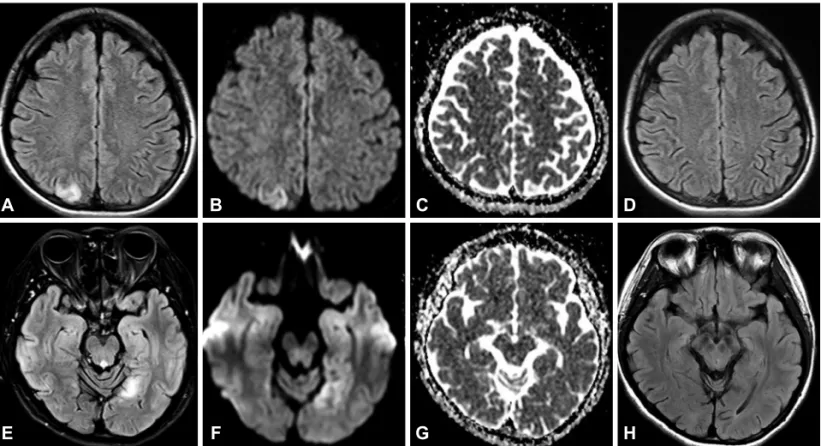
The diffusion-weighted imaging (DWI) and fluid-attenuated inversion recovery (FLAIR) MRI scans performed on the sec- ond day of hospitalization revealed a high signal intensity in the right parietal lobe (Fig. 1A, B, and C); the EEG exhibited rhythmic activities in the corresponding area.
Under the presumptive diagnosis of partial status epilepti- cus, she was treated with levetiracetam (1,000 mg/day) after receiving an injection of intravenous lorazepam. The visual aura subsided completely after 3 days, and the follow-up MRI performed on day 7 of hospitalization revealed disap- pearance of the previously observed abnormality (Fig. 1D).
The patient was maintained on low-dose levetiracetam (500 mg/day) for 2 years. Three years later the patient visited our outpatient clinic again for recurrence of visual aura lasting for 3 days, contralateral to the previous side. DWI and FLAIR MRI scans revealed a high signal intensity in the left occipi- tal lobe (Fig. 1E, F, and G); a follow-up EEG showed rhyth- mic activities in the corresponding area. A genetic study revealed the presence of a mitochondrial mutation (m.3243A>G), in- dicating the presence of MELAS syndrome.
Neurological problems are the most frequent clinical fea- tures of mitochondrial disorder, and recurrent strokes or tran- sient-ischemic-attack-like episodes are the most common mani- festations of MELAS syndrome. Stroke-like lesions usually appear as multifocal infarct-like areas with cortical involve- ment that has no matching vascular territory.4 The MRI abnor- malities in MELAS syndrome often disappear on follow-up MRI, as in our case, as a result of the reversible pathogenesis due to mitochondrial cytopathy or angiopathy.4,5 However,
Mitochondrial Encephalopathy, Lactic Acidosis, and Stroke-Like Episode Syndrome Presenting with Prolonged Visual Aura
Ileok Jung, So-Hee Park, Dong Wook Kim
Department of Neurology, Konkuk University School of Medicine, Seoul, Korea Open Access
Received August 6, 2014 Revised October 20, 2014 Accepted October 22, 2014 Correspondence
Dong Wook Kim, MD, PhD, Department of Neurology, Konkuk Uni- versity School of Medicine, 120-1 Neungdong-ro, Gwangjin-gu, Seoul 143-729, Korea
Tel +82-2-2030-7562, Fax +82-2-2030-5169 E-mail [email protected]
cc This is an Open Access article distributed under the terms of the Cre- ative Commons Attribution Non-Commercial License (http://creative- commons.org/licenses/by-nc/3.0) which permits unrestricted non-com- mercial use, distribution, and reproduction in any medium, provided the ori- ginal work is properly cited.
104 Copyright © 2015 Korean Neurological Association
Print ISSN 1738-6586 / On-line ISSN 2005-5013 http://dx.doi.org/10.3988/jcn.2015.11.1.104 LETTER TO THE EDITOR
J Clin Neurol 2015;11(1):104-105
Jung I et al.
www.thejcn.com 105 other common and clinically nonserious symptoms of MELAS
syndrome, including migraine-like headache, seizure, short stature, and even psychological problems such as depression and anxiety, can go unrecognized for up to 20 years before the onset of overt neurological impairment.6
The findings of the present study show that patients with MELAS syndrome can present with common neurological diseases, such as migraine with aura or occipital lobe epilep- sy.1-3 Our experience suggests that clinical suspicion is impor- tant for an early diagnosis of this particular metabolic disease.
Conflicts of Interest
The authors have no financial conflicts of interest.
REFERENCES
1. Bindoff LA, Engelsen BA. Mitochondrial diseases and epilepsy. Epi- lepsia 2012;53 Suppl 4:92-97.
2. Zeviani M, Di Donato S. Mitochondrial disorders. Brain 2004;127(Pt 10):2153-2172.
3. Loder E, Cardona L. Evaluation for secondary causes of headache:
the role of blood and urine testing. Headache 2011;51:338-345.
4. Tzoulis C, Bindoff LA. Serial diffusion imaging in a case of mito- chondrial encephalomyopathy, lactic acidosis, and stroke-like epi- sodes. Stroke 2009;40:e15-e17.
5. Renard D, Taieb G. Neurological picture. Cortical susceptibility- weighted imaging hypointensity after stroke-like episode in MELAS.
J Neurol Neurosurg Psychiatry 2014;85:1055-1056.
6. Sproule DM, Kaufmann P. Mitochondrial encephalopathy, lactic aci- dosis, and strokelike episodes: basic concepts, clinical phenotype, and therapeutic management of MELAS syndrome. Ann N Y Acad Sci 2008;1142:133-158.
A
E
B
F
C
G
D
H
Fig. 1. A, B, and C: Diffusion-weighted imaging (DWI) and fluid-attenuated inversion recovery (FLAIR) MRI scans during the first episode showing a high signal intensity in the right parietal lobe. E, F, and G: DWI and FLAIR MRI scans during the second episode showing a high signal intensity in the left occipital lobe. D and H: Both lesions had disappeared on the follow-up MRI scans.