Vol. 18, No. 3, December, 2011
□ 증 례 □1)
Introduction
Mitochondria, a cell organelle containing a DNA of its own, plays an important role in cel- lular energy production by synthesizing adenosine triphosphate (ATP). When a defect happens in its oxidative phosphorylation system (OXPHOS) pathway, we call it mitochondrial disease
1). The clinical symptoms or the onset age presented of mitochondrial disease tend to be quite variable.
Though it shows multiple system involvement, brain and skeletal muscle are affected most of the time along with the heart
2, 3).
The mitochondrial encephalomyopathy, lactic acidosis, and stroke-like episodes (MELAS)
Received : 28 September, 2011, Revised : 9 November, 2011 Accepted : 29 November, 2011
Correspondence : Young-Mock Lee, M.D, Ph.D.
Department of Pediatrics, Gangnam Severance Hospital, Severance Children's Hospital Yonsei University College of Medicine, Seoul, Korea
Tel : +82.2-2019-3354, Fax : +82.2-3461-9473 E-mail : [email protected]
syndrome was first described in 1984
4). It is characterized by symptoms of repeated episodes of hemiparesis, hemianopsia, migraine, nausea, or vomiting. The point mutation (A3243G) of tRNA gene in mitochondria has been reported in 80% of the cases
5). Cardiac involvement is also reported, it is but rarely known to be com- bined with Wolff-Parkinson-White (WPW) syn- drome
6). WPW syndrome is defiened as the presence of an accessory pathway (AP) and has a predisposition to the development of supra- ventricular tachydysrhythmias. Conduction over an AP circumvents conduction delay occurring within the atrioventricular node, which leads to early eccentric activation of ventricles and fusion complex
7).
Here we present a rare case of early onset MELAS patient with WPW syndrome.
A Case of Early Onset MELAS Patient with Wolff-Parkinson-White Syndrome
Jeong-A Kim, M.D., Jung Min Ahn, M.D., Young Mock Lee, M.D.
Hoon Chul Kang, M.D., Joon Soo Lee, M.D. and Heung Dong Kim, M.D.
Department of Pediatrics, Gangnam Severance Hospital, Severance Children s Hospital, ’ Yonsei University College of Medicine, Seoul, Korea
= Abstract =
Mitochondrial encephalomyopathy, lactic acidosis, and stroke-like episodes (MELAS) syndrome is one of the classic mitochondrial diseases characterized by symptoms of repeated episodes of hemiparesis with mitochondrial DNA mutation. We report a rare case of early onset MELAS patient confirmed by genetic analysis with Wolff-Parkin- son-White syndrome.
Key Words : Mitochondrial Myopathies, MELAS syndrome, Wolff-Parkinson-White syndrome
Case Report
A Six years and 2 months old female was transferred to Gangnam Severance Hospital for her left side weakness and gait disturbance. She had a birth history of prematurity and hyaline membrane disease which led her to have venti- lator care then. She could control her head at 4 month, walk holding on to furniture at 12 month, she produce meaningful words at 13 month.
Though she had a mild developmental delay, she was capable of independent walking, running, and had no problem in verbal communication.
She had no serious disability in her daily life and no specific neurological complication before the onset of the symptoms mentioned above. Her mental status was alert, vital sign was normal.
She could walk alone, and she didn t show muscle ’ weakness and other signs of neurologic deficit.
There was no specific finding in her family his- tory, including arrhythmia, heart disease, stroke, either.
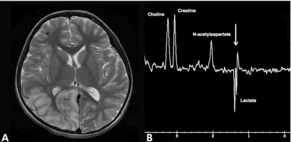
The brain MRI preformed after the admission
revealed diffuse cerebral atrophy and high signal in her right temporal, parietal, and occipital area on T2 weighted image that were suspected as infarction while the MR spectroscopy showed a lactate peak (Fig. 1).
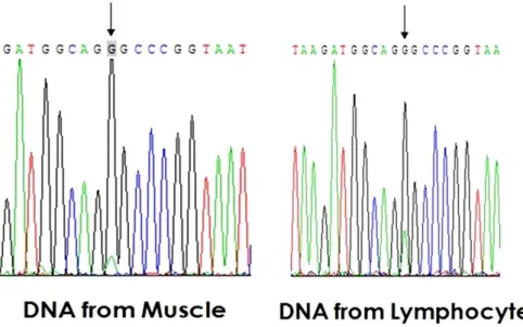
Laboratory tests, including those for muscle enzymes, lactate, pyruvate, and amino acid and organic acid assays were performed. The results were normal except an increased plasma lactate level 8.2 mmol/L (normal, 0.5-1.6 mmol/L). CK, CK-MB level was 96 U/L, 3.6 mcg/L, liver en- zyme (AST/ALT) was 28/14 U/L, hemoglobin was 11.9 g/dL. As for muscle biopsy, ragged red fiber was observed with Gomori trichrome stain, but no specific aberration was found with other immunohistochemical stain and electron microscopy. In the biochemical respiratory chain complex enzyme assay using muscle tissue, the enzyme activity was all decreased in the whole complex. More specifically, complex I enzyme activity was as low as less than 10% of the nor- mal control. Direct sequencing of mitochondrial DNA (mtDNA) in the blood and muscle speci- mens of the patient revealed mtDNA A3243G
Fig. 1. Brain MRI and MR spectroscopy of the patient. Brain MRI shows diffuse cerebral atrophy and high signal in right temporal, parietal and occipital area on T2 weighted image (A). MR spectroscopy shows lactate peak (B).
mutation confirming that she had MELAS, a specific type of mitochondrial disease (Fig. 2).
WPW syndrome was diagnosed by delta wave, short PR interval, and QRS widening on an elec- trocardiogram (Fig. 3). Echocardiography showed no anatomical abnormality with normal left ven- tricle contractility, either. The left ventricular
ejection fraction was checked as 73%.
Mitochondrial cocktail (Coenzyme Q10, thia- mine, carnitine and multivitamin) were prescribed for the patient after the diagnosis of mitochon- drial disease was made. Propranolol was also given to treat WPW syndrome. Left side weak- ness is still remaining and a mild decline of cog-
Fig. 2. mtDNA sequencing of the patient. Direct sequencing of mtDNA in blood and muscle specimens revealed mtDNA A3243G mutation.
Fig. 3. Electrocardiogram of the patient. Electrocardiogram shows delta wave, short PR interval and QRS widening compatible to Wolff-Parkinson-White syndrome.
nitive function is observed, but no new stroke- like episode has been broken yet for about 2 years. Also, though WPW syndrome is still ob- served in the series of follow-up electrocardio- gram, there had been no change of heart con- tractility.
Discussion
MELAS tend to show normal development at its early stage. That is up to about 90% of its patients. The onset usually occurs before the age of 40 and can be seen even at childhood, followed by gradual deterioration. Clinical symp- toms are highly variable though. Episodic head- aches with vomiting are the most common symp- toms in MELAS, observed in almost all the pa- tients. Also, seizures are noted in the majority
8-10)
. Our patient had a first neurological manife- station at the age of 6 mainly consisting of uni- lateral weakness. No headache or vomiting which are the common symptoms of MELAS was ob- served. This could be due to the fact that the patient was too young to express her symptoms more precisely, but it is also possible that an early onset type of MELAS present with some- what different clinical features. Though the girl had birth history of prematurity and hyaline membrane disease, there has been no specific proof that they are related with mitochondrial disease or MELAS.
Cardiac involvement including preexcitation, atrioventricular heart block, rhythm abnormalities and dilated or hypertrophic cardiomyopathy is reported in up to half of the patients with MELAS, though not as a predominant symptom
8, 9, 11, 12). Whether those abnormalities tend to progress to major cardiac abnormalities or not are still needed to be studied. WPW syndrome is even
harder to find among MELAS patients. Peter NS et al. reported abnormal mitochondria found in surgically resected pathyways in WPW synd- rome. Mutation in the PRKAG2 gene, which en- codes the r2 regulatory subunit of AMP-activated protein kinase, and the mutation may result in defects during cardiac morphogenesis, resulting development of accessory pathway
7). An abnor- mality in heart conduction was found, but cardiac contractility was normal in our patient as well.
This could mean that the severity, disease dura- tion, or mutant burden of MELAS can affect the variability of symptom within the same organ system.
The studies concerning WPW Syndrome in
patients with MELAS have been very limited
and in children even more. There was a report
by Hirano et al. that noted 6 patients with WPW
syndrome out of 43 with MELAS, and another
3 patients with cardiac conduction block out of
47 with MELAS
13, 14). Okajima et al. reported 3
cases with WPW syndrome among 11 pediatric
patients with MELAS syndrome
11). Sproule et
al. reported 4 of 30 patients (13%) with MELAS
who had a clinical history of, or electrocardio-
graphic findings that matches with WPW synd-
rome
6). In their reports, WPW syndrome preceded
MELAS syndrome by 15 and 21 years in 2 of
their patients. However, those studies were
done based on adult patients who showed their
symptoms mostly after adolescent period which
makes us hard to figure out the precise preva-
lence in the pediatric aged group. We also have
difficulty in identifying which came first that is
between electrocardiogram abnormality of WPW
syndrome and neurological manifestation in our
patient. Since the onset of symptom vary greatly
in MELAS, studies on WPW syndrome of pati-
ents diagnosed with MELAS during childhood
would be needed.
In this case, the patient have taken propranolol as chronic maintain therapy. And haven t deve ’ - loped any tachyarrhythmias. Individualized chronic maintenance therapy must be established based in the severity of the presentation. And other factors such as proximity of hospital, medication side effect, and doctor s experience. We don t ’ ’ consider cardiac surgery or electrophysiologic intervention not yet
15).
The prevalence of WPW syndrome appears much higher in patients with MELAS and the A3243G mutation than in the general population.
Because there is a possibility that cardiac invol- vement may manifest earlier than neurologic symptoms and the dysfunction may have im- portant therapeutic and prognostic consequences, we d like to recommend that all patients with ’ MELAS receive cardiac evaluations in order to detect abnormalities including cardiomyopathy and WPW syndrome.
Acknowledgments
This research was supported in part by a Basic Science Research Program through the National Research Foundation of Korea (NRF) funded by Ministry of Education, Science and Technology (7-2010-0268).
한 글 요 약
증후군을 Wolff-Parkinson-White
동반한 MELAS 증후군 환아 1 례
연세대학교 의과대학 소아과학교실
김정아 ㆍ 안정민 ㆍ 이영목 ㆍ 강훈철 ㆍ 이준수 ㆍ 김흥동
Mitochondrial encephalomyopathy, lactic aci- 증후 dosis, and stroke-like episodes (MELAS)
군은 반복되는 편측 마비가 특징적으로 대표적인 미 ,
토콘드리아 질환이다 저자들은 조기에 신경학적 이 .
상이 발현된 MELAS 증후군 환자 중 심장 전도 이
상과 관련하여 드물게 발견되는 Wolff-Parkinson-
증후군을 동반한 례를 경험하였기
White (WPW) 1
에 보고하는 바이다.
References
1) Schmiedel J, Jackson S, Schafer J, Reichmann H.
Mitochondrial cytopathies. J Neurol 2003;250:
267-77.
2) Koga Y, Akita Y, Takane N, Sato Y, Kato H. He- terogeneous presentation in A3243G mutation in the mitochondrial tRNA (Leu(UUR)) gene. Arch Dis Child 2000;82:407 411.–
3) Haas RH, Parikh S, Falk MJ, Saneto RP, Wolf NI, Darin N, et al. Mitochondrial medicine society s’ committee on diagnosis. The in-depth evaluation of suspected mitochondrial disease. Mol Genet Metab 2008;94:16-37.
4) Pavlakis SG, Phillips PC, DiMauro S, De Vivo DC, Rowland LP. Mitochondrial myopathy, encephalo- pathy, lactic acidosis, and strokelike episodes: a distinctive clinical syndrome. Ann Neurol 1984;
16:481-8.
5) Finsterer J. Genetic, pathogenetic, and phenotypic implications of the mitochondrial A3243G tRNALeu (UUR) mutation. Acta Neurol Scand 2007;116:1- 14.
6) Sproule DM, Kaufmann P, Engelstad K, Starc TJ, Hordof AJ, DeVivo DC. Wolff-Parkinson-White syndrome in Patients With MELAS. Arch Neurol.
2007 Nov;64:1625-7.
7) Peter NS, Rowland E, Bennett JG, Green CR, Anderson RH, Severs NJ. The Wolff-Parkinson- White syndrome: the cellular substrate for con- duction in the accessory atrioventricular pathway.
Eur Heart J 1994;15:981-7
8) Hirano M, Pavlakis SG. Mitochondrial myopathy, encephalopathy, lactic acidosis, and stroke-like episodes (MELAS): current concepts. J Child Neurol 1994;9:4-13.
9) Hirano M, Ricci E, Koenigsberger MR, Defendini R, Pavlakis SG, DeVivo DC, et al. MELAS: an original case and clinical criteria for diagnosis.
Neuromuscul Disord 1992;2:125-35.
10) Ciafaloni E, Ricci E, Shanske S, Moraes CT, Sil- vestri G, Hirano M, et al. MELAS: clinical fea- tures, biochemistry, and molecular genetics. Ann Neurol 1992;31:391-8.
11) Vydt TC, de Coo RF, Soliman OI, Ten Cate FJ, van Geuns RJ, Vletter WB, et al. Cardiac involve- ment in adults with m.3243A>G MELAS gene mutation. Am J Cardiol 2007;99:264-9.
12) Okajima Y, Tanabe Y, Takayanagi M, Aotsuka H.
A follow up study of myocardial involvement in patients with mitochondrial encephalomyopathy, lactic acidosis, and stroke-like episodes (MELAS).
Heart 1998;80:292-5.
13) Hirano M, Ricci E, Koenigsberger MR, Defendini R, Pavlaskis SG, DeVivo DC, et al. MELAS: an original case and clinical criteria for diagnosis.
Neuromuscul Disord 1992;2:125-35.
14) Pavlakis SG, Phillips PC, DiMauro S, De Vivo DC, Rowland LP. Mitochondrial myopathy, encephalo- pathy, lactic acidosis, and strokelike episodes: a distinctive clinical syndrome. Ann Neurol 1984;
16:481-8.
15) Hermosura T, Bradshow WT. Wolff-Parkinson- White syndrome in infants. Neonatal Netw 2010;
29:215-23.