IntroductIon
Mitochondria are organelles in cells that perform a variety of cellular metabolic functions, including the generation of most cellular energy in the form of adenosine triphosphate.1 Mito- chondrial disease is a clinically heterogeneous group of dis-
eases, in which mitochondrial dysfunction is caused by sever- al mutations of mitochondrial or nuclear DNA.2
The disease mitochondrial encephalomyopathy, lactic aci- dosis and stroke-like episodes (MELAS) is characterized by early onset of stroke-like episodes. MELAS syndrome is the most dominant subtype of mitochondrial disease.3,4 It is one of the most frequent maternally inherited mitochondrial dis- orders and has been shown to be associated with an A>G tran- sition at position 3243 of the tRNALeU(UUR) gene.5,6
The diagnosis of MELAS is based on a combination of clini- cal findings, biochemical testing, muscle pathology, and mo- lecular genetic testing. Typically, a blood sample is initially tested as part of the diagnostic evaluation in patients clinically suspected with MELAS.7 Meanwhile, the usefulness of muscle biopsy in terms of initial diagnostic evaluation remains un- known. This study aimed to study the usefulness of muscle bi- opsy in MELAS patients with mitochondrial DNA (mtDNA) 3243A>G mutation as an initial diagnostic tool.
The usefulness of Muscle Biopsy in Initial diagnostic Evaluation of Mitochondrial Encephalomyopathy, Lactic Acidosis, and Stroke-Like Episodes
Min-Seong Baek1*, Se Hoon Kim2*, and Young-Mock Lee1
Departments of 1Pediatrics and 2Pathology, Yonsei University College of Medicine, Seoul, Korea.
Purpose: The disease entity mitochondrial encephalomyopathy, lactic acidosis, and stroke-like episodes (MELAS) is character- ized by an early onset of stroke-like episodes. MELAS is the most dominant subtype of mitochondrial disease. Molecular genetic testing is important in the diagnosis of MELAS. The mitochondrial DNA (mtDNA) 3243A>G mutation is found in 80% of MELAS patients. Nevertheless, molecular analysis alone may be insufficient to diagnose MELAS because of mtDNA heteroplasmy. This study aimed to evaluate whether muscle biopsy is useful in MELAS patients as an initial diagnostic evaluation method.
Materials and Methods: The medical records of patients who were diagnosed with MELAS at the Department of Pediatrics of Gangnam Severance Hospital between January 2006 and January 2017 were reviewed. The study population included 12 patients.
They were divided into two subgroups according to whether the results of muscle pathology were in accordance with mitochon- drial diseases. Clinical variables, diagnostic evaluations, and clinical outcomes were compared between the two groups.
results: Of the 12 patients, seven were muscle pathology-positive for mitochondrial disease. No statistically significant difference in clinical data was observed between the groups that were muscle pathology-positive and muscle pathology-negative for mtD- NA 3243A>G mutation. Additionally, the patients with weakness as the initial symptom were all muscle pathology-positive.
conclusion: The usefulness of muscle biopsy appears to be limited to an initial confirmative diagnostic evaluation of MELAS.
Muscle biopsy can provide some information in MELAS patients with weakness not confirmed by genetic testing.
Key Words: MELAS, muscle biopsy, genetic diagnosis, morphological diagnosis
pISSN: 0513-5796 · eISSN: 1976-2437
Received: May 30, 2018 Revised: October 21, 2018 Accepted: October 23, 2018
Corresponding author: Young-Mock Lee, MD, PhD, Department of Pediatrics, Gangnam Severance Hospital, Yonsei University College of Medicine, 211 Eonju- ro, Gangnam-gu, Seoul 06273, Korea.
Tel: 82-2-2019-3350, Fax: 82-2-2019-4881, E-mail: [email protected]
*Min-Seong Baek and Se Hoon Kim contributed equally to this work.
•The authors have no potential conflicts of interest to disclose.
© Copyright: Yonsei University College of Medicine 2019
This is an Open Access article distributed under the terms of the Creative Com- mons Attribution Non-Commercial License (https://creativecommons.org/licenses/
by-nc/4.0) which permits unrestricted non-commercial use, distribution, and repro- duction in any medium, provided the original work is properly cited.
Yonsei Med J 2019 Jan;60(1):98-105 https://doi.org/10.3349/ymj.2019.60.1.98
MAtErIALS And MEthodS
Patients
The medical records of patients who were diagnosed with ME- LAS based on the diagnostic criteria of Yatsuga, et al.2 at the Department of Pediatrics of Gangnam Severance Hospital be- tween January 2006 and January 2017 were reviewed. Patients without mtDNA mutation were excluded from the analysis. The Institutional Review Board of Gangnam Severance Hospital in Seoul, Korea approved all procedures (3-2015-0156). Informed consent was obtained, and all methods were performed in ac- cordance with the relevant guidelines and ethics board regu- lations.
Study design
The study population was divided into two subgroups accord- ing to whether the results of muscle pathology were in accor- dance with mitochondrial diseases or not (n=7 vs. n=5, respec- tively). Clinical variables, diagnostic evaluations, and clinical outcomes were compared between the two groups (Fig. 1).
clinical characteristics, diagnostic investigations, and clinical outcomes for MELAS
Clinical data on age at onset of the first symptom, nature of the first symptom, age at diagnosis, period from the first symp- toms to the last visit, and organ involvement were collected.
Laboratory test results were also obtained, including serum lactic acid levels. The degree of serum lactic acidosis was de- fined as mild, moderate, or severe if the increase was more than the normal reference values at less than two-, three-, or more than three-fold, respectively. All patients were tested for genetic mutations involved in MELAS, including mtDNA 3243A>G mutation. Muscle biopsies from the quadriceps muscle, with which histologic, light microscopic, and electron microscopic examinations were performed. Specific findings
for mitochondrial diseases under a light microscope were de- fined as the presence of ragged red fibers (RRF) or abnormal staining. Abnormal mitochondrial morphology was defined under the electron microscope as pleoconia and megaconia.
Biochemical enzyme assay in the muscle was also performed to evaluate mitochondrial respiratory chain (MRC) enzyme activity. MRC complex defect was defined as a residual en- zyme activity <10% of the reference value. Data from magnetic resonance imaging and magnetic resonance spectroscopy study were also collected.
The clinical severity of patients was defined as follows: nor- mal, ambulatory, and independent for daily activities; mild, ambulatory, or independent for daily activities; moderate, wheelchair-bound, or partially dependent for daily activities;
and severe, bedridden, totally dependent for daily activities, or expired.
Statistical analysis
All analyses were performed using SPSS version 20.0 (IBM Corp., Armonk, NY, USA). Descriptive statistics were used in- cluding the median and range. Differences between subgroups were evaluated using the Mann-Whitney U test (Wilcoxon rank sum test) and Fisher’s exacts test. p values <0.05 were considered statistically significant.
rESuLtS
Patient characteristics and clinical features at the last visit
A total of 21 patients was recruited in this study. Among them, 12 were male and nine were female (Table 1). The initial pre- senting symptoms varied, with seizures (9 of 21, 42.9%) being the most common, followed by mental change (4 patients, 19%), weakness (4 patients, 19%), visual disturbance, and ataxia. The mean age of the first symptom onset was 14.9±9.7 years. Central nervous system involvement was found in all patients, and various organs were affected as shown in Table 1.
Besides central nervous system involvement, the ear was the most commonly involved organ in 16 (76.2%) patients. The majority of patients had multiple organ involvement includ- ing the gastrointestinal tract, endocrine system, eyes, and heart (71.4, 71.4, 66.7, and 57.1%, respectively).
diagnostic evaluations
All patients were diagnosed with MELAS, which was con- firmed by genetic testing, and they were all positive for mtDNA 3243A>G mutation. Increased serum lactic acid levels were observed in all patients. Magnetic resonance images of the brain revealed a variety of abnormal findings, including atro- phy or abnormal signal intensities in different areas, and al- most all patients (20 of 21 patients) had evidence of infarction at the time of the study. Magnetic resonance spectroscopy Fig. 1. Flowchart of patient inclusion. MELAS, mitochondrial encephalo-
myopathy, lactic acidosis, and stroke-like episodes; mtDNA, mitochondri- al DNA.
Clinically suspected MELAS patients (n=29)
mtDNA 3243 A>G mutation (+) (n=21)
Muscle biopsy done (n=12)
mtDNA mutation (-) (n=8)
Muscle biopsy not done (n=9)
Muscle pathology (+) (n=7)
Muscle pathology (-) (n=5)
study data were obtained in 19 patients. The presence of lactate peak was observed in 16 patients (84.2%). Muscle biopsy was performed in 12 patients (57.1%). Table 2 shows the abnormal
findings seen under light microscopy and electron microsco- py, as well as MRC enzyme activity: abnormal changes specif- ic to mitochondrial diseases under a light microscope were Table 1. Characteristics and Clinical Features at the Last Visit
Characteristics and clinical features Total (n=21)
Sex (male:female) (%) 12 (57.1):9 (42.9)
Age at onset of the first symptom (yr) 14.9±9.7 (0.6–37.3) Age at onset of the first seizure (yr) 15.6±9.6 (0.6–37.3) Time interval from first clinical presentation
to the last visit (yr) 7.2±4.2 (1.4–14.4)
Initial presenting symptoms, n (%)
Seizure 9 (42.9)
Mental change 4 (19.0)
Weakness 4 (19.0)
Visual disturbance 3 (14.3)
Ataxia 1 (4.8)
Clinical features at the last visit, n (%)
Central nervous system 21 (100)
Respiratory system
Frequent pneumonia 4 (19.0)
O2 dependency 1 (4.8)
Normal 16 (76.2)
Renal system
Abnormal kidney findings of ultrasonography 6 (28.6) Nephrotic syndrome or proteinuria 4 (19.0)
Normal 11 (52.4)
Gastrointestinal system
Gastroesophageal reflux disease 11 (52.3)
Enteral tube feeding 9 (42.9)
Diffuse liver disease 6 (28.6)
Gallbladder polyp or stone 4 (19.0)
Pancreatitis 2 (9.5)
Normal 6 (28.6)
Heart
Cardiomyopathy 10 (47.6)
Wolff-Parkinson-White syndrome 6 (28.6)
Normal 9 (42.9)
Eye
Optic atrophy 8 (38.1)
Retinopathy 4 (19.0)
Visual field defect 2 (9.5)
Ophthalmoplegia 1 (4.8)
Normal 7 (33.3)
Ear
Hearing impairment 14 (66.7)
Hearing loss 2 (9.5)
Normal 5 (23.8)
Endocrine system
Diabetes mellitus 13 (61.9)
Osteoporosis 4 (19.0)
Adrenal insufficiency 1 (4.8)
Normal 6 (28.6)
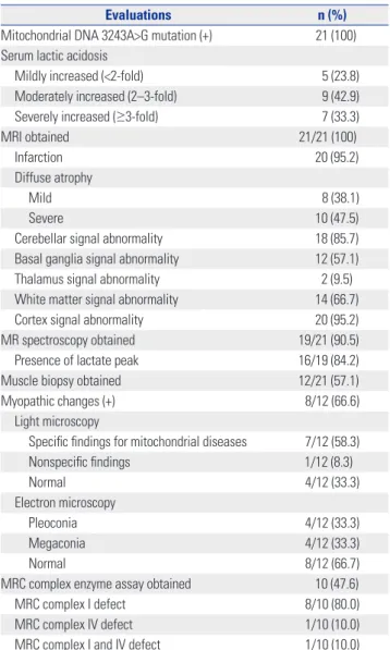
Table 2. Diagnostic Evaluations of MELAS (n=21)
Evaluations n (%)
Mitochondrial DNA 3243A>G mutation (+) 21 (100) Serum lactic acidosis
Mildly increased (<2-fold) 5 (23.8)
Moderately increased (2–3-fold) 9 (42.9)
Severely increased (≥3-fold) 7 (33.3)
MRI obtained 21/21 (100)
Infarction 20 (95.2)
Diffuse atrophy
Mild 8 (38.1)
Severe 10 (47.5)
Cerebellar signal abnormality 18 (85.7)
Basal ganglia signal abnormality 12 (57.1)
Thalamus signal abnormality 2 (9.5)
White matter signal abnormality 14 (66.7)
Cortex signal abnormality 20 (95.2)
MR spectroscopy obtained 19/21 (90.5)
Presence of lactate peak 16/19 (84.2)
Muscle biopsy obtained 12/21 (57.1)
Myopathic changes (+) 8/12 (66.6)
Light microscopy
Specific findings for mitochondrial diseases 7/12 (58.3)
Nonspecific findings 1/12 (8.3)
Normal 4/12 (33.3)
Electron microscopy
Pleoconia 4/12 (33.3)
Megaconia 4/12 (33.3)
Normal 8/12 (66.7)
MRC complex enzyme assay obtained 10 (47.6)
MRC complex I defect 8/10 (80.0)
MRC complex IV defect 1/10 (10.0)
MRC complex I and IV defect 1/10 (10.0)
MELAS, mitochondrial encephalomyopathy, lactic acidosis, and stroke-like episodes; MRC, mitochondrial respiratory chain.
Table 3. Clinical Outcomes at Last Visit
Clinical outcomes Total (n=21)
Number of stroke-like episodes 3.5±2.9 (0–10)
Delayed development or mental retardation, n (%) 16 (76.2) Clinical severity at last outpatient clinic, n (%)
Normal (ambulatory and independent for daily
activities) 6 (28.6)
Mild (ambulatory or independent for daily activities) 3 (14.3) Moderate (WC bound and/or partially dependent
for daily activities) 4 (19.0)
Severe (bedridden, totally dependent for daily
activities or expired) 8 (38.1)
WC, wheelchair.
seen in seven of the 12 patients (58.3%); electron microscopic changes with pleoconia or megaconia were noted in four of the 12 patients (33.3%). Biochemical enzyme assay in the
muscle tissue revealed deficiency of MRC complex I in nine of 10 patients (90%) (Table 2).
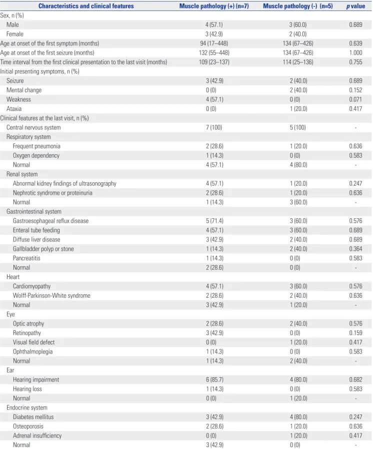
Table 4. Comparison of General Characteristics and Clinical Features between Subgroups
Characteristics and clinical features Muscle pathology (+) (n=7) Muscle pathology (-) (n=5) p value Sex, n (%)
Male 4 (57.1) 3 (60.0) 0.689
Female 3 (42.9) 2 (40.0)
Age at onset of the first symptom (months) 94 (17–448) 134 (67–426) 0.639
Age at onset of the first seizure (months) 132 (55–448) 134 (67–426) 1.000
Time interval from the first clinical presentation to the last visit (months) 109 (23–137) 114 (25–136) 0.755 Initial presenting symptoms, n (%)
Seizure 3 (42.9) 2 (40.0) 0.689
Mental change 0 (0) 2 (40.0) 0.152
Weakness 4 (57.1) 0 (0) 0.071
Ataxia 0 (0) 1 (20.0) 0.417
Clinical features at the last visit, n (%)
Central nervous system 7 (100) 5 (100) -
Respiratory system
Frequent pneumonia 2 (28.6) 1 (20.0) 0.636
Oxygen dependency 1 (14.3) 0 (0) 0.583
Normal 4 (57.1) 4 (80.0) -
Renal system
Abnormal kidney findings of ultrasonography 4 (57.1) 1 (20.0) 0.247
Nephrotic syndrome or proteinuria 2 (28.6) 1 (20.0) 0.636
Normal 1 (14.3) 3 (60.0) -
Gastrointestinal system
Gastroesophageal reflux disease 5 (71.4) 3 (60.0) 0.576
Enteral tube feeding 4 (57.1) 3 (60.0) 0.689
Diffuse liver disease 3 (42.9) 2 (40.0) 0.689
Gallbladder polyp or stone 1 (14.3) 2 (40.0) 0.364
Pancreatitis 1 (14.3) 0 (0) 0.583
Normal 2 (28.6) 0 (0) -
Heart
Cardiomyopathy 4 (57.1) 3 (60.0) 0.576
Wolff-Parkinson-White syndrome 2 (28.6) 2 (40.0) 0.636
Normal 3 (42.9) 1 (20.0) -
Eye
Optic atrophy 2 (28.6) 2 (40.0) 0.576
Retinopathy 3 (42.9) 0 (0) 0.159
Visual field defect 0 (0) 1 (20.0) 0.417
Ophthalmoplegia 1 (14.3) 0 (0) 0.583
Normal 1 (14.3) 2 (40.0) -
Ear
Hearing impairment 6 (85.7) 4 (80.0) 0.682
Hearing loss 1 (14.3) 0 (0) 0.583
Normal 0 (0) 1 (20.0) -
Endocrine system
Diabetes mellitus 3 (42.9) 4 (80.0) 0.247
Osteoporosis 2 (28.6) 1 (20.0) 0.636
Adrenal insufficiency 0 (0) 1 (20.0) 0.417
Normal 3 (42.9) 0 (0) -
clinical outcomes
The mean time interval from the first clinical presentation to the last visit at our institute was 7.2±4.2 years, and the number of stroke like episodes averaged during the study duration was 3.5±2.9 (range 0 to 10). Of the 21 patients, 16 patients (76.2%) exhibited delayed development or mental retardation, and 12 patients (57.1%) had moderate to severe impairment in daily
living functions. Our data showed that clinical severities were quite variable (Table 3).
Analysis of general characteristics, clinical features, diagnostic evaluations, and clinical outcomes by subgroup
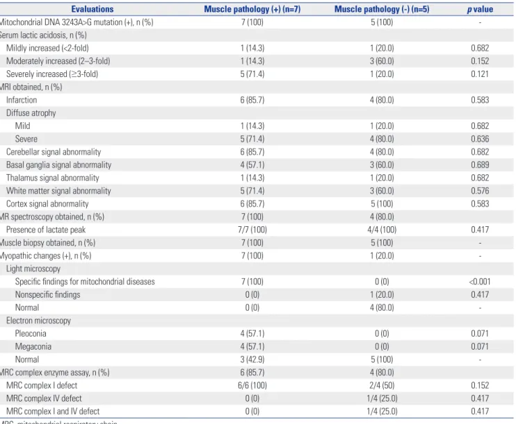
Patients with weakness as the initial symptom demonstrated Table 5. Comparison of Diagnostic Evaluations between Subgroups
Evaluations Muscle pathology (+) (n=7) Muscle pathology (-) (n=5) p value
Mitochondrial DNA 3243A>G mutation (+), n (%) 7 (100) 5 (100) -
Serum lactic acidosis, n (%)
Mildly increased (<2-fold) 1 (14.3) 1 (20.0) 0.682
Moderately increased (2–3-fold) 1 (14.3) 3 (60.0) 0.152
Severely increased (≥3-fold) 5 (71.4) 1 (20.0) 0.121
MRI obtained, n (%)
Infarction 6 (85.7) 4 (80.0) 0.583
Diffuse atrophy
Mild 1 (14.3) 1 (20.0) 0.682
Severe 5 (71.4) 4 (80.0) 0.636
Cerebellar signal abnormality 6 (85.7) 4 (80.0) 0.682
Basal ganglia signal abnormality 4 (57.1) 3 (60.0) 0.689
Thalamus signal abnormality 1 (14.3) 1 (20.0) 0.682
White matter signal abnormality 5 (71.4) 3 (60.0) 0.576
Cortex signal abnormality 6 (85.7) 5 (100) 0.583
MR spectroscopy obtained, n (%) 7 (100) 4 (80.0)
Presence of lactate peak 7/7 (100) 4/4 (100) 0.417
Muscle biopsy obtained, n (%) 7 (100) 5 (100) -
Myopathic changes (+), n (%) 7 (100) 1 (20.0) -
Light microscopy
Specific findings for mitochondrial diseases 7 (100) 0 (0) <0.001
Nonspecific findings 0 (0) 1 (20.0) 0.417
Normal 0 (0) 4 (80.0) -
Electron microscopy
Pleoconia 4 (57.1) 0 (0) 0.071
Megaconia 4 (57.1) 0 (0) 0.071
Normal 3 (42.9) 5 (100) -
MRC complex enzyme assay, n (%) 6 (85.7) 4 (80.0)
MRC complex I defect 6/6 (100) 2/4 (50) 0.152
MRC complex IV defect 0 (0) 1/4 (25.0) 0.417
MRC complex I and IV defect 0 (0) 1/4 (25.0) 0.417
MRC, mitochondrial respiratory chain.
Table 6. Comparison of Clinical Outcomes between Subgroups
Clinical outcomes at last visit Muscle pathology (+) (n=7) Muscle pathology (-) (n=5) p value
Number of stroke-like episodes 4.0±3.3 (1–10) 4.0±2.9 (2–9) 0.876
Delayed development or mental retardation, n (%) 7 (100) 4 (80.0) 0.417
Clinical severity at last outpatient clinic, n (%)
Normal (ambulatory and independent for daily activities) 1 (14.3) 1 (20.0) 0.682
Mild (ambulatory or independent for daily activities) 0 (0) 1 (20.0) 0.417
Moderate (WC bound or partially dependent for daily activities) 2 (28.6) 0 (0) 0.318
Severe (bedridden, totally dependent for daily activities or expired) 4 (57.1) 3 (60.0) 0.689
WC, wheelchair.
positive muscle pathology, although the difference was statis- tically insignificant (p=0.071). No significant trends were ob- served concerning general characteristics or clinical features according to muscle pathology, nor were there any significant differences in the severity of serum lactic acidosis or neuroim- aging studies and organ involvement (Tables 4 and 5). Divid- ing the patients into subgroups according to muscle patholo- gy, we found that differences in light microscopic changes were statistically significant, while electron microscopic changes were similar (p<0.001, p=0.071, respectively). No meaningful differences were found in terms of the parameters of the number, development, and clinical severity of stroke-like episodes (Table 6).
dIScuSSIon
Most of the current diagnostic criteria on mitochondrial dis- ease were developed prior to the recent expansion of molecu- lar genetic knowledge.8,9 These criteria are based on combina- tions of clinical, laboratory, pathologic, biochemical, and genetic findings.10 Given the complexity of mitochondrial dis- eases, their clinical manifestations are extremely heteroge- neous, and the various combinations of organ involvement have led to defining mitochondrial syndromes according to the presentations thereof: some are well known for their acro- nyms, such as MELAS and myoclonic epilepsy with RRF.1 How- ever, with outstanding accuracy, molecular diagnosis is gain- ing popularity, and with further development in diagnostic technology, muscle biopsy has generally come to be consid- ered less useful in MELAS patients confirmed by genetic test- ing. Our study is unique and meaningful because few studies have sought to assess the usefulness of muscle biopsy in mito- chondrial diseases, such as MELAS.
MELAS syndrome was first described in 1984 by Pavlakis, et al.4 Later, Hirano, et al.11 defined the three almost invariant criteria of MELAS: 1) stroke-like episode before the age of 40 years; 2) encephalopathy characterized by seizures, dementia, or both; and 3) lactic acidosis, presence of RRFs, or both. The diagnosis is confirmed if at least two of the following criteria were also present: 1) normal early development, 2) recurrent headaches, and 3) recurrent vomiting episodes. Recently, the MELAS study committee in Japan published other diagnostic criteria by which the diagnosis is considered definitive with at least two category A criteria (headaches with vomiting, sei- zures, hemiplegia, cortical blindness, and acute focal lesions in neuroimaging) and two category B criteria (high plasma or cerebrospinal fluid lactate, mitochondrial abnormalities in muscle biopsy, and a MELAS-related gene mutation).6 Molec- ular genetic testing is also important in the diagnosis of ME- LAS. The mtDNA 3243A>G mutation in the MT-TL1 gene en- coding tRNALeu(UUR) is found in 80% of MELAS patients.5,12 Additional mtDNA mutations have been shown to be associat-
ed with MELAS, such as the MT-TL1 pathogenic variants (mi- tochondrial DNA 3271 T>C and mitochondrial DNA 3252 A>G mutations).13 Currently, over 30 mtDNA gene mutations have been found to be associated with this syndrome (http://www.
ncbi.nlm.nih.gov/omim/540000). Our data were consistent with previous studies in terms of prevalence of the mtDNA 3243A>G mutation.2 Molecular genetic testing was performed in all patients, and they were all (100%) positive for mtDNA 3243A>G mutation. No other mutations were noted. The re- sults of the present study correspond with an earlier study that reported that the primary considerable and common gene is mtDNA 3243A>G mutation. The mtDNA 3243A>G mutation in the MT-TL1 gene has a wide phenotypic spectrum, includ- ing MELAS, chronic progressive external ophthalmoplegia, maternally inherited deafness and diabetes, and Leigh syn- drome. Other reported features include isolated myopathy, cardiomyopathy, seizures, migraine, ataxia, cognitive impair- ment, bowel dysmotility, and short stature.14,15 The extreme variability in the clinical phenotype is due to the proportion of mutated mtDNA or the number of wild-type mtDNA within tis- sues.16,17 Detection of mtDNA point mutation without quanti- fication alone has been generally accepted to be insufficient in the diagnosis of MELAS because of mtDNA heteroplasmy. The heteroplasmy of mtDNA makes the detection of mtDNA point mutation alone and without quantification insufficient to di- agnose MELAS.
Before the development of newer molecular genetic testing, muscle biopsy was thought to be the best method to obtain accurate diagnosis of mitochondrial diseases, MELAS in par- ticular. The diagnosis of MELAS is often confirmed by the pres- ence of RRF on succinate dehydrogenase histochemical stain, as a result of diseased mitochondrial aggregates in the subsar- colemmal areas of muscle fibers.10,18,19 Muscle biopsy also pro- vides muscle tissue samples for morphological, biochemical, and molecular studies. In the current study, no statistically sig- nificant difference was observed between MELAS patient har- boring 3243A>G point mutation who were positive for muscle pathology and those who were negative for muscle pathology.
On the basis of this finding, we deemed the usefulness of mus- cle biopsy to be limited at initial confirmative diagnosis of ME- LAS. Additionally, the patients with weakness as the initial symptom of MELAS were all positive for muscle pathology.
While it is assumed that muscle pathology can provide some information to MELAS patients with weakness, no statistically significant relationship was found between the groups that were positive for muscle pathology and negative for muscle pathology (p=0.071). Nevertheless, these findings may reflect the difficulty of obtaining a statistical correlation because of the limited number of patients in each subgroup. Moreover, myopathy has long been recognized and well described in MELAS and several other mitochondrial diseases.18 In a pro- spective cohort of MELAS patients in Japan, of 96 patients, 36 (37.5%) patients had muscle weakness at onset, and 40 (41.7%)
experienced muscle weakness over the entire course.2 The importance of muscle pathology in a patient suspected with MELAS syndrome should not be overlooked, particularly in a patient whose predominant clinical symptoms include mus- cle weakness.3
MELAS patients harboring point mutation involve a high fre- quency of RRF.20 However, no studies investigating the useful- ness of muscle biopsy in MELAS patients diagnosed by mo- lecular analysis have been conducted. Previous studies have reported that the accumulation of mitochondria in muscle fi- bers, which were composed of typical RRF, is found in up to 97% of MELAS patients.20,21 Nevertheless, several points should be considered in regards to the limitations of muscle biopsy.
The procedure is invasive and requires sedation when dealing with children. On occasion, the pathologist is only able to con- clude that the specimen provides findings consistent with an unspecified myopathy. This result may occur for a variety of reasons, including cases where the obtained tissue may not have been taken from a disease-fulminant region or the volume of muscle tissue is not enough.22 Moreover, a normal reference value of muscle tissue based on age still remains unknown.10,19 Notwithstanding, muscle biopsy has been performed in the routine analysis for mitochondrial disease when the diagnosis cannot be confirmed with genetic testing.10
This study was retrospective in nature, and it has some limi- tations. Because of disease rarity, the size of the study popula- tion was small, which may imply that the results are not suffi- cient for making generalized interpretations. From the results, the usefulness of muscle biopsy was shown to be limited in making the initial confirmative diagnosis of MELAS. Muscle biopsy was not able to provide additional information on ME- LAS patients when already diagnosed through genetic testing.
However, muscle biopsy could provide some information if genetic testing cannot confirm a diagnosis in clinically sus- pected MELAS patients with weakness, which may be a symp- tom of myopathy. In short, though a genetic test should be the first choice to diagnose MELAS, muscle biopsy should be con- sidered as an additional choice in clinically suspected MELAS patient with weakness not confirmed by genetic testing. In or- der to provide a more generalized guideline, studies on larger numbers of patients are needed to better delineate the useful- ness of muscle biopsy in MELAS. Mitochondrial disease is heterogeneous in general, and each of the mitochondrial syn- dromes tend to display their own specific clinical manifesta- tions. These characteristics warrant continuous study, and whether previous diagnostic tools are in fact the best methods for each specific type of syndrome needs to be verified.
AcKnoWLEdGEMEntS
This research was supported by a grant from the Korea Health Technology R&D Project through the Korea Health Industry Development Institute (KHIDI), funded by the Ministry of Health
& Welfare, Republic of Korea (grant number: HI16C0673).
orcId ids
Min-Seong Baek https://orcid.org/0000-0002-7132-362X Se Hoon Kim https://orcid.org/0000-0001-7516-7372 Young-Mock Lee https://orcid.org/0000-0002-5838-249X
rEFErEncES
1. Zeviani M, Di Donato S. Mitochondrial disorders. Brain 2004;127 (Pt 10):2153-72.
2. Yatsuga S, Povalko N, Nishioka J, Katayama K, Kakimoto N, Mat- suishi T, et al. MELAS: a nationwide prospective cohort study of 96 patients in Japan. Biochim Biophys Acta 2012;1820:619-24.
3. Goto Y, Horai S, Matsuoka T, Koga Y, Nihei K, Kobayashi M, et al.
Mitochondrial myopathy, encephalopathy, lactic acidosis, and stroke-like episodes (MELAS): a correlative study of the clinical features and mitochondrial DNA mutation. Neurology 1992;42(3 Pt 1):545-50.
4. Pavlakis SG, Phillips PC, DiMauro S, De Vivo DC, Rowland LP.
Mitochondrial myopathy, encephalopathy, lactic acidosis, and strokelike episodes: a distinctive clinical syndrome. Ann Neurol 1984;16:481-8.
5. Goto Y, Nonaka I, Horai S. A mutation in the tRNA(Leu)(UUR) gene associated with the MELAS subgroup of mitochondrial en- cephalomyopathies. Nature 1990;348:651-3.
6. El-Hattab AW, Adesina AM, Jones J, Scaglia F. MELAS syndrome:
clinical manifestations, pathogenesis, and treatment options. Mol Genet Metab 2015;116:4-12.
7. Kisler JE, Whittaker RG, McFarland R. Mitochondrial diseases in childhood: a clinical approach to investigation and management.
Dev Med Child Neurol 2010;52:422-33.
8. Haas RH, Parikh S, Falk MJ, Saneto RP, Wolf NI, Darin N, et al. Mi- tochondrial disease: a practical approach for primary care physi- cians. Pediatrics 2007;120:1326-33.
9. Koenig MK. Presentation and diagnosis of mitochondrial disor- ders in children. Pediatr Neurol 2008;38:305-13.
10. Parikh S, Goldstein A, Koenig MK, Scaglia F, Enns GM, Saneto R, et al. Diagnosis and management of mitochondrial disease: a consensus statement from the Mitochondrial Medicine Society.
Genet Med 2015;17:689-701.
11. Hirano M, Ricci E, Koenigsberger MR, Defendini R, Pavlakis SG, DeVivo DC, et al. Melas: an original case and clinical criteria for diagnosis. Neuromuscul Disord 1992;2:125-35.
12. Parsons T, Weimer L, Engelstad K, Linker A, Battista V, Wei Y, et al.
Autonomic symptoms in carriers of the m.3243A>G mitochon- drial DNA mutation. Arch Neurol 2010;67:976-9.
13. Morten KJ, Cooper JM, Brown GK, Lake BD, Pike D, Poulton J. A new point mutation associated with mitochondrial encephalo- myopathy. Hum Mol Genet 1993;2:2081-7.
14. Moraes CT, Ciacci F, Silvestri G, Shanske S, Sciacco M, Hirano M, et al. Atypical clinical presentations associated with the MELAS mutation at position 3243 of human mitochondrial DNA. Neuro- muscul Disord 1993;3:43-50.
15. Nesbitt V, Pitceathly RD, Turnbull DM, Taylor RW, Sweeney MG, Mudanohwo EE, et al. The UK MRC Mitochondrial Disease Pa- tient Cohort Study: clinical phenotypes associated with the m.3243A>G mutation--implications for diagnosis and manage- ment. J Neurol Neurosurg Psychiatry 2013;84:936-8.
16. Uusimaa J, Moilanen JS, Vainionpää L, Tapanainen P, Lindholm P, Nuutinen M, et al. Prevalence, segregation, and phenotype of the
mitochondrial DNA 3243A>G mutation in children. Ann Neurol 2007;62:278-87.
17. Manwaring N, Jones MM, Wang JJ, Rochtchina E, Howard C, Mitchell P, et al. Population prevalence of the MELAS A3243G mutation. Mitochondrion 2007;7:230-3.
18. Sproule DM, Kaufmann P. Mitochondrial encephalopathy, lactic acidosis, and strokelike episodes: basic concepts, clinical pheno- type, and therapeutic management of MELAS syndrome. Ann N Y Acad Sci 2008;1142:133-58.
19. Rollins S, Prayson RA, McMahon JT, Cohen BH. Diagnostic yield
muscle biopsy in patients with clinical evidence of mitochondrial cytopathy. Am J Clin Pathol 2001;116:326-30.
20. Goto Y. Clinical features of MELAS and mitochondrial DNA mu- tations. Muscle Nerve Suppl 1995;3:S107-12.
21. Lorenzoni PJ, Scola RH, Kay CS, Arndt RC, Freund AA, Bruck I, et al. MELAS: clinical features, muscle biopsy and molecular genet- ics. Arq Neuropsiquiatr 2009;67:668-76.
22. Joyce NC, Oskarsson B, Jin LW. Muscle biopsy evaluation in neu- romuscular disorders. Phys Med Rehabil Clin N Am 2012;23:609-31.