INTRODUCTION
Liver transplantation (LT) is an effective treatment for pa- tients with end-stage liver disease because of improved re- sults and broadening of indications. For pediatric patients,
LT with size-matched whole liver allografts from pediatric donors is considered as ideal due to lower complication rates and better survival compared with other variant types of LT [1]. However, the number of liver allografts recovered from pediatric donors is very limited in Korea. Further-
Whole liver deceased donor liver
transplantation for pediatric recipients:
single-center experience for 20 years
Jung-Man Namgoong
1, Shin Hwang
1, Dae-Yeon Kim
1, Tae-Yong Ha
1, Gi-Won Song
1, Dong-Hwan Jung
1, Gil-Chun Park
1, Kyung Mo Kim
2, Seak Hee Oh
21Department of Surgery, Asan Medical Center, University of Ulsan College of Medicine, Seoul, Korea
2Department of Pediatrics, Asan Medical Center, University of Ulsan College of Medicine, Seoul, Korea
Background: We investigated the incidence and outcomes of pediatric deceased donor liver transplantation (DDLT) using whole liver grafts in a high-volume liver transplantation (LT) center.
Methods: The study was a retrospective single-center analysis of whole LT in pediatric recipients. The study period was set as 20 years between January 2000 and December 2019. We defined pediatric recipients and donors to be aged ≤18 years.
Results: During the study period, there were 98 cases of pediatric DDLT, and 34 pa- tients (34.7%) received whole liver grafts. The age range of the deceased donors was 3 months to 56 years and that of pediatric recipients was 7 months to 17 years. Common primary diseases for LT were biliary atresia in 13, acute liver failure in four, Wilson dis- ease in four, congenital portal vein agenesis in three, and genetic metabolic diseases in three. Pediatric-to-pediatric and adult-to-pediatric whole LTs were 22 (64.7%) and 12 (35.3%), respectively. A good correlation was noted between the donor and the re- cipient’s body weight, and the recipient’s body weight and allograft’s weight. Graft and overall patient survival rates were 91.2% and 91.2% at 1 year, 88.0% and 88.0% at 3 years, and 88.0% and 88.0% at 5 years, respectively.
Conclusions: The results of this study revealed that Korean Network for Organ Sharing (KONOS) regulations with donor-recipient body weight matching exhibited good perfor- mance. Considering the reciprocal trades of liver organs among pediatric and adult do- nors and recipients, it is necessary to establish a policy for pediatric donor liver grafts to pediatric recipients on a priority basis.
Keywords: Donor age; Pediatric donor; Deceased donor liver transplantation; Infant;
Adolescent
Received September 1, 2020 Revised September 30, 2020 Accepted October 2, 2020 Corresponding author: Shin Hwang Department of Surgery, Asan Medical Center, University of Ulsan College of Medicine, 88 Olympic-ro 43-gil, Songpa- gu, Seoul 05505, Korea
Tel: +82-2-3010-3930 Fax: +82-2-3010-6701 E-mail: [email protected]
© The Korean Society for Transplantation This is an Open Access article distributed under the terms of the Creative Commons Attribution Non-Commercial License (http://creativecommons.org/licenses/
by-nc/4.0/) which permits unrestricted non-commercial use, distribution, and reproduction in any medium, provided the original work is properly cited.
pISSN 2671-8790
eISSN 2671-8804
more, the body size of pediatric patients is widely variable from infant to adolescent, thus donor-recipient body size matching is much more complex compared with adult-to- adult deceased donor liver transplantation (DDLT).
Because of low incidence of pediatric deceased donors and complex donor-recipient body size matching, split LT and living donor LT have been more frequently performed for pediatric patients than LT with whole liver grafts [2].
Small-sized whole liver grafts from young pediatric do- nors have usually been allocated to young pediatric re- cipients, and liver grafts from adolescent donors have been allocated to both adolescent and adult recipients, and those from adult donors have also been reciprocally allocated to adolescent recipients [2-6]. There exists only
limited detailed information on pediatric DDLT in Korea, thus, there is an essential need to analyze the status of pediatric DDLT using whole liver grafts [2,6]. We herein in- vestigated the incidence and outcomes of pediatric DDLT using whole liver grafts in a high-volume LT center.
METHODS
The study protocol was approved by the Institutional Re- view Board of Asan Medical Center (IRB No. 2020-0857), which waived the requirement for informed consent due to the retrospective nature of this study. This study was performed in accordance with the ethical guidelines of the World Medical Association Declaration of Helsinki 2013.
Study Design
The study was a retrospective single-center analysis of DDLT in pediatric recipients. The study period was set as 20 years between January 2000 and December 2019.
We defined pediatric recipients and pediatric donors to be aged ≤18 years. There were 348 cases of pediatric LTs including living donor LT in 250 and DDLT in 98 (split LT in 64 and whole liver graft LT in 34). The recipients of whole HIGHLIGHTS
• The study was a retrospective single-center analysis of whole liver transplantation in 34 pediatric recipients.
• Considering the reciprocal trades of liver organs among pediatric and adult donors and recipients, it is neces- sary to establish a policy for pediatric donor liver grafts to pediatric recipients on a priority basis.
Fig. 1. Illustration of the modified piggyback technique to make a large
cavocaval anastomosis. After clamping of the suprahepatic and retro- hepatic inferior vena cava (IVC), the orifices of the right, middle, and left hepatic vein trunks are opened altogether to make a large single orifice.
A 4–5-cm-long longitudinal incision is made at the ventral surface of the retrohepatic IVC to enlarge the anastomotic vein orifice. A 4-cm-long longitudinal incision is also made at the dorsal surface of the graft IVC.
These triangular-shaped orifices at the recipient and graft IVCs are well matched, thus being tolerant to extrinsic compression.
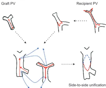
Graft PV Recipient PV
Side-to-side unification
Fig. 2. Illustration of the side-to-side unification technique used for portal
vein (PV) reconstruction. A deep longitudinal incision is made at the 6
o’clock direction of the graft PV and the 12 o’clock direction of the recip-
ient PV. Running sutures are used to unify these two PVs. This technique
creates an enlarged conduit from the superior mesenteric vein-splenic
vein confluence to the hilar PV confluence.
liver grafts were followed up until June 2020. The im- munosuppressive regimens for pediatric recipients were similar to those for adult recipients [7,8].
Surgical Technique for Whole LT
The standard techniques for whole LT have been used for pediatric recipients in principle. There are four technical points unique for pediatric DDLT. For recipients with body weight less than 40 kg, a modified piggyback technique involving a large cavocaval anastomosis was primarily used to secure graft outflow vein reconstruction. This method effectively prevents graft hepatic outflow obstruc- tion under the situation of extrinsic compression of the inferior vena cava due to large-for-size graft implantation (Fig. 1). For recipients with body weight less than 20 kg and portal vein hypoplasia, the side-to-side unification venoplasty technique was used for portal vein reconstruc- tion as it enables accomplishment of the effective size of the anastomotic cross-sectional area and a streamlined configuration without axial rotation (Fig. 2) [9]. For con- genital portal vein hypoplasia or agenesis, portal vein in- terposition with deceased donor femoral vein or external iliac vein was used. Surgical microscopy was used for hepatic artery reconstruction regardless of the diameter of the artery.
Statistical Analysis
The numerical data are presented as mean±standard de- viation. The continuous variables were compared using Student t-test. The incidence variables were compared using the chi-square test and Fisher’s exact test. The
survival rates were estimated using the Kaplan-Meier method and compared using a log-rank test. A P-value of
<0.05 was considered statistically significant. Statistical analyses were performed using IBM SPSS ver. 22.0 (IBM Corp., New York, NY, USA).
RESULTS Donor and Recipient Profiles
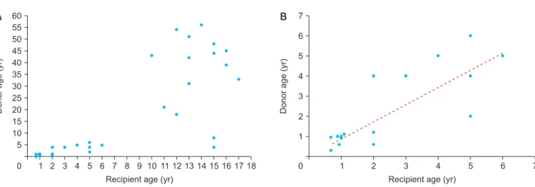
During the 20-year study period, there were a total of 1056 cases of DDLT. Of them, 98 cases (9.2%) involved pediatric patients. The number of pediatric whole LT was 34 out of 98 (34.7%). The correlation in the age of the 34 deceased donors (range, 3 months–56 years) and 34 pedi- atric recipients (range, 7 months–17 years) is presented in Fig. 3. Male to female ratio was 20:14 in donors and 16:18 in recipients. Detailed recipient and donor profiles are summarized in Table 1.
The primary diseases for LT in 34 pediatric recipi- ents were biliary atresia in 13, acute liver failure in four, Wilson disease in four, congenital portal vein agenesis with congenital portosystemic venous shunt in three, genetic metabolic diseases in three (each one case of Alagille syndrome, glycogen storage disease, and or- nithine transcarbamylase deficiency), Caroli disease in one, progressive familial intrahepatic cholestasis in one, hepatoblastoma in one, and retransplantation after LDLT in four cases. One patient with glycogen storage disease had undergone hepatocyte transplantation 3 years before
60 55 50 45 40 35 30 25 20 15 10 5
18
Donorage(yr)
Recipient age (yr)
0 1 2 3 4 5 6 7 8 9 10 11 12 13 14 15 16 17
7 6 5 4 3 2 1
7
Donorage(yr)
Recipient age (yr)
0 1 2 3 4 5 6
A B
Fig. 3. Scatter plots showing the age distribution of recipients and donors. Distribution of all the 34 recipients (A) and younger subgroups with recipient
age ≤6 years (B). A dotted line denotes correlation.
LT. Eight deceased donors were managed at our institu- tion and the remaining 26 donors were managed at other institutions.
Donor and Recipient Matchings Regarding Age, Body Weight and Graft Size
The age distribution of the deceased donors and pediatric recipients is depicted in Fig. 3A. The recipients were cate- gorized into subgroups depending on the age: between 7
months and 6 years and between 10 years and 17 years.
In the younger subgroup, the age of the donors and re- cipients exhibited good correlation (donor age=0.86×re- cipient age, r
2=0.83, P<0.001) (Fig. 3B), and all the donors were pediatric donors. On the contrary, in the older sub- group, out of 15, only three were pediatric donors and the remaining 12 donors were adult donors. Thus, pediat- ric-to-pediatric and adult-to-pediatric whole LTs were 22 (64.7%) and 12 (35.3%), respectively (Table 1).
The body weight distribution of the deceased donors and pediatric recipients is depicted in Fig. 4. The body weight of donors and recipients exhibited good correlation (donor body weight=0.93×recipient body weight, r
2=0.92, P<0.001). The Korean Network for Organ Sharing (KONOS) regulation regarding donor-recipient body weight match ratio of 1:2–2:1 was met in 33 of 34 cases. The distri- bution of the recipient’s body weight and liver allograft’s weight is depicted in Fig. 5. A good correlation was ob- served between the recipient’s body weight and allograft’s weight (liver graft weight (g)=21.8×recipient body weight (kg), r
2=0.89, P<0.001). The distribution of the recipient’s body weight and graft-recipient weight ratio (GRWR) is depicted in Fig. 6. The recipient’s body weight and GRWR demonstrated coarse correlation (GRWR=8.92×recipient body weight (kg)
–0.35, r
2=0.53, P<0.001). The correlation curve was close to the theoretical upper limit of GRWR (4 for infants and 2.5 for adolescents or adults) according to the recipient’s age or body weight.
Operation Profiles
For pediatric recipients with a body weight of less than 40 kg, the abovementioned modified piggyback techniques were primarily used for graft outflow vein reconstruction.
For older adolescent patients, the standard technique of DDLT with interposition of the graft inferior vena cava was used. The mean warm, cold, and total ischemic times were 275.8±153.4 minutes (range, 71–839 minutes), 46.1±9.9 minutes (range, 30–74 minutes), and 335.3±221.8 minutes (range, 101–1,397 minutes), respectively. There was no in- cidence of major vascular complications, except for portal vein stenosis in infant-to-infant whole LT in three cases [7].
Survival Outcomes
During a mean follow-up period of 73.1±51.5 months, four patients passed away. In-hospital mortality within 2 months occurred in two cases due to portal vein compli- cation-associated graft failure. One patient who had DDLT as retransplantation died at 6 months due to progressive
Table 1. Comparison of recipient and donor profilesVariable
Pediatric-to- pediatric transplantation
Adult-to- pediatric transplantation
P-value
No. of patients 22 12 -
Recipient sex (male:female) 8:14 8:4 0.09
Recipient age (yr) 4.2±4.4 13.8±2.1 <0.001
Primary disease NA
Biliary atresia 12 1
Acute liver failure 0 4
Wilson disease 0 4
Metabolic disease 3 0
Congenital portal vein agenesis
3 0
Retransplantation 2 2
Others 2 1
Recipient ABO blood group NA
A 8 4
B 2 2
O 6 5
AB 6 1
Preoperative laboratory finding
Total bilirubin (mg/dL) 11.3±11.1 20.6±13.6 0.05
Albumin (g/dL) 2.9±0.6 3.1±0.7 0.51
Serum creatinine (mg/dL) 0.34±0.21 0.83±0.46 <0.001 Prothrombin time (INR) 1.24±0.34 2.77±1.22 <0.001
PELD/MELD score 10.5±7.3 27.0±8.6 <0.001
Donor sex (male:female) 14:8 6:6 0.44
Donor age (yr) 3.4±3.9 42.3±10.1 <0.001
Graft weight (g) 576.1±315.2 1,222.3±492.9 <0.001 Graft-recipient weight ratio 3.69±1.66 2.68±0.91 0.03 Ischemic time
Cold 297.8±262.6 286.1±131.9 0.88
Warm 46.2±9.2 45.7±11.7 0.84
Values are presented as mean±standard deviation.
NA, not available; INR, international normalization ratio; PELD, pediatric
end-stage liver disease; MELD, model for end-stage liver disease.
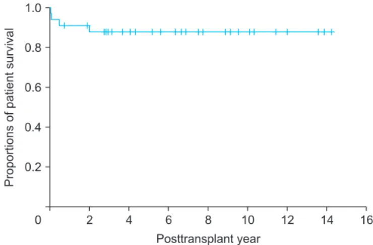
graft failure. One patient died at 23 months due to progres- sive graft failure after repeated episodes of acute cellular rejection. Graft and overall patient survival rates were 91.2% and 91.2% at 1 year, 88.0% and 88.0% at 3 years, and 88.0% and 88.0% at 5 years, respectively (Fig. 7).
DISCUSSION
The present study included 22 cases of pediatric-to-pedi- atric and 12 cases of adult-to-pediatric DDLT using whole
liver grafts. During the 20-year study period, there were 348 cases of pediatric LTs including living donor LT in 250 and DDLT in 98 with inclusion of split LT in 64 and whole liver graft LT in 34. Thus, the proportion of whole liver graft LT was 9.8% in pediatric LT cases. There were also a total of 1,056 cases of DDLT, thus pediatric whole liver graft LT occupied 3.2% of all DDLTs. These proportions indicate that whole liver graft LT is the least common form of pedi- atric LT. The body weight of the deceased donors and pe- diatric recipients were well matched, thus high correlation between the recipient’s body weight and allograft’s weight and reasonable GRWR were observed.
We previously demonstrated that, of the 31 pediatric
0 90 80 70 60 50 40 30 20 10
90
Donorbodyweight(kg)
Recipient body weight (kg)
10 20 30 40 50 60 70 80
Recipient-donor body weight ratio=1:2
Recipient-donor body weight ratio=2:1
Fig. 4. A scatter plot showing body weight distribution of recipients and
donors. The red dotted line denotes correlation. Two green dotted lines indicate the range of donor body weight matching according to the Korean Network for Organ Sharing (KONOS) regulations. All cases except for one were located within the eligible range of recipient-donor weight matching.
0 2,000 1,800 1,600 1,400 1,200 1,000 800 600 400 200
90
Livergraftweight(g)
Recipient body weight (kg)
10 20 30 40 50 60 70 80
Fig. 5. A scatter plot showing the distribution of the recipient’s body and
whole liver graft’s weight. The red dotted line denotes correlation.
0 1.0
0.8
0.6
0.4
0.2
16
Proportionsofpatientsurvival
Posttransplant year
2 4 6 8 10 12 14
Fig. 7. Kaplan-Meier analysis of the overall patient survival outcomes fol-
lowing whole liver transplantation in pediatric recipients.
0 8 7 6 5 4 3 2 1
90 Recipient body weight (kg)
10 20 30 40 50 60 70 80
Theoretical upper limit of GRWR
GRWR
Fig. 6. Scatter plot showing the distribution of the recipient’s body and