267
Correspondence to: Jae-Won Joh, Department of Surgery, Samsung Medical Center, Sungkyunkwan University School of Medicine, 50, Irwon-dong, Gangnam-gu, Seoul 135-710, Korea. Tel: 02-3410- 3466, Fax: 02-3410-0040, E-mail: [email protected]
Received May 28, 2010, Accepted July 7, 2010
Aspergillosis in Liver Transplant Recipients: A Single Center Experience
Department of Surgery, Samsung Medical Center,
Sungkyunkwan University School of Medicine, Organ Transplant Center, Seoul, Korea
Jong Man Kim, M.D., Ph.D., Choon Hyuck David Kwon, M.D., Ph.D., Jae-Won Joh, M.D., Ph.D., Sanghyun Song, M.D., Milljae Shin, M.D., Sung Joo Kim, M.D., Ph.D.,
Seung Heui Hong, Bok Nyeo Kim, Suk-Koo Lee, M.D., Ph.D.
Purpose: The aim of this study was to review the epidemiology, clinical features, diagnosis, and effect of treatments
of aspergillosis infections in adult liver transplantation patients.Methods: We retrospectively studied the cases of aspergillosis among 625 (164 deceased donor liver transplantation
(DDLT), 461 living donor liver transplantation (LDLT)) adult liver transplantation recipients (≥18 yrs old) operated between May 1996 to May 2008.Results: Fourteen cases of aspergillosis infection were detected, which consisted of 9 cases of invasive aspergillosis,
5 cases of aspergilloma in maxillary sinuses which occurred before the transplantation. One patient with invasive aspergillosis, who had received liver transplantation overseas and had post-operation care in our center, was also included. Among the 8 cases (1.28%) of invasive aspergillosis (excluding one case operated overseas), 6 cases (3.7%) were DDLT and 2 cases (0.4%) LDLT (P-value<0.05). Among the 6 patients with early onset of invasive aspergillosis, 5 patients (1.8%) had fluconazole and only 1 patient (0.3%) had itraconazole as prophylactic antifungal agent (P-value<0.05). The three cases with localized lesions in the lung survived after lobectomy, but the disseminated and inoperable cases died.Conclusion: The incidence of invasive aspergillosis was lower in LDLT cases and prophylactic itraconazole might
be more effective than fluconazole. Paranasal aspergilloma, detected before transplantation had no relation with posttransplant invasive aspergillosis infection. Patients who had localized lesion in the lung, and underwent surgery had better survival than who could not. (J Korean Surg Soc 2010;79:267-274)Key Words: Liver transplantation, Invasive aspergillosis, Paranasal aspergilloma, Antifungal agents, Lung lobec-
tomyINTRODUCTION
Invasive aspergillosis is an uncommon infection, but is a serious and devastating complication after liver transplantation (LT). The incidence of invasive aspergillosis varies depending on the organ transplant involved are liver (1∼8%), kidney (0∼4%), lung (3∼14%) and heart (1∼
15%).(1,2) The mortality of invasive aspergillosis in liver
transplantation recipients exceeds 90%.(3)
Presently most infections of invasive aspergillosis occur late in the postoperative period, as compared with those in the early 1990s.(4-6) Risk factors for invasive asper- gillosis in liver transplantation recipients include poor graft dysfunction, retransplantation, post transplant renal dys- function, cytomegalovirus infection and a history of fulmi- nant hepatic failure before the transplantation.(3,5-8) The aim of this study was to review the epidemiology, clinical features, diagnosis, and effect of treatments of aspergillosis infections in adult LT patients.
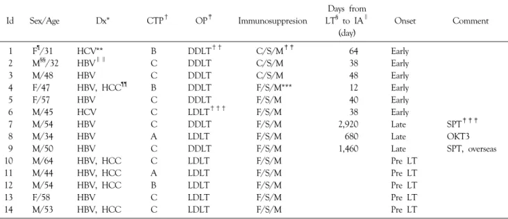
Table 1. Clinical characteristics of recipients with aspergillosis after transplantation
Id Sex/Age Dx* CTP† OP‡ Immunosuppresion
Days from LT§ to IA∥
(day)
Onset Comment
1 F¶/31 HCV** B DDLT†† C/S/M‡‡ 64 Early
2 M§§/32 HBV∥∥ C DDLT C/S/M 38 Early
3 M/48 HBV C DDLT C/S/M 48 Early
4 F/47 HBV, HCC¶¶ B DDLT F/S/M*** 12 Early
5 F/57 HBV C DDLT F/S/M 40 Early
6 M/45 HCV C LDLT††† F/S/M 38 Early
7 M/54 HBV C DDLT F/S/M 2,920 Late SPT‡‡‡
8 M/34 HBV A LDLT F/S/M 680 Late OKT3
9 M/50 HBV C DDLT F/S/M 1,460 Late SPT, overseas
10 M/64 HBV, HCC C LDLT F/S/M Pre LT
11 M/44 HBV, HCC A LDLT F/S/M Pre LT
12 M/54 HBV, HCC B LDLT F/S/M Pre LT
13 F/58 HBV C LDLT F/S/M Pre LT
14 M/53 HBV, HCC C LDLT F/S/M Pre LT
*Dx = diagnosis; †CTP = Child-Turchotte-Pugh score; ‡OP = operation; §LT = liver transplantation; ∥IA = invasive aspergillosis; ¶F = female; **HCV = hepatitis C virus; ††DDLT = deceased donor liver transplantation; ‡‡C/S/M = cyclosporine/steroid/mycofenolate mofetil;
§§M = male; ∥∥HBV = hepatitis B virus; ¶¶HCC = hepatocellular carcinoma; ***F/S/M = FK506/steroid/mycofenolate mofetil; †††LDLT
= living donor liver transplantation; ‡‡‡SPT = steroid pulse therapy.
METHODS 1) Patients
Six hundred twenty five adult patients (≥18 yrs old) who underwent LT at Samsung Medical Center between May 1996 to May 2008 were reviewed. Four hundred and sixty one cases (73.8%) were living donor liver trans- plantation (LDLT) and 164 cases (26.2%) were deceased donor liver transplantation (DDLT). Fourteen cases (11 males and 3 females) of aspergillosis were detected from the computerized database of the hospital’s information system among our LT recipients during this period. Sex, age, primary diagnosis, LT type (DDLT/LDLT), immuno- suppression, time interval between LT and aspergillosis infection are shown in Table 1.
Prophylactic antifungal agents were used for all LT recipients and the agent was changed during the study interval. Fluconazole (100 mg/day o.d.) as single antifungal prophylactic therapy was administered between 1996 and 2004 (275 patients) and itraconazole (100 mg b.i.d) was used for 30 days postoperative period since 2004 (350
patients). Prophylactic antibiotics (cefotaxime and ampicillin/
sulbactam) were administered for 4 days in postoperative period. Trimethoprim/sulfamethoxazole was administered as prophylaxis against Pneumocystitis jirovecii infection every weekend until six months. Cytomegalovirus antigen detec- tion was performed at regular intervals. One cytomegalo- virus pp65 antigen positive cell/400,000 white blood cells was defined as positive for cytomegalovirus antigenemia.
Pre-emptive intravenous ganciclovir was administered if cytomegalovirus pp65 antigen positive cell value was greater than 10 positive cells per 400,000 white blood cells regardless of clinical manifestation. Selective bowel decontamination using erythromycin and gentamycin was performed a day before liver transplantation until oral intake was established after LT. All LT patients received an immunosuppressive regimen consisting of FK 506 or cyclosporine, mycofenolate mofetil, and corticosteroid. The optimal blood level of FK 506/cyclosporin were adjusted to 10∼15 ng/ml and 200
∼300 ng/ml respectively during the first one month and reduced to 5∼10 ng/ml / 100∼200 ng/ml thereafter.
Intravenous methylprednisolone 500 mg was administered during anhepatic phase, and tapered gradually during 7
days. Steroid was gradually withdrawn in most patients by 3 months after the LT if liver function was stable.
Acute rejection episodes were diagnosed by clinical and serum biochemical signs, and confirmed by fine needle liver biopsy. Moderate or severe acute rejection was treated with bolus infusion of methylprednisolone, and the immuno- suppressive drug dose was increased. Steroid resistant rejection was treated with a 10∼14 day course of OKT3 monoclonal antibody. Antifungal agents, amphotericin B, capsofungin, and voriconazole were used as single or combined protocol.
2) Method
Urine, sputum, drains, bile, wounds, and blood in all recipients after LT were examined routinely twice a week as surveillance for infection. Invasive aspergillosis was defined as per criteria for proven or probable invasive aspergillosis described by European Organization for Re- search and Treatment of Cancer/Invasive Fungal Infec- tions Co-operative Group and the National Institute of Allergy and Infectious Disease Mycoses Study Group.(9)
3) Statistical analysis
Fisher’s exact tests were used to compare categorical variables such as DDLT versus LDLT and itraconazole versus fluconazole. A P-value of ≤0.05 was considered significant.
The data analyses were performed with SPSS 17.0 (SPSS Inc., Chicago, IL, USA).
RESULTS
Fourteen LT recipients were identified with aspergillosis infection - 13 patients operated in Samsung Medical Center and 1 patient with overseas surgery and post-operative care at Samsung Medical Center. Eight patients (1.28%) developed invasive aspergillosis after liver transplantation and 5 patients were diagnosed as aspergilloma of maxillary sinus before the LT. The preoperative diagnoses in this study consisted of liver cirrhosis (n=9) and hepatocellular carci- noma (n=5). Among 8 patients who had transplantation in our hospital and developed aspergillosis afterward, 6 cases
(3.7%) were DDLT and 2 cases (0.4%) were LDLT (P-value
<0.05).
From September 2004 to January 2008, new building was under construction and the main hospital was renova- ting in Samsung Medical Center. One case (0.003%) of 311 recipients developed invasive aspergillosis in trans- plant units during this period.
The mean time from LT to invasive aspergillosis was 40 days (12∼64 days) in early onset group (n=6) and 1,686.7 days (680∼2,920 days) in late onset group (n=3). Among the 6 patients with early onset of aspergillosis infection - 5 patients (1.8%) had fluconazole (5 out of 275) as pro- phylactic antifungal agent. Among the other 350 recipients who received itraconazole prophylaxis, only 1 patient (0.3%) developed invasive aspergillosis. Patients who received oral itraconazole rather than fluconazole after liver trans- plantation had statistically significant lower incidence of invasive aspergillosis (P-value<0.05). Patients with late onset had history of steroid pulse therapy (n=2) or OKT3 (n=1) treatment for acute rejection. Immunosuppressive drug doses were reduced to half in patients during the antifungal therapy for invasive aspergillosis.
Prophylactic antifungal agents, symptoms, sites of infec- tion, methods of diagnosis, type of aspergillosis infection, treatments, and outcomes are presented in Table 2.
The median levels of white blood cell, AST, ALT, total bilirubin, INR and serum creatinine at the time of invasive aspergillosis diagnosis were 7,000/μl (range, 1,600∼
26,310/μl), 30 U/L (range, 12∼4,230 U/L), 107 U/L (range, 14∼1,520 U/L), 1.9 mg/dl (range, 0.4∼57.9 mg/dl), 1.3 (range, 1.17∼3.52) and 0.92 mg/dl (range, 0.6
∼2.1 mg/dl), respectively.
Pulmonary aspergillosis was seen in 8 patients and presenting symptoms were hemoptysis, pleuritic chest wall pain, cough, dyspnea, and fever. Chest X-ray and chest CT showed mainly nodular infiltration and cavitation. ‘Halo signs’ characteristic of invasive aspergillosis was seen in one patient. Percutaneous biopsy (if tolerated) was performed in patients whose radiologic findings showed nodular lesions in the lung, otherwise a bronchoalveolar lavage and transbronchial lung biopsy was performed.
Proven pulmonary aspergillosis (n=5) showed cavitary lesions in the lung which were confirmed by biopsy. Pro- bable pulmonary aspergillosis patients (n=3) had severe clinical signs, and were diagnosed by chest CT, brocho- alveolar lavage or sputum culture. Two patients with the lung and brain lesion were diagnosed as disseminated type.
Three patients of the proven cases (n=5) had localized lesion in one lobe of lung. They had treated with lobec- tomy and antifungal agents. All patients were survived.
Other six patients had multiple cavitary lesions in the lung were inoperable. Two patients with the lung and brain lesion were diagnosed as disseminated type. These patients were managed by antifungal agents only, and all died.
Three patients with brain aspergillosis were seen as multiple brain abscesses in brain CT and brain magnetic resonance imaging. Brain involvement showed symptoms such as seizure, motor weakness or drowsiness. Multiple brain abscesses in two patients were drained and biopsied.
One of these patients had multiple skin lesions of asper- gillosis which was confirmed by excision biopsy. Mortality in patients with brain involvement was 100%.
Amphotericin B was administered in all cases of invasive aspergillosis. One patient who showed sustained fever and no improvement of radiologic findings in spite of admini- stration of amphotericin B and right lower lobectomy was changed to a combination of caspofungin and voriconazole.
Two patients received voriconazole because pleural fluid culture from chest tube drainage kept proved to be aspergillosis infection. Due to worsening of clinical symp- toms, amphotericin B was changed to caspofungin and itraconazole to each patient.
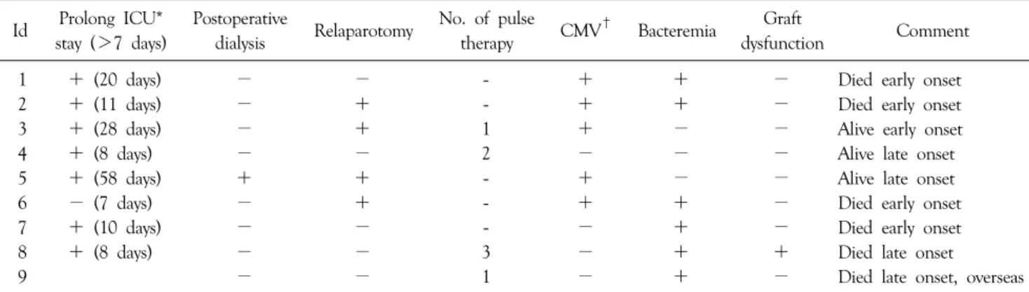
All the nine patients with invasive aspergillosis had associated risk factors (Table 3). Serious bacterial infections and prolonged duration of wide spectrum antibiotic therapy in six recipients, prolonged intensive care unit stay in seven, postoperative hemodialysis in one, relaparotomy due to postoperative bleeding in two, wound dehiscence in two, acute rejection in four, cytomegalovirus infection in five and poor graft function in one patient.
Five cases had asymptomatic fungus ball in maxillary sinus which was detected before LT by ostiomeatal unit Table 2. Aspergillosis: prophylaxis, symptoms, sites, diagnosis, type, treatment, and outcome IdProphylaxisSymptomsSite(s)MethodsTypeTreatmentsOutcome 1FluconazoleSeizure, motor weaknessBrain, lungMRI, CT, Bx*ProvenAmphotericin B, brain abscess drainageDied 2FluconazoleDrowsinessBrain, skinMRI, CT, BxProvenAmphotericin B, brain abscess drainageDied 3FluconazoleHemoptysisLungCT, BxProvenAmphotericin B, Rt. lower lobectomyAlive 4FluconazoleFever, sputum, coughLungCT, Bx, galactomannanProvenAmphotericin B, Lt. upper lobectomy,Alive antigen test capsofungin, voriconazole 5FluconazoleHemoptysisLungCT, BxProvenAmphotericin B, capsofunginDied 6ItraconazoleHemoptysisLungPleural fluid culture, BxProvenAmphotericin B, voriconazole,Alive Lt. upper lobectomy 7FluconazoleCough, sputumLungSputum, CTProbableAmphotericin B, itroaconazoleDied 8FluconazolePleuritic chest wall pain,Lung, brainCT, BAL† ProbableAmphotericin B, voriconazoleDied motor weakness 9FluconazoleHemotysis, pleuritic chest wall pain,LungCT, BALProbableAmphotericin BDied cough, dyspnea 10FluconazoleLT‡ work upMaxillary sinusCT, BxExcisionAlive 11ItraconazoleLT work upMaxillary sinusCT, BxExcisionAlive 12ItraconazoleLT work upMaxillary sinusCT, BxExcisionAlive 13ItraconazoleLT work upMaxillary sinusCT, BxExcisionAlive 14ItraconazoleLT work upMaxillary sinusCT, BxExcisionAlive *Bx = biopsy;† BAL = brochoalveolar lavage;‡ LT = liver transplantation.
Table 3. Risk factors for invasive aspergillosis in recipients after liver transplantation Id Prolong ICU*
stay (>7 days)
Postoperative
dialysis Relaparotomy No. of pulse
therapy CMV† Bacteremia Graft
dysfunction Comment
1 + (20 days) − − - + + − Died early onset
2 + (11 days) − + - + + − Died early onset
3 + (28 days) − + 1 + − − Alive early onset
4 + (8 days) − − 2 − − − Alive late onset
5 + (58 days) + + - + − − Alive late onset
6 − (7 days) − + - + + − Died early onset
7 + (10 days) − − - − + − Died early onset
8 + (8 days) − − 3 − + + Died late onset
9 − − 1 − + − Died late onset, overseas
*ICU = intensive care unit; †CMV = cytomegalovirus infection.
(OMU) CT. These patients had functional endoscopic sinus surgery in otolaryngology department before the trans- plantation. Antifungal agents were not prescribed before LT and none developed invasive aspergillosis after the transplant.
DISCUSSION
The common route of invasive aspergillosis is the respiratory tract by spore inhalation. Brain is the second most common place of invasive aspergillosis with an incidence of 10% in transplant population with respiratory tract aspergillosis, most of which occur during the first 3 months after transplantation.(10)
Incidence of invasive aspergillosis (six cases proved and two cases probable type, excluding the case done abroad) was 1.28% in our hospital. Others have documented inci- dence between 1.2% in LDLT to 5.6% in DDLT.(2,11) The incidence of invasive aspergillosis in this study was 3.7% in DDLT and 0.4% in LDLT recipients and the difference between DDLT and LDLT was statistically significant (P-value<0.05). It may be true because living donor recipients were evaluated thoroughly before trans- plantation, but deceased donors were not and operated in emergency.
Invasive aspergillosis usually developed within the first 90 post-transplantation days in 1990s.(3) However, recently other studies reported that late onset invasive aspergillosis was increased and developed in almost half of all cases after
three months of transplantation.(5,6,8,12) The precise reason for the later occurrence of invasive aspergillosis in liver transplant recipients is unclear.(2)
Antifungal prophylaxis to prevent invasive aspergillosis remains an unsettled issue. Other study showed that low-dose amphotericin prophylaxis, targeted at high risk patients, might represent an advance in the management of these patients, and is associated with a very low inci- dence of break through invasive infection.(13)
A recent meta-analysis of antifungal prophylaxis in liver transplantation recipients demonstrated no beneficial effect on invasive aspergillosis.(14) But, in recent observational study, 91% of the sites in North America employed antifungal prophylaxis; 28% used universal prophylaxis and 72% targeted it toward high risk patients. Echinocandin (37.5%), a lipid formulation of amphotericin B (25%), and voriconazole or itraconazole (12.5%) were usually used for antifungal prophylaxis.(15)
All recipients with invasive aspergillosis infection received prophylactic treatment in present study. Fluconazole was administered as prophylaxis from May 1994 to March 2004 and incidence of invasive aspergillosis was 1.8%. We changed prophylactic antifungal agent to itraconazole since April 2004, only one case (0.3%) occurred afterward. In two prospective trials, with fluconazole as the comparator, itraconazole recipients had fewer invasive fungal infections, particularly aspergillosis.(16,17) Itraconazole may have better prophylactic outcome in high risk patients, but further study is needed.
Exposure to potential sources of molds should be mini- mized and special attention is needed during hospital construction or renovation as Aspergillus outbreaks have been reported in transplant units during hospital const- ruction.(18,19) Our hospital has Hepafilter (Cambridge, Cheongju, Korea) and HVAC (Heating, Ventilation, Air Conditioning) in ICU for microbiological control. During construction and renovation, only one recipient developed invasive aspergillosis. Prophylactic itraconazole, Hepafilter, and HVAC may have been effective for prevention.
Three cases in this study showed brain involvement in disseminated invasive aspergillosis with multiple brain abscesses. Singh et al.(6) reported that from 1990 to 1995 in patients with disseminated invasive aspergillosis, central nervous system infections were observed in 46%, but from 1998 to 2002 none had this complication. Disseminated infection beyond the lungs has been reported in more than 50% of cases in the past.(1) Two cases in this study occurred before 1998 and one case was in 2005. Aspergillosis infection in brain showed very poor prognosis, and all died.
A recently retrospective study with brain aspergillosis resulted in significantly better survival in patients undergoing sur- gery and voriconazole.(20)
Antifungal therapy had been initiated on suspicion of the diagnosis of aspergillosis infection in this report. Most patients in this study have received amphotericin B as first-line antifungal agent. Amphotericin B is no longer considered as the first choice drug for invasive aspergillosis since the results of a recent randomized trial that demon- strated an improved outcome and a survival benefit with voriconazole.(21) Voriconazole is the most commonly used drug as monotherapy, and voriconazole plus capsofungin as combination therapy are widely used for invasive asper- gillosis in North America.(15) Voriconazole and capsofungin were used in our cases due to worsening of clinical symptoms and no improvement of radiologic findings.
In addition to antifungal therapy, surgical intervention has also been shown to improve outcome.(22) In patients with hematological diseases undergoing surgical intervention of invasive pulmonary aspergillosis, the overall survival at 6 months was 65%.(23) Removal of lung lesions and
surgical debridement may be recommended with two purpose: the already mentioned prevention of disease relapse when additional immunosuppression is required and prevention and treatment of massive bleeding.(24) Localized pulmonary aspergillosis should be treated with complete surgical resection.(4) In our center, all patients with localized lung lesion of invasive aspergillosis, survived after lobectomy. The characteristics of patients who under- went lobectomy were no ICU stay and no bacterial infec- tion at the time of operation. But, patients who had multiple cavitary lesions in lung were inoperable and died.
Takeda et al.(25) noted that pulmonary resection should be performed before the patient’s condition gets worse, and when the fungal mass in the lung is small. But, excessive pulmonary resection should be avoided in patients with limited respiratory function.(26) Lung resection should be performed as reduction surgery even if aspergillus is dissemi- nated to other organs, followed by salvage treatment with voriconazole.
Five cases with fungus ball in maxillary sinus were detected in OMU CT as pretransplant evaluation. The patients had LT 2 weeks to 2 months after removal surgery of fungus ball in maxillary sinus. Available evidence indicates that paranasal surveillance cultures are valuable for early dia- gnosis of invasive aspergillosis in patients with leukemia, but not in posttransplant recipients.(27) Surgery is most often curative and no local or systemic antifungal therapy is required. Close follow-up is mandatory in immuno- suppressed patients.(28) Aspergillus infection in paranasal sinus did not occur after liver transplantation in this report. Aspergilloma in maxillary sinus detected before LT is not a risk factor for invasive aspergillosis and should not be thought as a contraindication of LT if adequately treated.
Mortality rate in liver transplant with invasive asper- gillosis was 66.7% in our series. Most studies report mor- tality rates from 85 to 92%(1,3,5) but in recent studies better outcomes (60%) are noted as a result of lower inci- dence of disseminated infection and use of efficacious, new antifungal agents.(2)
In conclusion, the incidence of invasive aspergillosis was lower in LDLT recipient than in DDLT. Itraconazole as
prophylactic antifungal agent showed more effectiveness than fluconazole in clinical cases. Paranasal aspergilloma detected before transplantation had no relation with post- transplant invasive aspergillosis infection. Patients who had localized lung lesion and had lobectomy before dissemi- nation had good prognosis. Our study has several limitations, such as small retrospective study, and more studies and sampling number are needed to reveal the risk factors and to choose more effective prophylactic antifungal therapy.
REFERENCES
1) Paterson DL, Singh N. Invasive aspergillosis in transplant recipients. Medicine (Baltimore) 1999;78:123-38.
2) Singh N, Paterson DL. Aspergillus infections in transplant reci- pients. Clin Microbiol Rev 2005;18:44-69.
3) Singh N, Arnow PM, Bonham A, Dominguez E, Paterson DL, Pankey GA, et al. Invasive aspergillosis in liver transplant reci- pients in the 1990s. Transplantation 1997;64:716-20.
4) Singh N. The changing face of invasive aspergillosis in liver transplant recipients. Liver Transpl 2002;8:1071-2.
5) Fortún J, Martín-Dávila P, Moreno S, De Vicente E, Nuño J, Candelas A, et al. Risk factors for invasive aspergillosis in liver transplant recipients. Liver Transpl 2002;8:1065-70.
6) Singh N, Avery RK, Munoz P, Pruett TL, Alexander B, Jacobs R, et al. Trends in risk profiles for and mortality associated with invasive aspergillosis among liver transplant recipients. Clin Infect Dis 2003;36:46-52.
7) Collins LA, Samore MH, Roberts MS, Luzzati R, Jenkins RL, Lewis WD, et al. Risk factors for invasive fungal infections complicating orthotopic liver transplantation. J Infect Dis 1994;170:644-52.
8) Gavalda J, Len O, San Juan R, Aguado JM, Fortun J, Lumbreras C, et al. Risk factors for invasive aspergillosis in solid-organ transplant recipients: a case-control study. Clin Infect Dis 2005;41:52-9.
9) De Pauw B, Walsh TJ, Donnelly JP, Stevens DA, Edwards JE, Calandra T, et al. Revised definitions of invasive fungal disease from the European Organization for Research and Treatment of Cancer/Invasive Fungal Infections cooperative group and the National Institute of Allergy and Infectious Diseases Mycoses Study Group (EORTC/MSG) consensus group. Clin Infect Dis 2008;46:1813-21.
10) Selby R, Ramirez CB, Singh R, Kleopoulos I, Kusne S, Starzl TE, et al. Brain abscess in solid organ transplant recipients receiving cyclosporine-based immunosuppression. Arch Surg 1997;132:304-10.
11) Osawa M, Ito Y, Hirai T, Isozumi R, Takakura S, Fujimoto Y, et al. Risk factors for invasive aspergillosis in living donor liver
transplant recipients. Liver Transpl 2007;13:566-70.
12) Singh N, Limaye AP, Forrest G, Safdar N, Muñoz P, Pursell K, et al. Late-onset invasive aspergillosis in organ transplant recipients in the current era. Med Mycol 2006;44:445-9.
13) Shah T, Lai WK, Gow P, Leeming J, Mutimer D. Low-dose amphotericin for prevention of serious fungal infection follow- ing liver transplantation. Transpl Infect Dis 2005;7:126-32.
14) Cruciani M, Mengoli C, Malena M, Bosco O, Serpelloni G, Grossi P. Antifungal prophylaxis in liver transplant patients: a systematic review and meta-analysis. Liver Transpl 2006;12:
850-8.
15) Singh N, Wagener MM, Cacciarelli TV, Levitsky J. Antifungal management practices in liver transplant recipients. Am J Transplant 2008;8:426-31.
16) Marr KA, Crippa F, Leisenring W, Hoyle M, Boeckh M, Balajee SA, et al. Itraconazole versus fluconazole for prevention of fungal infections in patients receiving allogeneic stem cell transplants. Blood 2004;103:1527-33.
17) Winston DJ, Maziarz RT, Chandrasekar PH, Lazarus HM, Goldman M, Blumer JL, et al. Intravenous and oral itraconazole versus intravenous and oral fluconazole for long-term antifungal prophylaxis in allogeneic hematopoietic stem-cell transplant recipients. A multicenter, randomized trial. Ann Intern Med 2003;138:705-13.
18) Panackal AA, Dahlman A, Keil KT, Peterson CL, Mascola L, Mirza S, et al. Outbreak of invasive aspergillosis among renal transplant recipients. Transplantation 2003;75:1050-3.
19) Vonberg RP, Gastmeier P. Nosocomial aspergillosis in outbreak settings. J Hosp Infect 2006;63:246-54.
20) Schwartz S, Ruhnke M, Ribaud P, Corey L, Driscoll T, Cornely OA, et al. Improved outcome in central nervous system aspergillosis, using voriconazole treatment. Blood 2005;106:
2641-5.
21) Herbrecht R, Denning DW, Patterson TF, Bennett JE, Greene RE, Oestmann JW, et al. Voriconazole versus amphotericin B for primary therapy of invasive aspergillosis. N Engl J Med 2002;347:408-15.
22) Caillot D, Casasnovas O, Bernard A, Couaillier JF, Durand C, Cuisenier B, et al. Improved management of invasive pulmonary aspergillosis in neutropenic patients using early thoracic computed tomographic scan and surgery. J Clin Oncol 1997;15:
139-47.
23) Matt P, Bernet F, Habicht J, Gambazzi F, Gratwohl A, Zerkowski HR, et al. Predicting outcome after lung resection for invasive pulmonary aspergillosis in patients with neutro- penia. Chest 2004;126:1783-8.
24) Del Bono V, Mikulska M, Viscoli C. Invasive aspergillosis:
diagnosis, prophylaxis and treatment. Curr Opin Hematol 2008;15:586-93.
25) Takeda K, Morioka D, Matsuo K, Endo I, Sekido H, Moroboshi T, et al. A case of successful resection after long-term medical treatment of invasive pulmonary aspergillosis following living
donor liver transplantation. Transplant Proc 2007;39:3505-8.
26) Regnard JF, Icard P, Nicolosi M, Spagiarri L, Magdeleinat P, Jauffret B, et al. Aspergilloma: a series of 89 surgical cases. Ann Thorac Surg 2000;69:898-903.
27) Denning DW, Evans EG, Kibbler CC, Richardson MD, Roberts MM, Rogers TR, et al. Guidelines for the investigation
of invasive fungal infections in haematological malignancy and solid organ transplantation. British Society for Medical Mycology. Eur J Clin Microbiol Infect Dis 1997;16:424-36.
28) Grosjean P, Weber R. Fungus balls of the paranasal sinuses: a review. Eur Arch Otorhinolaryngol 2007;264:461-70.