Hand-assisted Laparoscopic Donor Surgery for Living Donor Pancreas and Kidney Transplantation: A Single Center Experience
Jeong Sub Kim, M.D.
1, Cheol Woong Jung, M.D.
1, Heungman Jun, M.D.
1and Kwan-Tae Park, M.D.
2Department of Surgery, Korea University Anam Hospital, Korea University College of Medicine
1, Seoul, Korea, Department of Surgery, National Medical University of Mongolia
2, Ulaanbaatar, Mongolia
Background: In this era of donor shortage, use of organs from living donors has increased significantly. Nonetheless, less than 1% of pancreas transplantations involve living donors, despite the immunological benefits, reduced cold ischemic time, and de- creased waiting time. One reason for the paucity of donors is the high morbidity after open surgery. Using hand-assisted laparo- scopic donor surgery (HALDS) can be a favorable technique for living donors.
Methods: Using HALDS, we performed three Simultaneous pancreas-kidney transplantations (SPKs) involving living donors. Two donors were women; one was a man.
Results: Their mean age was 34.3±4.7 years, and their body mass index was 23.2±2.36 kg/m
2. The mean operation time was 241±19.0 minutes and the mean cold-ischemic time of the kidney was 42.7±9.8 minutes, while that of the pancreas was 64.3±5.2 minutes. One donor developed a pancreatic fistula, which was controlled using conservative management. The donors’ pancreatic and renal functions were well preserved postoperatively.
Conclusions: HALDS for SPKs can be performed without significant complications if the surgeon has sufficient skill.
Key Words: Hand-assisted laparoscopy, Simultaneous pancreas-kidney transplantation, Living donors
중심 단어: 수부보조 복강경 수술, 신-췌장 동시이식, 생체 공여자Received September 8, 2016 Revised December 11, 2016 Accepted December 13, 2016
Corresponding author: Cheol Woong Jung
Department of Surgery, Korea University Anam Hospital, Korea University College of Medicine, 73 Inchon-ro, Seongbuk-gu, Seoul 02841, Korea
Tel: 82-2-920-6385, Fax: 82-2-920-6568 E-mail: [email protected]
INTRODUCTION
The incidence and prevalence of type 1 diabetes (T1D) have been increasing, and diabetic complications continue to be a major cause of morbidity and mortality in people with T1D(1). Simultaneous pancreas-kidney transplantation (SPK) is considered the fundamental treatment for patients with end-stage renal disease (ESRD) resulting from T1D(2). In this regard, most pancreas transplantations (PTs) still de-
pend on deceased donors, even though pancreases from liv- ing donors have several advantages, such as immunological benefits, decreased cold-ischemia time, and reduced waiting time(3,4).
The first living-donor PT was performed in June 1979 at the University of Minnesota(5). Nonetheless, although the pancreas was the first extrarenal solid organ that was successfully transplanted from a living donor, PTs from liv- ing donors have not become as widespread as kidney or liver transplantation, because the procedure is technically diffi- cult and associated with significant morbidity in both donor and recipient(5,6). To improve donor safety, hand-assisted laparoscopic donor surgery (HALDS) was introduced in 1998. This method combines laparoscopic technique with quicker and safer organ retrieval by one hand through small incision(7). The first laparoscopic, living-donor distal pan- createctomy was performed in 1999—surgeons wished to de-
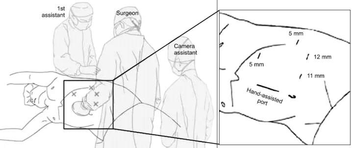
Fig. 1. Patient position and port site.
MATERIALS AND METHODS
Three hand-assisted laparoscopic donor pancreatectomy and nephrectomy procedures were performed at the Korea University Anam Hospital between January 2012 and December 2013, as part of living-donor SPK. We retro- spectively reviewed the patient characteristics, clinical out- come in donors, and surgical perspectives.
1. Donor selection criteria
Gruessner et al.(9) reported the following donor selection criteria: insulin response to glucose or arginine >300% of basal insulin (insulin secretion test), glycated hemoglobin (HbA1c) level <6%, basal fasting insulin levels <20
IU/mL, plasma glucose levels <150 mg/dL in the 75 g oral glucose tolerance test (OGTT), glucose disposal rate >1%
in the intravenous glucose tolerance test, and body mass in- dex <27 kg/m2. Additionally, all donors must be at least 10 years older than the recipient’s age at diabetes onset. We
eral decubitus position on a flexed operating table. The in- cision for the hand-assisted port was made in the midline—
just above the umbilicus. The first 11-mm port was inserted at the midclavicular line—on the margin of the rectus mus- cle—under guidance from the hand. The pneumoperitoneum was created through that port, and the laparoscopic camera was inserted. Another 12 mm port was placed lateral to the previous port. Two 5-mm trocars were inserted: one along the mid-axillary line—between the costal margin and the iliac crest—and the other at the costal margin on the mid- clavicular line (Fig. 1).
3. Surgical procedures
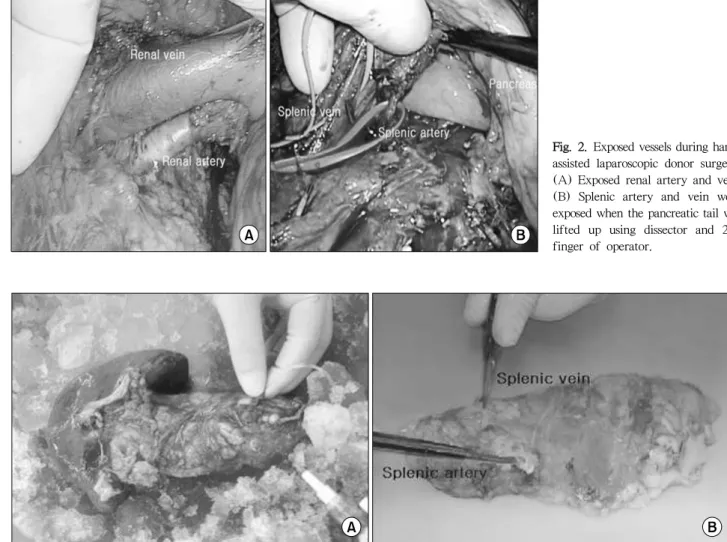
Firstly, the colonic attachments to the lateral abdominal wall, as well as the splenic flexure of the left colon, were completely mobilized until the left renal vein and the tail of the pancreas were exposed. The left adrenal and gonadal veins were separated, and the left renal vein and artery were dissected (Fig. 2A). The lateral attachment of the kidney to the retroperitoneum and upper pole of Gerota’s fascia
Fig. 2. Exposed vessels during hand- assisted laparoscopic donor surgery.
(A) Exposed renal artery and vein.
(B) Splenic artery and vein were exposed when the pancreatic tail was lifted up using dissector and 2nd finger of operator.
Fig. 3. Benching of the pancreas graft. (A) Cold perfusion with preservation solution right after the pancreas brought out from the living donor. (B) Pancreas graft after completing benching.
was then dissected. At this point, the inferior poles of the kidney and ureter were dissected—the dissection proceeded along the course of the ureter. Next, we checked the recipi- ent’s status regarding implantation of kidney; when the re- cipient was somewhat ready to receive the kidney, the proc- ess of removing the kidney from the donor began. The renal artery and vein were divided, and the kidney was retrieved through the hand-assisted port.
Subsequently, we focused on retrieving the pancreas.
Firstly, the lower margin of the pancreas tail was dissected—
all the way to superior mesenteric vein (SMV); the inferior mesenteric vein was then divided. Next, the upper margin of the pancreas was dissected. The gastrocolic ligament was divided, and the splenic artery was identified. The surgeon then lifted the pancreas tail using their index finger, and
the crotch area of the splenic vein and artery was dissected.
Finally, the splenic artery and vein were isolated, and the vessels were tagged using elastic vessel loops (Fig. 2B).
When the venous anastomosis of kidney graft in recipient’s side was ready to be performed, the neck of the pancreas was then resected at the level of the portal vein using an endostapler. Subsequently, the splenic vein was cut at the junction of the SMV and splenic artery—at the level of the celiac artery. The pancreatic segment was retrieved through the hand-assisted port and delivered to the bench table for flushing (Fig. 3A). All dissections were performed as gently as possible to avoid pancreatitis (Fig. 3B)(10).
4. Postoperative follow-up for living donors Postoperative pancreatic and renal functions of donors
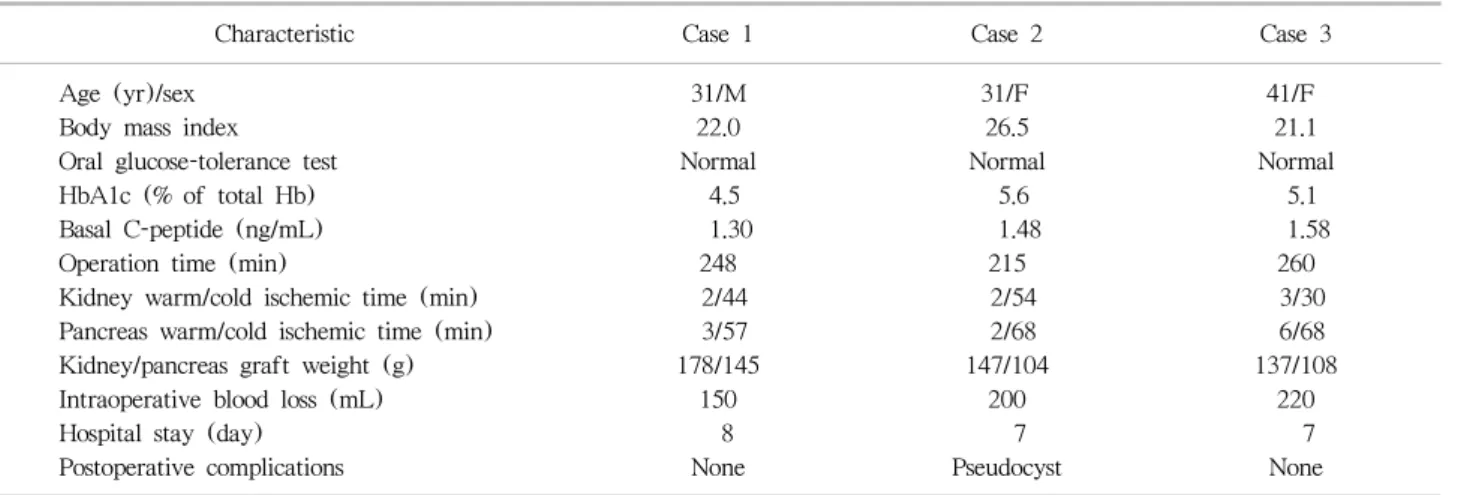
Table 1. Characteristics and clinical outcomes of donors
Characteristic Case 1 Case 2 Case 3
Age (yr)/sex 31/M 31/F 41/F
Body mass index 22.0 26.5 21.1
Oral glucose-tolerance test Normal Normal Normal
HbA1c (% of total Hb) 4.5 5.6 5.1
Basal C-peptide (ng/mL) 1.30 1.48 1.58
Operation time (min) 248 215 260
Kidney warm/cold ischemic time (min) 2/44 2/54 3/30
Pancreas warm/cold ischemic time (min) 3/57 2/68 6/68
Kidney/pancreas graft weight (g) 178/145 147/104 137/108
Intraoperative blood loss (mL) 150 200 220
Hospital stay (day) 8 7 7
Postoperative complications None Pseudocyst None
Abbreviations: HbA1c, glycated hemoglobin; Hb, hemoglobin.
scan was sometimes performed depending on patient’s symptoms in hospital or outpatient clinic for evaluating postoperative complications.
RESULTS
All the donors’ preoperative characteristics met the donor selection criteria (Table 1). More specifically, the OGTT, HbA1c levels, and basal C-peptide levels, all of which re- flect the pancreatic function of donors, were appropriate for pancreas donation. There was one man and two women among the donors; their mean age was 34.3±4.7 years, and their body mass index was 23.2±2.36 kg/m2. The mean op- eration time was 241±19.0 minutes; the cold ischemic time for the kidney was 42.7±9.8 minutes, while that for the pancreas was 64.3±5.2 minutes. No significant bleeding re- quiring blood transfusion occurred during the operation. All donors stayed in hospital for 7 or 8 days after surgery. One donor developed a pancreatic pseudocyst, which was con- trolled using conservative management. The donors’ pancre- atic and renal functions were well preserved after surgery.
was performed for the first time in June 1979 at the University of Minnesota, and the first living donor SPK was performed in March 1994 at the same institution(5,12).
However, even though the pancreas was the first extrarenal solid organ for which a living donor was successfully used, and despite the benefits of living-donor over cadav- eric-donor PT (immunological benefits, decreased cold is- chemic time, and reduction of waiting time), less than 1%
of PTs involve living donors(3-5). Open pancreas donation involves high morbidity and a prolonged postoperative re- covery period on the part of the donor; therefore, the pro- cedure has not become widely accepted(13). In 1999, to de- crease the morbidity associated with open pancreas dona- tion, the first laparoscopic donor distal pancreatectomy was performed(8). As with laparoscopic nephrectomy, laparo- scopic pancreatectomy is more cost-effective than open sur- gery because both the hospital stay and recovery time are shorter(8). The HALDS procedure, introduced in 1998, combines laparoscopic technique with quicker and safer or- gan retrieval—the organ is removed using one hand through a small incision(7). Because the surgeon uses their hands to handle the tissue during HALDS, the procedure preserves
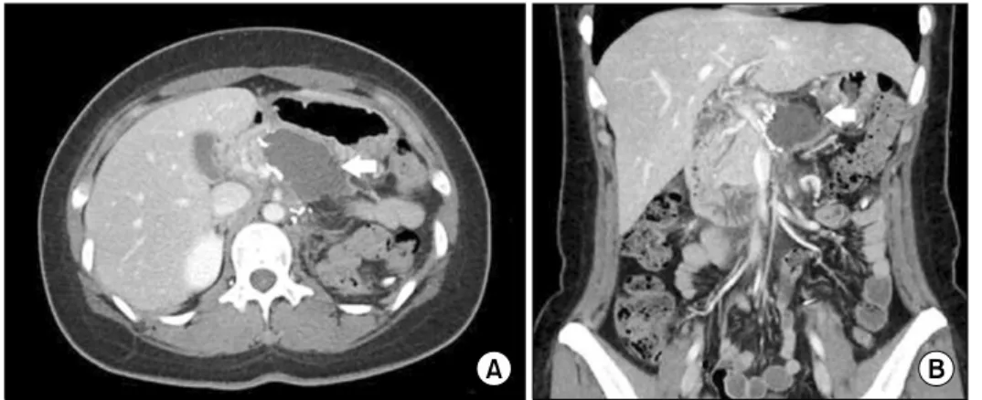
Fig. 4. Abdominopelvic computer tomography. (A) Horizontal view.
(B) Coronal view. Arrows indicate a cystic mass (pseudocyst) at the pan- createctomy site.
tactile sensation; thus, the tissue planes are more easily de- fined, and the intraperitoneal hand can be used for retraction. The surgeon can also control urgent bleeding by using their hand to apply pressure; this prevents short- and long-term harm to graft function(14). The most important advantage of hand assistance is an increase in safety; in ad- dition, it also reduces warm ischemic time, which is an im- portant factor influencing graft outcome(15).
Donor safety still is one of the major concerns of living donor PTs. Surgical complications occur in fewer than 5%
of cases, and no donor mortality has been reported(4).
Pancreatitis, pancreatic leak or fistula, and pancreatic pseu- docyst are uncommon complications(6). Furthermore, such pancreatic complications can be reduced using selective liga- tion of the main pancreatic duct, as well as by oversewing the cut pancreatic surface(6). Nonetheless, when pancreatic complications do occur, percutaneous interventions or surgi- cal management are frequently required(6). In our own study, we experienced one case of pseudocyst. The donor visited the outpatient clinic 3 weeks after living-donor SPK, complaining of indigestion and abdominal pain that wors- ened with deep breathing. Abdominal CT scan showed a 5×7 cm cystic mass at the pancreatectomy site (Fig. 4). An endoscopic ultrasound-guided transmural internal drainage was performed, and a subsequent CT scan confirmed that the pseudocyst had been completely removed, and there was no recurrence at postoperative 1 year follow-up CT scan imaging.
Metabolic complications are also a significant issue in liv- ing donors(16). The Minnesota group reported 10 of 115 donors increased HbA1c levels and three of them required
insulin treatment(4). Choi et al.(17) also reported two of 20 donors developed hyperglycemia which was controlled with oral hypoglycemic agents. Therefore, thorough donor selection should be strictly carried out according to the se- lection criteria described above to maximize donor safety.
CONCLUSION
HALDS can be performed without significant complica- tions, provided that the surgeon has sufficient skill. However, pancreatic complication should be observed carefully. In ad- dition, thorough donor selection is mandatory to maximize donor safety.