pISSN: 0378-6471⋅eISSN: 2092-9374
http://dx.doi.org/10.3341/jkos.2014.55.8.1202
Original Article
생쥐를 이용한 스테로이드 유발 고안압 모델
Steroid-Induced Ocular Hypertension Model in the Mice
김유라⋅강완석⋅권의용⋅조남천⋅이동욱
You Ra Kim, MD, Wan Seok Kang, MD, Eui Yong Kweon, MD, Nam Chun Cho, MD, Dong Wook Lee, MD
전북대학교 의학전문대학원 전북대학교병원 안과학교실
Department of Ophthalmology, Chonbuk National University Hospital, Chonbuk National University Medical School, Jeonju, Korea
Purpose: To determine whether rat eyes develop increases in intraocular pressure (IOP) in response to a topically applied corti- costeroid and to investigate the relationship between ocular hypertension and apoptosis of retinal ganglion cells.
Methods: IOP was monitored by rebound tonometry in a group of 10 rats that received topically administered dexamethasone in both eyes (experimental) and in another group of 5 rats that received artificial tears (control) three times daily for 4 weeks after the establishment of baseline IOP values. Only eyes that increased by more than 50% compared with the basal IOP were ad- ministered once per day for 5 weeks. After 8 weeks, selective immunofluorescence stain for retinal ganglion cells and terminal deoxynucleotidyl transferase dUTP nick end labeling (TUNEL) stain were conducted.
Results: Among 20 experimental eyes, 11 eyes (55%) showed a greater than 50% increase in IOP compared with basal IOP.
After 8 weeks, the mean IOPs for the experimental and control groups were 11.8 ± 1.4 mm Hg and 18.5 ± 1.0 mm Hg, re- spectively (p < 0.01). The counts of central retinal ganglion cells (RGCs) were 2718 ± 240 and 2612 ± 443, respectively (p = 0.294). The results of the TUNEL stain also showed no differences.
Conclusions: Rat eyes exhibit a steroid-induced ocular hypertensive response with no local complications. However, maintain- ing ocular hypertension increased by 50% for two months was not enough to detect changes in RGCs.
J Korean Ophthalmol Soc 2014;55(8):1202-1207
Key Words: Ocular hypertension, Retinal ganglion cell, Steroid-induced glaucoma
■Received: 2013. 11. 9. ■ Revised: 2014. 2. 11.
■Accepted: 2014. 7. 12.
■Address reprint requests to Dong Wook Lee, MD
Department of Ophthalmology, Chonbuk National University Hospital, #20 Geonji-ro, Deokjin-gu, Jeonju 561-712, Korea Tel: 82-63-250-1996, Fax: 82-63-250-1960
E-mail: [email protected]
ⓒ2014 The Korean Ophthalmological Society
This is an Open Access article distributed under the terms of the Creative Commons Attribution Non-Commercial License (http://creativecommons.org/licenses/by-nc/3.0/) which permits unrestricted non-commercial use, distribution, and reproduction in any medium, provided the original work is properly cited.
녹내장은 안압 상승을 비롯한 여러 가지 원인에 의하여 망막신경절세포와 그 축삭의 변성이 발생하며 이에 따라 시야의 결손이 나타나게 되는 시신경병증이다.1 녹내장 발 생의 여러 가지 위험요인 중 안압은 우리가 조절할 수 있는 유일한 주요 위험인자이다. 또한 안압이 정상인 녹내장 환 자에서조차 안압을 떨어뜨리는 것은 녹내장의 진행을 늦추 는 것으로 알려졌다.2
지금까지 알려진 실험 동물모델을 대상으로 안압을 상승 시키기 위해 사용했던 방법으로는 상공막정맥을 폐쇄시키거 나,3 레이저 섬유주 성형술을 시행하거나,4 앞방에 작은 구슬 을 주입하여 방수유출로를 폐쇄하는 방법,5 혹은 스테로이드 를 투여하는 것6-14 등이 여러 차례에 걸쳐 소개되었다.
스테로이드는 일부 감수성이 있는 사람에게서 투여 방법 에 관계 없이 안압을 상승시키는 것으로 알려졌다. 스테로 이드에 의한 안압의 상승 기전은 정확히 알려진 바는 없지 만 일반적으로 섬유주의 형태학적, 생화학적 변화에 의한 방수유출의 감소에 의한 것으로 보고되었다.15,16
Armaly17는 정상인에 덱사메타손을 점안하여 약 5-6%의 사람에서 안압이 상승하는 것을 관찰하였다. 사람이 아닌 동물에서도 스테로이드 투여에 의한 안압상승을 관찰할 수
있는데, 소에게 스테로이드 안약을 점안하여 100%에서 안압상 승을 보고한 바 있으며,6 양에서도 같은 결과가 보고된 바 있 다.7 그 외에도 고양이나 원숭이, 토끼, 쥐와 같은 여러 동물들 에서도 스테로이드 투여에 의한 안압 상승이 보고되었다.8-13
스테로이드 투여에 의한 안압의 상승은 다른 방법에 비 해 점안하거나 안구 주위에 주사하는 경우 더 많은 상승효 과가 있는 것으로 알려졌으며,18 투약한 스테로이드의 항염 증 작용의 강도에 클수록 안압의 상승정도가 더 높은 것으 로 알려졌다.19
이에 저자들은 개체의 변이가 비교적 적고 사람과 돌연 변이나 질병이 유사하고 비교적 짧은 시간에 유전적 질환 이 발생하며, 안압상승, 망막신경절세포의 손상에 영향을 주는 유전적 위험인자가 잘 알려졌으며, 사람과 방수역동 및 시각계 조직이 유사하고20 24시간 안압변동이 비슷하다 는21 여러 장점을 가진 생쥐를 대상으로 하여 고안압을 유 발할 수 있는 여러 가지 방법 중 비침습적이며 비교적 쉬운 방법인 덱사메타손을 투여하여 고안압을 유도하고, 이로 인한 망막신경절세포의 손상 정도를 알아보고자 하였다.
대상과 방법
안압 상승 모델 제작
실험동물은 생쥐(C57BL/6, 3개월, 암컷, 20-25g)를 사용 하였으며, 모든 동물과 관련된 실험은 전북대학교병원의 IRB 승인과 동물실험에 관한 ARVO 규정에 따라 시행되었 다. 실험군(n=20)에서 덱사메타손 점안액(Maxidex®, Alcon Korea, Korea)을 하루 3회 4주간 점안하였다. 기저 안압 대비 50% 이상 상승한 눈만을 대상으로 추가 4주간 하루 1회씩 안약을 점안하였다. 대조군(n=10)은 실험군과 연령이 일치하 는 생쥐를 대상으로 하였으며, 실험군과 같은 횟수로 인공누 액을 점안하였다. 안압은 숙련된 한 명의 실험자에 의해 한 손으로 쥐의 목과 몸을 움직이지 않게 고정한 후 국소마취 없이 Rebound tonometry (iCare®, Tiolat, Helsinki, Finland) 를 사용하여 일주일에 3회에 걸쳐 동일한 시간에 측정하였 으며, 매 측정 시마다 총 5회 측정하여 평균값을 취하였다.
전층 망막 면역조직화학 분석
안압 상승 후 8주째 생쥐의 안구를 적출한 후 망막을 분 리하였다. 분리한 망막은 30% 수크로스를 포함한 PBS완충 액에 24시간 동안 4°C에서 처리 후 3% donkey serum과, 1% bovine serum albumin, 1% fish gel과 0.1%의 triton X-100이 혼합된 PBS완충액에 1시간 동안 blocking하였다.
그리고 goat polyclonal anti-Brn3a 항체(1:500; Santa Cruz Biotechnology, Santa Cruz, CA), 망막신경절세포에 대한 특
이항체가 포함된 용액에서 4°C에서 3일간 처리하였다. PBS로 3차례 망막을 씻어낸 후에 이차항체인 donkey anti-goat IgG antibody (Invitrogen, Carlsbad, CA)에 24시간 동안 처리한 후 PBS로 3차례 망막을 씻어낸 후에 편광현미경(Olympus America Inc., Center Valley, PA)으로 관찰하였다.
망막신경절세포의 정량적 분석
Whole mount된 망막표본을 20배 편광현미경으로 중심 부, 중간부, 주변부로 나누어 하나의 망막당 각각 0.28 mm2 의 24개 부위를 촬영하였다. 염색된 망막신경절세포수는 총 2회에 개수하고 그 평균을 구했다.
Western blot analysis
안구를 적출하여 얻은 망막조직을 buffer로 분해시켜 얻 어진 망막조직(10 µg; n=3 retinas/groups)을 PAGE gel에 전 기 영동하여 polyvinylidenedifluoride membrane에 이동시 켰다. 망막조직을 이동시킨 membrane을 5% nonfat dry milk와 0.1% Tween-20이 섞인 PBS완충액에 한 시간 동안 blocking하였다. Blocking 후 rabbit polyclonal anti-Bax an- tibody (1:500; Santa Cruz Biotechnology), mouse monoclonal anti-phosphorylated Bad (pBad, 1:2000; Cell Signaling)와 배 양을 하였다. 배양한 membrane을 peroxidase-conjugated anti IgG (1:5000: Bio-Rad, Hercules, CA, USA)를 이용하여 반 응시키고, chemiluminescence detection (ECL Plus; GE Healthcare Bio-Science, Piscataway, NJ) kit system을 사용 하여 감광시켜 발현을 확인하였다. Western blot에서 얻은 anti-Bax antibody, pBad의 발현은 ImageJ (National Institute of Health)를 이용하여 분석하였으며, actin의 발현된 양을 기준으로 발현양을 표준화하였다.
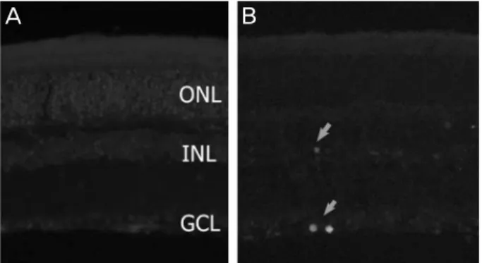
TUNEL 염색
대조군과(n=3) 고안압군(n=3) 생쥐의 적출한 안구를 냉 동절삭 망막 절편을 만들어 proteinase K (pH 7.4)에 37°C에 서 10분 동안 고정한 후 PBS로 세척한 후 그 절편을 TdT와 nucleotide가 혼합된 완충액에 37°C에서 60분간 처리하였다 (In situ Cell Death Detection kit, Roche Applied Science, Indianapolis, IN). 0.2 mm의 망막절편의 신경절세포층(GCL), 속핵층(INL), 바깥핵층(ONL)의 세 층에서 TUNEL 양성 세포의 수를 현미경 8시야에서 분석하였다.22
통계분석
본 실험에서의 모든 측정치는 평균 ± 표준편차로 표기하 였다. 통계학적 분석은 두 군의 비교는 two-sample wilcox- on test와 unpaired, two-tailed Student’s t-test를 사용하였다.
Baseline
Dexamethasone /hyalein instillation 3 times
a day
4 weeks
IOP check &
selection
8 weeks RGC counting
Baseline 8 weeks
CNT DEXA
IOP (mm Hg)
20 15 10 5 0
n = 10 10.6 1.2± n = 20
10.1 1.1±
n = 20 18.5 1.0±
n = 10 11.8 1.4±
Figure 1. Dexamethasone instillation and induction of intra-
ocular pressure (IOP). (A) Diagram for induction of IOP.Dexamethasone instillation began after baseline IOP check three times a day. After 4 weeks, in eyes with 50% elevated IOP than baseline, dexamethasone instillation was done 1 time a day for 4 weeks more. (B) IOP elevation in the mouse eyes at baseline and 8 weeks after dexamethasone instillation. CNT = control group; DEXA = dexamethason instillation group;
RGC = retinal ganglion cell.
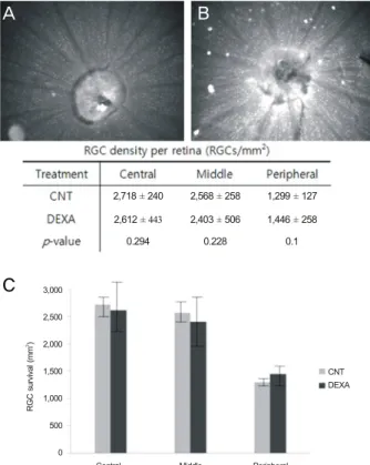
Central Middle Peripheral
CNT DEXA
RGC survival (mm)2
3,000 2,500 2,000 1,500 1,000 500 0
2,718 240± 2,612 ± 443 0.294
2,568 258± 2,403 506± 0.228
1,299 127± 1,446 258±
0.1
Figure 2. Retinal ganglion cell (RGC) survival in control eyes and
treated with dexamethasone eyes. Dexamethasone was instilled 3 times a day for 4 weeks in dexa group. After that, instilled once a day for 4 more weeks in eyes with elevated intraocular pressure (IOP) 50% than baseline. Retinal whole-mount immunohistochemistry for Brn3a in control group (A) and experimental group (B). (C) Quantitative analysis of RGC loss. Values are mean. There were no significant changes in RGC survival between control and dex- amethasone treated C57BL mice. Significant at p < 0.05. DEXA= dexamethason instillation group.
p<0.05인 경우를 통계학적으로 유의한 것으로 판단하였다.
결 과
안압상승
실험군 20안과 대조군 10안의 기저 안압은 각각 10.1 ± 1.1 mmHg와 10.6 ± 1.2 mmHg였다(p= 0.325). 스테로이드 안약을 첫 4주간 점안 후 20안의 실험군 중 기저 안압에 비 해 50% 이상 안압이 상승한 수는 11안(55%)으로 이들만을 대상으로 실험을 진행하고 안압이 50% 이상 상승하지 않 은 생쥐는 실험에서 제외하였다. 안압이 상승한 11안의 실 험생쥐의 8주째 평균 안압은 18.5 ± 1.0 mmHg였고, 인공눈 물을 점안한 대조군 생쥐 10안의 평균 안압은 11.8 ± 1.4 mmHg이었다(Fig. 1) (p<0.01).
안압 상승에 의한 망막신경절세포수 정량
8주째에 망막표본을 편광현미경으로 중심부, 중간부, 주 변부로 나누어 촬영하여 망막신경절세포수를 분석하였다.
중심부의 망막신경절세포수는 대조군 2,718 ± 240개와 실험 군 2,612 ± 443개로 측정되었고(p=0.294), 중간부의 망막신 경절세포수는 2,568 ± 258개와 실험군 2,403 ± 506개로 측정 되었으며(p=0.228), 주변부의 망막신경절세포수는 1,299 ± 127개와 실험군 1,446 ± 258개(p=0.1)로 모든 부분에서 두 군 간 망막신경절세포수의 통계학적 차이는 보이지 않았다 (Fig. 2).
안압상승에 의한 망막신경절세포의 아포프토시스 확인 (TUNEL stain)
0.2 mm의 망막절편의 TUNEL 양성 세포의 수를 분석한 결과, 대조군에서는 TUNEL염색에 양성을 보이는 세포가 관찰되지 않았으나, 안압 상승을 유도한 실험군에서는 망 막신경절세포층과 일부 속핵층에서 국소적으로 TUNEL 양 성 반응이 관찰되었다. 하지만 두 군 간의 유의한 차이는 보이지 않았다(p=0.183) (Fig. 3).
안압 상승 생쥐 망막에서 anti-Bax antibody, pBad의 발현 측정(Western blot analysis)
안압 상승 후 8주째 망막조직에서 아포프토시스관련 단백 질인 Bax와 pBad의 발현결과를 Western blot 방법을 통해 알 아보았다. 그 결과 대조군에 비해 실험군에서 anti-Bax anti- body, pBad의 발현이 증가하는 것을 관찰하였다(Fig. 4).
A B
A
B
C
Figure 3. Increased intraocular pressure (IOP) mediated apop-
totic cell death. (A) There was no TUNEL-positive cells in the retina of control mice treated with artificial tears. (B) However, increased IOP retinas showed TUNEL-positive apoptotic cell death in the GCL and INL. Arrows: TUNEL-positive cell.TUNEL = terminal deoxynucleotidyl transferase dUTP nick end labeling; ONL = outer nuclear layer; INL = inner nuclear layer; GCL = ganglion cell layer.
CNT DEXA CNT DEXA
DEXA Control
Bax pBad
pBad Bax
Actin
Figure 4. Increased intraocular pressure (IOP) induced apopto-
sis in dexamethasone instilled eyes. Dexamethasone was in- stilled 3 times a day for 4 weeks in experimental group. After that, instilled 1 time a day for 4 more weeks in eyes with ele- vated IOP 50% than baseline. Bax and pBad protein expression significantly increased in retina treated with dexamethasone.Values are mean. Significant at *p < 0.05. CNT = control group; DEXA = dexamethason instillation group.
고 찰
스테로이드 점안을 통해 안압을 상승시키는 여러 연구 중 실험대상을 양으로 한 연구에서는 18안 모두에서 대조 군에 비해 15.8 mmHg가 상승하여 평균 27.5 mmHg 보였 으며,7 고양이를 대상으로 한 연구에서는 대조군에 비해 24 안 모두에서 약 4.5 mmHg의 상승을 보였고,8 원숭이에서는 약 45% (10안/22안)에서 기저안압에 비해 약 10.6 mmHg의 안압의 상승이 관찰되었다.9 그러나 토끼를 대상으로 한 연 구에서는 스테로이드에 대한 안압 상승반응이 다양하게 나
타났다. 일부에서는 고안압이 생성되지 않기도 하였으며, 다른 한편에서는 안압이 일시적으로만 상승하였다.10,23-27 토끼에서의 이러한 결과는 아마도 인간이나 포유류에서 관 찰되는 방수유출로 중 쉴렘관이 발달되지 않았기 때문으로 추정된다.28
본 연구에서 저자들은 안압 상승을 유발할 수 있는 여러 가지 방법들 중 비교적 비침습적 방법인 스테로이드 안약 을 점안하는 방법을 통해 고안압을 유도하였으며, 실험대 상으로는 개체의 변이가 비교적 적고 방수역동이 사람과 유사한 생쥐를 사용하였으며, 이에 따른 신경절세포수의 변화와 망막에서 아포프토시스 발현 여부를 살펴보았다.
스테로이드에 의한 안압의 상승에 대한 기전으로는 섬유주 미세구조의 기계적인 변화로 액틴 스트레스 섬유(actin stress fi- ber)의 재형성과 섬유주의 스테로이드 수용체에 의한 가역적인 액 틴(actin) 그물조직의 형성으로 인한 섬유주 미세구조의 기계적인
변화와29,30 스테로이드에 의해 뮤코다당체(glycosaminoglycans),
탄력소(elastin), 섬유결합소(fibronectin) 등이 증가하여 세포 외 기질물질이 쉴렘관 연접부에 축적되어 방수유출로의 미 세구조가 변화하고,31-33 단백분해효소 활성의 감소와 탐식 작용이 줄어들어 섬유주 내의 물질들의 분해가 감소됨에 따른 축적이 증가하고,34-36 섬유주 내의 세포경유통로 혹은 세포주위통로에 의한 방수의 흐름이 줄어드는 것 등37,38에 의해 방수유출로의 저항이 증가하기 때문으로 알려졌다.
스테로이드의 전신적 사용은 사람은 물론 동물에서도 혈 압을 상승시키는 것으로 알려졌다.39-42 이러한 혈압의 상승 은 덱사메타손 치료 후에 안압이 상승하는 다른 원인으로 생각해 볼 수 있다. 그러나 Whitlock et al14은 쥐에서 안압 의 상승과 혈압의 상승 간에 유의한 상관관계가 없었음을 발표한 바 있으며, 더구나 정상혈압을 가진 쥐에서 10 mg/Kg의 phenylephrine을 전신적으로 투여한 후 안압에 영 향을 주지 않았다는 Cavalli et al43의 발표도 있다. 본 연구 에서는 실험 생쥐에 대한 혈압의 관찰이 시행되지 않아 그 연관관계를 확인할 수 없었다는 한계점이 있다.
본 연구에서 저자들은 스테로이드 점안으로 생쥐에서 안 압 상승이 유발됨을 확인하였으며, 이와 함께 안압 상승에 의하여 망막신경절세포의 변화 여부까지 확인한 최초의 연 구이다. 이번 연구에서 실험군에서 대조군에 비해 망막신 경절세포의 수는 전체적으로 감소한 양상을 보였으나 두 군 간에 유의한 상관관계를 보이지는 않았다. 세포의 아포 프토시스를 관찰할 수 있는 TUNEL 염색에서는 대조군에 비해 실험군에서 더 많은 TUNEL 염색 양성의 세포가 관찰되 었지만 이 또한 유의성을 보이지는 않았다. 이처럼 본 연구에 서는 안압의 상승, 망막신경절세포의 수의 감소와 TUNEL 염색의 결과 사이의 상호관계가 큰 연관성이 없는 것으로
A B
관찰되었다. 이러한 결과를 보여주는 원인으로 본 연구에 서 시행한 안압측정이 부적절했을 가능성을 고려할 수 있 다. 즉, 저자들이 측정한 일주일 세 번의 평균 안압이 정확 한 생쥐의 평균 안압이 아니라 좀 더 높게 측정된 것일 가 능성이 있으며, 목덜미를 손가락으로 고정하고 측정하다 보니 정확한 안압이 반영되지 않을 가능성도 있다. 또한 실 험에서 사용된 생쥐의 나이는 3개월로 상대적으로 어려서 안압변화에 대한 저항성이나 시신경손상의 회복이 노령의 생쥐에 비해 유리할 가능성도 고려할 수 있다.
그러나 Western blot analysis에서는 아포프토시스를 유 발하는 단백질인 Bax와 pBad의 발현이 실험군에서 대조군 에 비해 증가되었음을 확인되었다. 이러한 결과는 기저 안 압에 비해 50% 상승된 안압을 2개월간 유지하는 것만으로 는 유의한 망막신경절세포수의 변화는 관찰할 수 없었지만, 안압의 상승이 망막신경절세포에 영향을 줄 수 있다는 가 능성을 시사한다. 또한 아포프토시스를 유발하는 단백질이 실험군에서 증가한 것을 고려한다면 고안압이 생긴 생쥐의 망막에서 현재 우리가 실험가능한 방법으로는 망막신경절 세포의 미세한 손상을 아직 발견할 수 없다는 한계도 생각 해 볼 수 있으며, 고안압에 의한 망막신경절 세포의 손상이 전체 망막에 균일하게 진행되는 것이 아니라 국소적인 차 이가 있을 수 있다는 점도 생각해 볼 수 있다. 또한 Leung et al44에 의하면 시신경 압착 후 3달 이후에도 일부 망막신 경절세포의 세포체가 그대로 보존되어 면역형광염색에 염 색되어 나타나거나 생체 촬영에서 손상이 없는 것으로 나 타난다는 보고도 있어 저자들이 관찰한 8주의 기간이 망막 신경절세포의 손상이 일어나기에 상대적으로 시간이 짧았 을 가능성도 생각해볼 수 있다.
따라서 앞으로 더 많은 수의 생쥐를 대상으로 연속적인 안압을 측정하여 장기적인 연구를 구상하고 표본으로 사용 하는 망막의 수를 더 많이 사용하여 실험을 진행할 수 있는 추가 연구와 전신적인 부작용에 대한 연구가 필요할 것으 로 생각한다.
REFERENCES
1) Jonas JB, Budde WM. Diagnosis and pathogenesis of glaucoma- tous optic neuropathy: morphological aspects. Prog Retinal Eye Res 2000;19:1-40.
2) Kass MA, Heuer DK, Higginbotham EJ, et al. The ocular hyper- tension treatment study―a randomized trial determines that topical ocular hypotensive medication delays or prevents the onset of pri- mary open angle glaucoma. Arch Ophthalmol 2002;120:701-13.
3) Kim JH, Park CK. Expression of glutamate receptors in ex- perimental rat model of chronic gluacoma. J Korean Ophthalmol 2006;47:468-76.
4) Shimazawa M, Nkamura S, Miwa M, et al. Establishment of the oc-
ular hypertension model using the common marmoset. Exp Eye Res 2013;111:1-8.
5) Frankfort BJ, Khan AD, Tse DY, et al. Elevated intraocular pres- sure causes inner retinal dysfunction before cell loss in a mouse model of experimental glaucoma. Invest Ophthalmol Vis Sci 2013;54:762-70.
6) Gerometta R, Podos SM, Candia OA, et al. Steroid-induced ocular hypertension in normal cattle. Arch Ophthalmol 2004;122:1492-7.
7) Gerometta R, Podos SM, Danias J, Candia OA. Steroid-induced ocular hypertension in normal sheep. Invest Ophthalmol Vis Sci 2009;50:669-73.
8) Bhattacherjee P, Paterson CA, Spellman JM, et al. Pharmacological validation of a feline model of steroid-induced ocular hypertension.
Arch Ophthalmol 1999;117:361-4.
9) Fingert JH, Clark AF, Craig JE, et al. Evaluation of the myocilin (MYOC) glaucoma gene in monkey and human steroid-induced ocular hypertension. Invest Ophthalmol Vis Sci 2001;42:145-52.
10) Knepper PA, Breen M, Weinstein HG, Blacik JL. Intraocular pres- sure and glycosaminoglycan distribution in the rabbit eye: effect of age and dexamethasone. Exp Eye Res 1978;27:567-75.
11) Lorenzetti OJ. Effects of corticosteroids on ocular dynamics in rabbits. J Pharmacol Exp Ther 1970;175:763-72.
12) Sawaguchi K, Nakamura Y, Sakai H, Sawaguchi S. Myocilin gene expression in the trabecular meshwork of rats in a steroid-induced ocular hypertension model. Ophthalmic Res 2005;37:235-42.
13) Zhan GL, Miranda OC, Bito LZ. Steroid glaucoma: cortico- steroidinduced ocular hypertension in cats. Exp Eye Res 1992;54:
211-8.
14) Whitlock NA, McKnight B, Corcoran KN, et al. Increased intra- ocular pressure in mice treated with dexamethasone. Invest Ophthalmol Vis Sci 2010;51:6496-503.
15) Jones R 3rd, Rhee Dj. Corticosteroid-induced ocular hypertension and glaucoma: a brief review and update of the literature. Curr opin Ophthalmol 2006;17:163-7.
16) Kersey JP, Broadway DC. Corticosteroid-induced glaucoma: a re- view of the literature. Eye (Lond) 2006;20:407-16.
17) Armaly MF. Statistical attributes of the steroid hypertensive re- sponse in the clinically normal eye, I: the demonstration of three levels of response. Invest Ophthalmol 1965;4:187-97.
18) Godel V, feiler-Ofry V, Stein R. Systemic steroid and ocular fluid dynamics. II. Systemic versus topical steroids. Acta Ophthalmol 1972;50:664-76.
19) Mindel JS, Tavitian HO, Smith H Jr, Walker EC. Comparative ocu- lar pressure elevation by medrysone, fluorometholone, and dex- amethasone phosphate. Arch Ophthalmol 1980;98:1577-8.
20) May CA, Lutjen-Drecoll E. Morphology of the murine optic nerve.
Invest Ophthalmol Vis Sci 2002;43:2206-12.
21) Aihara M, Lindsey JD, Weinreb RN. Twenty-four-hour pattern of mouse intraocular pressure. Exp Eye Res 2003;77:681-6.
22) Lee DW, Kim KY, Shim MS, et al. Coenzyme Q10 ameliorates oxi- dative stress and prevents mitochondrial alteration in ischemic reti- nal injury. Apoptosis 2014;19:603-14.
23) Jackson R, Waitzman M. Effects of some steroids on aqueous hu- mor dynamics. Exp Eye Res 1965;4:112-23.
24) Levene RZ, Rothberger MD, Rosenberg S. Corticosteroid glauco- ma in the rabbit. Am J Ophthalmol 1974;78:505-10.
25) Podos SM. Animal models of human glaucoma. Trans Am Acad Ophthalmol Otol 1976;81:632-5.
= 국문초록 =
생쥐를 이용한 스테로이드 유발 고안압 모델
목적: 생쥐에서 스테로이드 점안이 고안압 녹내장 모델로서의 가능성이 있는지를 알아보기 위해 2개월간 스테로이드 점안 후 생쥐의 안압과 망망신경절세포수 변화를 알아보았다.
대상과 방법: 생쥐에서 스테로이드 녹내장을 유발시키기 위해 덱사메타손 점안액을 하루 3회 4주간 점안하였다. 안압이 기저 안압 대비 50% 이상 상승한 눈만을 대상으로 추가 4주간 하루 1회씩 점안을 하였다. 안압은 Rebound tonometry로 측정하였다. 8주 후 망막신 경절세포의 선택적 면역형광염색과 전체 망막 층의 TUNEL 염색으로 실험군과 대조군 간의 변화를 관찰하였다.
결과: 실험군 20안 중 11안(55%)에서 덱사메타손 점안 4주째에 기저 안압 대비 50% 이상의 안압 상승을 보였다. 8주째 고안압군과 대조군의 평균 안압은 각각 11.8 ± 1.4 mmHg와 18.5 ± 1.0 mmHg였다(p<0.01). 8주째 중심부의 망막신경절세포수는 각각 2,718
± 240개와 2,612 ± 443개로 유의한 차이를 보이지 않았다(p=0.294). 두 군 간 TUNEL 양성 세포수의 차이도 나타나지 않았다. 그러 나 아포프토시스와 관련된 Bax와 pBad의 발현은 실험군에서 증가하였다(p<0.05).
결론: 스테로이드 안약을 이용한 생쥐의 고안압 실험 모델은 쉽고 비교적 비침습적인 방법으로 국소적 합병증 없이 고안압을 유도하였 다. 그러나 2개월간의 기저 안압 대비 50% 상승된 안압을 유지하는 것으로는 유의한 망막신경절세포수의 변화를 관찰할 수 없었으며, 좀 더 장기적인 연구 결과와 전신 부작용에 대해서는 추가 연구가 필요할 것으로 생각한다.
<대한안과학회지 2014;55(8):1202-1207>
26) Touvinen E, Liesmaa M, Eslla R. The influence of corticosteroids on intraocular pressure in rabbits. II. The influence of massive sub- conjunctival doses of dexamethasone and betamethasone. Acta Ophthalmol 1966;44:901-5.
27) Hester DE, Trites PN, Peiffer RL, Petrow V. Steroid-induced ocu- lar hypertension in the rabbit: a model using subconjunctival injections. J Ocul Pharmacol 1987;3:185-9.
28) Cole DF. Ocular fluid. In: Davson H, ed. The Eye, 3rd ed. Orlando, Fla: Academic press, 1984;269-390.
29) Clark AF, Brotchie D, Read AT, et al. Dexamethasone alters F-actin architecture and promotes cross-linked actin network formation in human trabecular meshwork tissue. Cell Motil Cytoskeleton 2005;60:83-95.
30) Clark AF, Wilson K, McCartney MD, et al. Glucocorticoid-in- duced formation of cross-linked actin networks in cultured human trabecular meshwork cells. Invest Ophthalmol Vis Sci 1994;35:
281-94.
31) Wilson K, McCartney MD, Miggans ST, Clark AF. Dexamethasone induced ultrastructural changes in cultured human trabecular meshwork cells. Curr Eye Res 1993;12:783-93.
32) Yue BY. The extracellular matrix and its modulation in the tra- becular meshwork. Surv Ophthalmol 1996;40:379-90.
33) Johnson DH, Bradley JM, Acott TS. The effect of dexamethasone on glycosaminoglycans of human trabecular meshwork in perfu- sion organ culture. Invest Ophthalmol Vis Sci 1990;31:2568-71.
34) Matsumoto Y, Johnson DH. Dexamethasone decreases phag- ocytosis by human trabecular meshwork cells in situ. Invest Ophthalmol Vis Sci 1997;38:1902-7.
35) Wordinger RJ, Clark AF. Effects of glucocorticoids on the tra-
becular meshwork: towards a better understanding of glaucoma.
Prog Retin Eye Res 1999;18:629-67.
36) Zhang X, Ognibene CM, Clark AF, Yorio T. Dexamethasone in- hibition of trabecular meshwork cell phagocytosis and its modu- lation by glucocorticoid receptor beta. Exp Eye Res 2007;84:
275-84.
37) Xiong X, Miao J, Xi Z, et al. Regulatory effect of dexamethasone on aquaporin-1 expression in cultured bovine trabecular meshwork cells. J Huazhong Univ Sci Technolog Med Sci 2005;25:735-7.
38) Underwood JL, Murphy CG, Chen J, et al. Glucocorticoids regu- late transendothelial fluid flow resistance and formation of inter- cellular junctions. Am J Physiol 1999;277:C330-42.
39) Goodwin JE, Zhang J, Geller DS. A critical role for vascular smooth muscle in acute glucocorticoid-induced hypertension. J Am Soc Nephrol 2008;19:1291-9.
40) Mangos GJ, Whitworth JA, Williamson PM, Kelly JJ. Glucocorticoids and the kidney. Nephrology (Carlton) 2003;8:267-73.
41) Fardet L, Flahault A, Kettaneh A, et al. Corticosteroid-induced clinical adverse events: frequency, risk factors and patient’s opinion.
Br J Dermatol 2007;157:142-8.
42) Kelly JJ, Mangos G, Williamson PM, Whitworth JA. Cortisol and hypertension. Clin Exp Pharmacol Physiol Suppl 1998;25:S51-6.
43) Cavalli A, Lattion AL, Hummler E, et al. Decreased blood pressure response in mice deficient of the alpha1b-adrenergic receptor. Proc Natl Acad Sci U S A 1997;94:11589-94.
44) Leung CK, Weinreb RN, Li ZW, et al. Long-term in vivo imaging and measurement of dendritic shrinkage of retinal ganglion cells.
Invest Ophthalmol Vis Sci 2011;52:1539-47.