ISSN 0378-6471 (Print)⋅ISSN 2092-9374 (Online)
https://doi.org/10.3341/jkos.2017.58.3.289
Original Article
국한성 맥락막혈관종에서 발생한 장액망막박리에 대한 치료결과
Treatment Outcome of Serous Macular Detachment in Circumscribed Choroidal Hemangioma
권정민1⋅이승민1⋅권한조2⋅박성후2,3⋅이지은2,3⋅변익수1,3
Jeong Min Kwon, MD1, Seung Min Lee, MD1, Han Jo Kwon, MD2, Sung Who Park, MD2,3, Ji Eun Lee, MD, PhD2,3, Ik Soo Byon, MD1,3
양산부산대학교병원 안과학교실 의생명융합연구소1, 부산대학교병원 안과학교실 의생명연구소2, 부산대학교 의학전문대학원 안과학교실3 Research Institute for Convergence of Biomedical Science and Technology, Department of Ophthalmology,
Pusan National University Yangsan Hospital1, Yangsan, Korea
Medical Research Institute, Department of Ophthalmology, Pusan National University Hospital2, Busan, Korea Department of Ophthalmology, Pusan National University School of Medicine3, Yangsan, Korea
Purpose: To determine the clinical outcome of anti-vascular endothelial growth factor (anti-VEGF) and photodynamic therapy (PDT) for circumscribed choroidal hemangioma (CCH) with serous retinal detachment.
Methods: The medical records of patients having CCH with serous retinal detachment in macula were retrospectively reviewed.
CCH and serous retinal detachment were evaluated via fundus photography, optical coherence tomography, indocyanine green angiography, and ultrasonography.
Results: A total of 9 eyes were enrolled in this study. The average follow-up period was 19.2 months. The mean visual acuity was 0.51 ± 0.52 (logMAR) and the mean maximum diameter and thickness of the tumor were 6,154.4 ± 2,019.9 μm and 2,224.4 ± 862.1 μm, respectively. Of the 6 eyes receiving anti-VEGF (mean number of injections: 3.16) as the first treatment for serous reti- nal detachment, 5 had sustained or recurred intraretinal/subretinal fluid (IRF/SRF) and needed additional PDT and transpupillary thermotherapy. In the 3 eyes that received PDT (mean number of treatments: 1.3) as an initial treatment, IRF/SRF was com- pletely resolved. Finally, 8 eyes achieved complete resolution of SRF and IRF; however visual recovery was limited.
Conclusions: PDT, even with a small number of treatments, can alleviate IRF/SRF in CCH, while anti-VEGF did not.
J Korean Ophthalmol Soc 2017;58(3):289-295
Keywords: Circumscribed choroidal hemangioma, Serous retinal detachment, Photodynamic therapy
■Received: 2016. 9. 1. ■ Revised: 2017. 1. 25.
■Accepted: 2017. 3. 1.
■Address reprint requests to Ik Soo Byon, MD
Department of Ophthalmology, Pusan National University Yangsan Hospital, #20 Geumo-ro, Mulgeum-eup, Yangsan 50612, Korea
Tel: 82-55-360-2592, Fax: 82-55-360-2161 E-mail: [email protected]
* This work was supported by clinical research grant from Pusan National University Yangsan Hospital.
* This study was presented as an e-poster at the 115th Annual Meeting of the Korean Ophthalmological Society 2016.
ⓒ2017 The Korean Ophthalmological Society
This is an Open Access article distributed under the terms of the Creative Commons Attribution Non-Commercial License (http://creativecommons.org/licenses/by-nc/3.0/) which permits unrestricted non-commercial use, distribution, and reproduction in any medium, provided the original work is properly cited.
맥락막혈관종(choroidal hemangioma)은 드문 양성 혈관 종양으로 선천적으로 발생하여 안구가 성장함에 따라 같이 성장한다.1 맥락막 침범 정도에 따라 국한성과 미만성으로 나뉘며 국한성 맥락막혈관종은 일반적으로 후극부의 시신 경유두 이측에서 주로 관찰된다.1,2
전형적인 국한성 맥락막혈관종은 오렌지색을 띠며, 종양 이 크거나 황반 아래에 발생하지 않는다면 성인이 될 때까 지 증상이 잘 나타나지 않아 대개 20대에서 40대 사이에 진단된다.2 종양이 황반을 침범하거나 장액망막박리가 발 생하면 시력저하를 일으키게 되며, 동반되는 낭포황반부종,
Table 1. Patients’ characteristics
Sex Age(years)
Follow up (months)
Treatment (2nd treatment)
BCVA (logMAR) SRF IRF Tumor size (μm)
Initial Final Initial Final Initial Final Width Thickness
1 F 54 26 Anti-VEGF, (PDT) 0.4 0.3 + - - - 7,810 1,450
2 M 44 43 Anti-VEGF, (TTT) 0.2 0.4 + - - - 5,400 2,760
3 F 49 33 Anti-VEGF, (PDT) 0.1 0 + - - - 4,310 1,320
4 M 34 18 Anti-VEGF, (PDT) 0.4 0.1 + - - - 2,840 1,210
5 M 53 11 Anti-VEGF, (PDT) 1.4 1.4 + + + + 5,020 1,760
6 F 68 9 Anti-VEGF 1.4 1.7 + - - - 8,620 2,980
7 M 60 14 PDT 0.1 0.2 + - - - 5,360 2,060
8 F 56 12 PDT 0.4 0.1 + - + - 8,120 3,650
9 M 44 7 PDT 0.2 0.2 + - - - 7,910 2,830
BCVA = best corrected visual acuity; SRF = subretinal fluid; IRF = intraretinal fluid; F = female; M = male; Anti-VEGF = anti-vascular endothelial growth factor; PDT = photodynamic therapy; TTT = transpupillary thermotherapy.
망막색소상피변화 등에 의해서도 시력저하가 발생할 수 있 다.1,3-7
장액망막박리가 발생한 맥락막혈관종에서는 레이저광응고 술, 냉동요법, 국소방사선치료, 경동공온열요법, 광역학요법 등 이 치료에 사용되어 왔으며,8 최근에는 항혈관내피성장인자도 소개되었다.9 하지만 국한성 맥락막혈관종에 대한 국내외의 보 고가 많지 않아 장액망막박리의 치료 예후에 대하여 잘 알려지 지 않은 실정이다. 이에 저자들은 국한성 맥락막혈관종에서 발 생한 장액망막박리의 치료결과에 대해 알아보고자 한다.
대상과 방법
2010년부터 2016년까지 부산대학교병원과 양산부산대학 교병원에서 국한성 맥락막혈관종으로 진단받은 16명 환자 의 의무기록과 영상을 후향적으로 분석하였다. 본 연구는 양산부산대학교병원 의학연구윤리 심의위원회(institutional review board, IRB)의 승인하에 진행되었으며(승인번호:
05-2016-108), 헬싱키선언(Declaration of Helsinki)을 준수 하였다. 국한성 맥락막혈관종의 진단은 안저검사에서 오랜 지-적색의 종괴가 관찰되며, 안초음파 A 스캔에서 종양표 면에는 높은 반향이 나타나고 내부에는 중등도-고반향의 내반사파(50-100%)가 관찰되는 경우로 하였다.
의무기록을 이용하여 대상환자의 나이, 성별, 최대교정시 력을 조사하였다. 종양의 위치, 크기, 두께와 장액망막박리에 서 망막내액, 망막하액의 존재는 안초음파(AVISO, Quantel Medical, Clermont-Ferrand, France), 빛간섭단층촬영(Cirrus OCT, Carl Zeiss Meditec, Dublin, CA, USA), 형광안저혈 관조영 및 인도사이아닌그린혈관조영(FF450 plus IR, Carl Zeiss Meditec, Dublin, CA, USA), (Spectralis HRA, Heidelberg Engineering GmbH, Heidelberg, Germany)을 시 행하여 조사하였다. 종양의 크기와 두께는 안초음파 영상 에서 측정된 최대지름으로 하였다.
치료방법
장액망막박리가 동반된 환자는 술자의 판단에 따라 항혈 관내피성장인자 유리체내주사, 광역학치료, 경동공온열치료 를 이용하여 치료하였다. 항혈관내피성장인자 유리체내주사 는 0.5% proparacaine 점안액(Alcaine®, Alcon Laboratories, Fort Worth, TX, USA)으로 점안마취를 한 다음, 5% povi- done-iodine을 사용하여 눈알결막과 눈주변 피부를 소독하였 다. 각막 윤부에서 3.5 mm 떨어진 곳에 30 gauge 주사바늘을 이용하여 베바시주맙(Avastin®, Roche, Basel, Switzerland) 1.25 mg/0.05 mL를 주입하였다.
광역학요법은 환자의 체표면적당 6 mg의 Verteporfin (Visudyne®, Novartis Ophthalmics AG, Basel, Switzerland) 을 10분 동안 정맥내주사한 뒤 주사 시점에서 15분 후에 689 nm 파장의 레이저를 표준치료방법(power = 50 J/cm2, intensity 600 mW/cm2, duration = 83 sec)을 이용하여 조사 하였다.10 레이저 조사범위는 인도사이아닌그린혈관조영에 서 측정된 종양의 크기보다 500 μm 더 큰 직경으로 시행하 였다.
경동공온열요법은 동공이 완전히 산대된 상태에서 점안 마취를 한 뒤 세극등 현미경을 이용하여 810 nm 파장의 적 외선 다이오드 레이저(OcuLight®, Iris Medical Instruments, Mountain View, CA, USA)를 지속적으로 조사하였다. 레 이저 조사반경은 종양의 최대직경에 따라 시행하였다. 레 이저 조사는 종양의 모든 범위를 포함하여 이루어졌으며, 레이저 세기는 400-600 mW의 범위에서 치료하였다.
치료 전후의 시력과 망막내/하액의 변화를 조사하였다.
통계를 위해 스넬렌 시력표를 이용하여 측정한 시력을 logMAR 시력으로 변환하였다. 치료 전후의 시력변화를 위 해 Wilcoxon signed rank test를 사용하였으며, p<0.05인 경 우를 통계적으로 유의하다고 하였다.
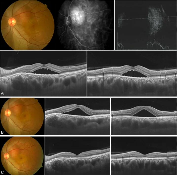
A
B
C
Figure 1. Representative case 4 having an exudative retinal detachment in circumscribed choroidal hemangioma (CCH). (A) At the
presentation, fundus photograph showed that an orange colored CCH at the supero-nasal macula in the left eye. Indocyanine green angiography and ultrasonography showed CCH at the same location. Optical coherence tomography showed subretinal fluid (SRF) at the macula. The central macular thickness was 542 μm and visual acuity was 0.4 (logMAR). The maximum diameter and thick- ness of the tumor was 2,840 μm and 1,210 μm, respectively. (B) Six months after intravitreal bevacizumab injection, SRF did not change. Visual acuity was 0.4. (C) Two months after photodynamic therapy, SRF disappeared, macualr thickness decreased to 204 μm and visual acuity increased to 0.1. Exduative retinal detachment did not recur during 9 months follow-up period.결 과
총 16명이 국한성 맥락막혈관종으로 진단되어 경과관찰 하였다. 12안에서 황반부에 맥락막혈관종이 위치하였으며, 모두 단안에 종양이 있었다. 10안에서 황반부 장액망막박 리가 발생하여 치료를 시행받았다. 2안은 망막하액과 함께
망막내액이 동반되어 있었다. 1안은 치료 후 경과관찰이 되 지 않아 최종적으로 9안이 본 연구에 포함되었다. 장액망막 박리가 발생한 환자는 남자가 5명(55.6%), 여자가 4명 (44.4%)이었고, 평균 연령은 51.3 ± 10.0세(34-68세)였다.
치료 전 평균 시력은 0.51 ± 0.52 (0.10-1.40), 종양의 지름 은 평균 6,154.4 ± 2,019.9 μm (범위, 2,840-8,620), 두께는
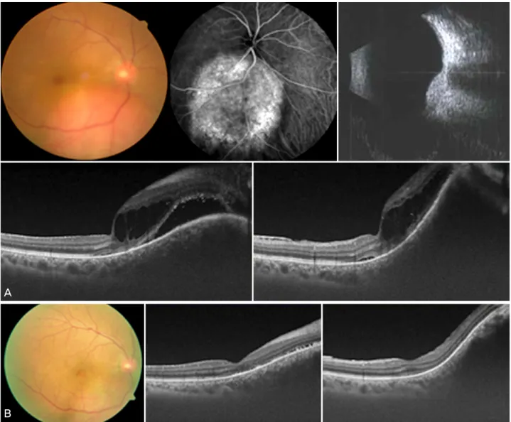
A
B
Figure 2. Representative case 9 having an exudative retinal detachment in circumscribed choroidal hemangioma (CCH). (A) At the
presentation, fundus photograph showed that an orange colored CCH at the infero-nasal macula in the right eye. Indocyanine green angiography and ultrasonography showed CCH at the same location. Optical coherence tomography showed intraretinal and sub- retinal fluid (intraretinal fluid [IRF]/subretinal fluid [SRF]) at the macula. The central macular thickness was 400 μm and visual acuity was 0.2 (logMAR). The maximum diameter and thickness of the tumor was 7,910 μm and 2,830 μm, respectively. (B) Two months after photodynamic therapy, IRF/SRF disappeared, central macular thickness decreased to 220 μm but visual acuity did not changed. Exudative retinal detachment did not recur during 10 months follow-up period.평균 2,224.4 ± 862.1 μm (범위, 1,210-3,650)였다. 치료 후 경과관찰 기간은 19.2 ± 12.3개월(9-43개월)이었다(Table 1).
장액망막박리에 대한 첫 치료로 9안 중 6안은 항혈관내 피성장인자 유리체내주사를 평균 3.16회(2-6회) 시행받았 다. 유리체내주사는 4주 간격으로 경과관찰하면서 망막하 액이 증가하면 다음 주사를 시행하였다. 1안은 항혈관내피 성장인자 유리체내주사를 2회 시행한 후 완전한 망막하액 의 소실을 얻을 수 있었다. 1안은 항혈관내피성장인자 유리 체내주사 후 망막하액의 소실이 있었으나 2개월 뒤 재발하 였다. 추가적인 항혈관내피성장인자 유리체내주사를 하였 으나 반응이 없어 3회의 경동공온열치료를 시행하고 망막
내/하액이 완전히 소실되었다. 4안은 주사 후 4주째 경과관 찰에서 망막내/하액의 변화가 없거나 오히려 증가하여 추 가적인 광역학치료를 시행하였으며, 그중 3안에서 완전한 망막내/하액의 소실을 얻었으나 1안에서는 남았다(Fig. 1).
첫 치료로 9안 중 3안은 광역학치료를 시행받았다. 평균 1.33 회(1-2회)의 치료를 통해 모두 완전한 망막내/하액의 소실을 얻었으며, 경과관찰기간 동안 재발이 없었다(Fig. 2).
최종적으로 장액망막박리로 치료받은 전체 9안 중 8안은 망막내/하액이 소실되었으며 1안은 남았다. 치료방법에 따 른 결과는 첫 치료로 항혈관내피성장인자로 치료받은 6안 중 1안(16.7%)에서, 광역학치료를 받은 3안(100.0%)은 모
두 망막내/하액의 완전한 소실을 이룰 수 있었다. 항혈관내 피성장인자 주사에 재발하거나 반응이 없던 환자 5안에서 시행한 추가치료에서는, 광역학치료를 받은 4안 중 3안 (75.0%)에서, 경동공온열치료를 받은 1안에서 망막내/하액 의 완전한 소실을 이룰 수 있었다.
최대교정시력(logMAR)은 치료 전 평균 0.51에서 치료 후 0.49로 유의한 시력변화는 없었다(p=0.732). 2안(22.2%) 에서 2줄 이상의 시력상승이 있었으며, 2안(22.2%)에서 2 줄 이하의 시력저하가 있었다.
고 찰
국한성 맥락막혈관종(circumscribed choroidal hemangio- ma)이 황반부를 침범하거나 장액망막박리가 발생하여 시 력저하를 일으키는 경우에는 레이저광응고술, 국소방사선 치료, 양성자선을 이용한 방사선치료, 정위적 방사선치료, 경동공온열요법, 광역학요법, 항혈관내피성장인자 유리체 내주사 등을 이용하여 치료하게 된다.8,11-16 하지만 맥락막 혈관종에서 발생한 장액망막박리의 치료방법에 대해 무작 위 전향적 연구가 시행된 적이 없어, 각각의 치료방법의 장·
단점을 술자가 판단하여 환자의 상태에 맞추어 시행하고 있는 실정이다.
본 연구에서는 국한성 맥락막혈관종양에서 환자에서 발 생한 장액망막박리에 대하여 항혈관내피성장인자 유리체 내주사, 광역학치료, 경동공온열치료를 이용하여 치료하였 다. 광역학치료가 다른 치료에 비해 망막내/하액의 감소에 있어서 우수한 결과를 보였다.
항혈관내피성장인자는 혈관투과성을 감소시키고 신생 혈관을 퇴축시키는 효과가 있어 당뇨망막병증과 망막분지 정맥폐쇄와 같은 망막혈관질환이나 삼출성 나이관련 황변 변성에서의 맥락막신생혈관 같은 혈관증식성 질환의 치료 에 이용되고 있다.17 눈속 종양세포도 혈관내피성장인자를 분비하여 내피세포의 치밀결합 구조에 영향을 끼쳐 혈관투 과성을 증가시키고 황반부종이나 장액망막박리를 일으키 게 되는데,18,19 항혈관내피성장인자는 이러한 기전을 억제할 수 있어 혈관증식성 눈속종양(vasoproliferative intraocular tumor)의 치료에 효과가 있다고 보고되었다.20 Kwon et al21 은 항혈관내피성장인자 유리체내주사를 시행한 9안 중에서 3안에서만 망막내/하액의 소실을 얻을 수 있었으며 Mandal et al9은 망막하액은 소실되었으나 망막내액은 모두 남았다 고 하였다. 본 연구에서도 항혈관내피성장인자 주사치료만 으로는 완전한 망막내/하액의 소실을 얻는 데 부족하였는 데, 항혈관내피성장인자의 눈속 반감기가 짧아 효과적인 치료농도를 유지하지 못한 것이 원인일 수 있다. 또한 눈속
에 주입된 약제의 농도와 주사간격은 삼출성 나이관련 황 반변성과 당뇨황반부종 등의 치료에 사용되는 용량을 이용 하였는데, 맥락막혈관종에서 발생한 장액망막박리의 치료 에는 부족했을 수도 있다.
광역학치료는 689 nm 파장의 레이저에 의해 활성화된 광감작물질이 활성산소 및 자유유리기를 발생하여 광화학 반응을 일으키게 되고 망막조직을 피해서 선택적으로 병변 의 혈관내피세포 손상을 일으키게 되고 혈관폐쇄를 유도하 게 된다.22 따라서 황반부 중심을 침범한 병변의 경우에도 치료의 적응이 된다. Barbazetto and Schmidt-Erfurth23과, Schmidt-Erfurth et al24이 맥락막혈관종 혈관들은 혈관벽 두께에 비해 많은 양의 혈액이 존재하여 광역학치료가 종 양의 퇴축에 효과적이라고 보고한 이후로, 여러 연구들에 서 맥락막혈관종에서 발생한 장액망막박리의 치료와 시력 개선에 있어서 우수한 결과들이 보고되었다.11,25-28 본 연구 에서도 광역학치료를 받은 3안은 모두 망막내/하액이 소실 되었으며, 이전 다른 치료에 반응이 없어 추가적인 광역학 치료를 시행받은 4안 중 3안에서도 완전한 망막내/하액의 소실을 이룰 수 있었다. 하지만 시력은 치료 전후 차이가 없었는데, 광역학치료가 맥락막모세혈관의 폐쇄에 의한 황 반부 허혈과 위축으로 인해 시력개선에 제한점이 될 수 있
으며,23,25 광역학치료 전 시력저하가 심한 환자에서는 시력
예후가 불량하다고 보고된 바 있다.29 한편, 항혈관내피성 장인자와 광역학치료를 함께 시행하여 광역학치료의 횟수 를 줄이고 종양의 퇴축을 달성할 수 있다고 보고된 적이 있 어,30 심한 장액망막박리가 있는 환자에서는 병합치료도 고 려해 볼 수 있겠다.
경동공온열치료는 810 nm 파장의 다이오드 레이저를 오 랜 시간 노출하여 종양 내 온도를 상승시킴으로써 비가역 적인 단백질변성, 세포막파괴, 혈관통로경화를 야기하여 국 한성 맥락막혈관종의 망막내/하액을 효과적으로 감소시킬 수 있다.12,31 하지만 경동공온열치료는 크기가 큰 종양(직경 10 mm 이상이거나 두께 4 mm 이상)과 중심와 아래에 위 치하거나 시신경유두와 인접해 있는 종양에는 시행할 수 없는 단점이 있다.12,31 게다가 반복적인 경동공온열치료는 낭포성황반부종, 망막앞섬유증, 국소적인 홍채위축, 망막혈 관폐쇄의 위험을 증가시킬 수도 있다.12 본 연구에서는 항 혈관내피성장인자 치료 후 재발한 1안에서 종양이 중심와 밖에 위치하여, 추가적인 경동공온열치료를 시행하여 장액 망막박리가 호전되었다.
본 연구는 후향적 연구로서 치료대상이 적으며, 치료 후 안초음파를 시행하지 못하여 종양의 크기변화를 측정하지 못한 제한점이 있으나 국한성 맥락막혈관종에서 발생한 장 액망막박리에 대하여 항혈관내피성장인자 주사와 광역학
치료를 시행하여 장기결과를 알아볼 수 있었다.
결론적으로 국한성 맥락막혈관종에서 장액망막박리의 치료에 있어서 항혈관내피성장인자 단독주사는 반복적인 치료에도 불구하고 장기간 경과에서 망막내/하액이 남거나 재발하는 경향이 있었으나, 광역학치료는 비교적 적은 치료 횟수로 망막내/하액의 소실을 얻을 수 있었다. 향후 더 많은 환자들을 대상으로 국한성 맥락막혈관종에서 발생한 장액망 막박리의 치료에 대한 연구가 필요할 것으로 생각된다.
REFERENCES
1) Augsburger JJ, Shields JA, Moffat KP. Circumscribed choroidal hemangiomas: long-term visual prognosis. Retina 1981;1:56-61.
2) Chisholm IH, Blach RK. Choroidal hemangioma, a diagnostic and therapeutic problem. Trans Ophthalmol Soc U K 1973;93:161-9.
3) Ferry AP. Lesions mistaken for malignant melanoma of the posteri- or uvea. A clinicopathologic analysis of 100 cases with oph- thalmoscopically visible lesions. Arch Ophthalmol 1964;72:463-9.
4) Gass JDM. Differential Diagnosis of Intraocular Tumors: a Stereoscopic Presentation, 1st ed. St. Louis: Mosby, 1974; 113-38.
5) Kamal A, Watts AR, Rennie IG. Indocyanine green enhanced trans- pupillary thermotherapy of circumscribed choroidal haemangioma.
Eye (Lond) 2000;14 Pt 5:701-5.
6) Schmidt-Erfurth U, Hasan T. Mechanisms of action of photo- dynamic therapy with verteporfin for the treatment of age-related macular degeneration. Surv Ophthalmol 2000;45:195-214.
7) Witschel H, Font RL. Hemangioma of the choroid. A clinicopatho- logic study of 71 cases and a review of the literature. Surv Ophthalmol 1976;20:415-31.
8) Mashayekhi A, Shields CL. Circumscribed choroidal hemangioma.
Curr Opin Ophthalmol 2003;14:142-9.
9) Mandal S, Naithani P, Venkatesh P, Garg S. Intravitreal bev- acizumab (avastin) for circumscribed choroidal hemangioma.
Indian J Ophthalmol 2011;59:248-51.
10) Vicuna-Kojchen J, Banin E, Averbukh E, et al. Application of the standard photodynamic treatment protocol for symptomatic cir- cumscribed choroidal hemangioma. Ophthalmologica 2006;220:351-5.
11) Singh AD, Kaiser PK, Sears JE, et al. Photodynamic therapy of cir- cumscribed choroidal haemangioma. Br J Ophthalmol 2004;88:
1414-8.
12) Gündüz K. Transpupillary thermotherapy in the management of circumscribed choroidal hemangioma. Surv Ophthalmol 2004;49:
316-27.
13) Kivelä T, Tenhunen M, Joensuu T, et al. Stereotactic radiotherapy of symptomatic circumscribed choroidal hemangiomas. Ophthalmology 2003;110:1977-82.
14) Sanborn GE, Augsburger JJ, Shields JA. Treatment of circum- scribed choroidal hemangiomas. Ophthalmology 1982;89:1374-80.
15) Ritland JS, Eide N, Tausjø J. External beam irradiation therapy for choroidal haemangiomas. Visual and anatomical results after a dose of 20 to 25 Gy. Acta Ophthalmol Scand 2001;79:184-6.
16) Frau E, Rumen F, Noel G, et al. Low-dose proton beam therapy for circumscribed choroidal hemangiomas. Arch Ophthalmol 2004;
122:1471-5.
17) Nicholson BP, Schachat AP. A review of clinical trials of an- ti-VEGF agents for diabetic retinopathy. Graefes Arch Clin Exp Ophthalmol 2010;248:915-30.
18) Noma H, Funatsu H, Yamasaki M, et al. Pathogenesis of macular edema with branch retinal vein occlusion and intraocular levels of vascular endothelial growth factor and interleukin-6. Am J Ophthalmol 2005;140:256-61.
19) Wang W, Dentler WL, Borchardt RT. VEGF increases BMEC monolayer permeability by affecting occludin expression and tight junction assembly. Am J Physiol Heart Circ Physiol 2001;280:
H434-40.
20) Kenawy N, Groenwald C, Damato B. Treatment of a vaso- proliferative tumour with intravitreal bevacizumab (Avastin). Eye (Lond) 2007;21:893-4.
21) Kwon HJ, Kim M, Lee CS, Lee SC. Treatment of serous macular detachment associated with circumscribed choroidal hemangioma.
Am J Ophthalmol 2012;154:137-45.
22) Soucek P, Cihelková I. Evaluation of subretinal fluid absorption by optical coherence tomography in circumscribed choroidal he- mangioma after photodynamic therapy with verteporfin. Neuro Endocrinol Left 2004;25:109-14.
23) Barbazetto I, Schmidt-Erfurth U. Photodynamic therapy of choroi- dal hemangioma: two case reports. Graefes Arch Clin Exp Ophthalmol 2000;238:214-21.
24) Schmidt-Erfurth UM, Michels S, Kusserow C, et al. Photodynamic therapy for symptomatic choroidal hemangioma: visual and ana- tomic results. Ophthalmology 2002;109:2284-94.
25) Porrini G, Giovannini A, Amato G, et al. Photodynamic therapy of circumscribed choroidal hemangioma. Ophthalmology 2003;
110:674-80.
26) Jurklies B, Bornfeld N. The role of photodynamic therapy in the treatment of symptomatic choroidal hemangioma. Graefes Arch Clin Exp Ophthalmol 2005;243:393-6.
27) Boixadera A, García-Arumí J, Martínez-Castillo V, et al.
Prospective clinical trial evaluating the efficacy of photodynamic therapy for symptomatic circumscribed choroidal hemangioma.
Ophthalmology 2009;116:100-5.
28) Jurklies B, Anastassiou G, Ortmans S, et al. Photodynamic therapy using verteporfin in circumscribed choroidal haemangioma. Br J Ophthalmol 2003;87:84-9.
29) Verbraak FD, Schlingemann RO, de Smet MD, Keunen JE. Single spot PDT in patients with circumscribed haemangioma and near normal visual acuity. Graefes Arch Clin Exp Ophthalmol 2006;
244:1178-82.
30) Sagong M, Lee J, Chang W. Application of intravitreal bev- acizumab for circumscribed choroidal hemangioma. Korean J Ophthalmol 2009;23:127-31.
31) García-Arumí J, Ramsay LS, Guraya BC. Transpupillary thermo- therapy for circumscribed choroidal hemangiomas. Ophthalmology 2000;107:351-6.
= 국문초록 =
국한성 맥락막혈관종에서 발생한 장액망막박리에 대한 치료결과
목적: 장액망막박리가 발생한 국한성 맥락막혈관종에서 항혈관내피성장인자 주사와 광역학치료 결과를 알아보고자 하였다.
대상과 방법: 국한성 맥락막혈관종 환자 중에서 황반부 장액망막박리가 발생하여 치료가 필요했던 환자들의 치료방법과 결과를 의무 기록을 통해 후향적으로 조사하였다. 종양의 진단과 치료경과는 안저사진, 인도사이아닌그린 혈관조영술, 빛간섭단층촬영, 안초음파 검사를 시행하여 분석하였다.
결과: 총 9명 9안을 치료 후 평균 19.2개월 동안 경과관찰하였다. 평균 시력은 0.51 ± 0.52 (logMAR), 종양의 최대지름은 평균 6,154.4
± 2,019.9 μm, 두께는 평균 2,224.4 ± 862.1 μm였다. 첫 치료로 항혈관내피성장인자 유리체내주사를 받은 6안 중 5안은 평균 3.16회 의 치료에 반응이 없거나 재발하여, 추가적인 광역학치료와 경동공온열치료가 필요하였다. 광역학치료를 받은 3안은 평균 1.3회의 치료 후 망막내/하액이 소실되고 재발은 없었다. 최종적으로 8안에서 완전한 망막내/하액의 소실을 얻을 수 있었으나, 평균시력은 치료 전과 차이가 없었다.
결론: 국한성 맥락막혈관종에서 발생한 장액망막박리에 대하여 항혈관내피성장인자 유리체내주사의 효과는 제한적이었으며, 광역학 치료가 적은 치료횟수로 망막하액의 소실을 얻을 수 있었다.
<대한안과학회지 2017;58(3):289-295>