1416
대한안과학회지 2017년 제 58 권 제 12 호 J Korean Ophthalmol Soc 2017;58(12):1416-1419 ISSN 0378-6471 (Print)⋅ISSN 2092-9374 (Online)
https://doi.org/10.3341/jkos.2017.58.12.1416
Case Report
감염성 심내막염의 첫 징후로 나타난 다발성 망막하 및 망막내 출혈
Multiple Subretinal and Intraretinal Hemorrhages as a First Sign of Infective Endocarditis
정규철1,2⋅윤창기1,2⋅김현웅3
Gyu Chul Chung, MD1,2, Chang Ki Yoon, MD1,2, Hyun Woong Kim, MD, PhD3 인제대학교 의과대학 부산백병원 안과학교실1, 인제대학교 부산백병원 안과질환 T2B 기반구축센터2,
인제대학교 의과대학 해운대백병원 안과학교실3
Department of Ophthalmology, Busan Paik Hospital, Inje University College of Medicine1, Busan, Korea T2B Infrastructure Center for Ocular Disease, Inje University Busan Paik Hospital2, Busan, Korea Department of Ophthalmology, Haeundae Paik Hospital, Inje University College of Medicine3, Busan, Korea
Purpose: To report a patient with multiple subretinal and intraretinal hemorrhages in the absence of retinal/choroidal lesions, di- agnosed with infective endocarditis (IE).
Case summary: We describe the case of a 44‐year‐old male with an acute decrease of vision in his right eye. Ophthalmic evalua- tion revealed multiple subretinal and intraretinal hemorrhages, but no choroidal or other retinal lesions. A systemic examination revealed a pansystolic murmur and blood cultures with echocardiography were suggestive of IE.
Conclusions: Thorough systemic evaluations are important when patients present with subretinal and intraretinal hemorrhages in the absence of other retinal/choroidal lesions.
J Korean Ophthalmol Soc 2017;58(12):1416-1419
Keywords: Infective endocarditis, Retinal hemorrhage, Roth spot
■Received: 2017. 7. 20. ■ Revised: 2017. 9. 22.
■Accepted: 2017. 11. 20.
■Address reprint requests to Hyun Woong Kim, MD, PhD Department of Ophthalmology, Inje University Haeundae Paik Hospital, #875 Haeun‐daero, Haeundae‐gu, Busan 48108, Korea Tel: 82‐51‐797‐0119, Fax: 82‐51‐797‐0298
E‐mail: [email protected]
*Conflicts of Interest: The authors have no conflicts to disclose.
ⓒ2017 The Korean Ophthalmological Society
This is an Open Access article distributed under the terms of the Creative Commons Attribution Non-Commercial License (http://creativecommons.org/licenses/by-nc/3.0/) which permits unrestricted non-commercial use, distribution, and reproduction in any medium, provided the original work is properly cited.
망막하출혈은 여러 망막질환에서 관찰될 수 있지만, 나 이관련황반변성이나 눈히스토플라스마증후군, 망막동맥대 혈관류, 고도근시안, 외상 등에서 나타나게 된다.1 심내막염 의 심장 외 징후들로는 손톱밑선상출혈, 점상출혈, 로트반 점, 오슬러 결절, 제인웨이 병변, 보우만 병변 등이 있다.2 심내막염에 의해 눈에 생길 수 있는 합병증들로는 감염성 색전형성, 내인성안내염, 국소 농양, 혈관염 등이 있다.3 이
런 합병증들이 생길 경우 환자의 시력은 심각하게 손상된 다. 본 저자들은 안저에 다른 이상 없이 다발성 망막하 및 망막내 출혈을 보이는 환자가 최종적으로 감염성 심내막염 으로 진단된 증례를 경험하였고, 감염성 심내막염의 눈 증 상으로 다발성 망막하 및 망막내 출혈을 보인 증례는 이전 에 보고된 바 없어 이에 증례를 보고하는 바이다.
증례보고
44세 남자 환자가 우안에 갑작스러운 시력저하를 호소하 며 병원을 방문하였다. 또한 병원 방문 2주 전부터 양쪽 무릎, 발목, 손목, 손가락 마디에 간헐적 관절통이 생겼다고 호소하 였고, 피로감, 미약한 두통 및 흉통도 동반되었다고 하였다.
환자는 3개월 전 허리 척추의 추간판탈출증으로 수술적 치료 를 받은 과거력이 있었다. 그 외 고혈압, 당뇨 등 다른 병력은
1417 - 정규철 외 : 심내막염에서 다발성 망막 출혈 -
A
B
C
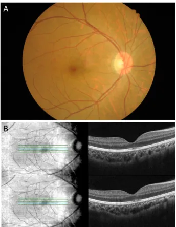
Figure 1. Fundus photograph and Optical coherence tomog-
raphy images at the fist visit. (A) Fundus photograph of the right eye at the time of presentation. Multiple subretinal and in- traretinal hemorrhages in the juxtafoveal area are apparent.Optical coherence tomography (OCT) images and red-free pho- tographs of the right eye obtained at the time of presentation.
Subretinal and intraretinal hemorrhages observed on OCT im- ages corresponded with those on red-free photographs. (B) One line scan shows that the external limiting membrane (ELM) is intact, even though a hyper-reflective lesion is present between the ellipsoid zone and the retinal pigment epithelium layer. (C) A different line scan shows a disrupted ELM and hyper-re- flective lesions that extend from the ganglion cell layer to the ellipsoid zone. The light blue line on the fundus shows the posi- tion at which the shown OCT line scan was obtained.
A B
C D
E F
Figure 2. Fluorescein angiography (FA) and indocyanine green
angiography (ICGA) images. (A, B) These images are arterial phase of FA and ICGA. (C-F) Both focal and round-shaped, juxtafoveal, hypofluorescent lesions are visible in venous phase and late phase. Hypofluroescent lesions indicate choroidal fluo- rescence blockage.없었고, 안구에 외상, 질환, 수술 등의 과거력도 없었다.
병원 방문 당시 교정시력은 우안 20/125, 좌안 20/20으로 나타났다. 세극등 현미경에서 양안 모두에서 1단계의 전방 세포가 관찰되었다. 안저검사에서는 우안에 중심 오목 근 처로 다발성 망막하 및 망막내 출혈이 관찰되었고, 빛간섭 단층촬영(optical coherence tomography, OCT) 검사에서 신 경절세포층에서부터 속분절타원체구역까지의 고반사 병변 이 나타났고, 그 주위로 외망막층 또한 손상되어 있었다 (Fig. 1). 형광안저조영술 및 인도시아닌그린혈관조영술에 서 맥락막 형광 차단 소견을 보였으나, 혈관신생 등 다른 이상은 관찰되지 않았다(Fig. 2). 내과 검사에서 범수축기심
잡음, 백혈구증가증, 적혈구침강속도 및 C-반응단백질증가 소견을 보였고, 2쌍의 혈액배양검사에서 Streptococcus an- ginosus 양성 소견을 보였다. 심초음파 검사에서 심한 승모 판역류 및 승모판 주위 증식 소견을 보였다(Fig. 3).
위 소견을 바탕으로 감염성 심내막염으로 진단하였고, 1 달간 입원하여 정맥내 ceftriaxone® (4 g/일) (CJ Health Care Corp., Seoul, Korea) 치료를 받았다. 내과적 치료 이후에 환 자는 승모판 치환 수술을 받았으며 그 이후에 전신적인 증 상(관절통, 피로, 두통 등)은 호전되었다. 1달간의 내과적 치 료 이후에 시력은 호전되었고(우안: 20/50, 좌안: 20/16), 망 막하 및 망막내 출혈도 호전되었다(Fig. 4).
고 찰
감염성 심내막염의 유병률은 2000년도에 10만 명당 11명
1418
- 대한안과학회지 2017년 제 58 권 제 12 호 -
Figure 3. Transesophageal echocardiography showed multiple
vegetations on the mitral valve (★). Accessory mitral valve leaflet prolapse (medial and middle portions) with severe ec- centric mitral valve regurgitation (☆) was also apparent.A
B
Figure 4. Ocular imaging obtained 1 month after the medical
treatment. (A) The red-free photograph of the right eye shows a substantial decrease in hemorrhage size. (B) Optical coher- ence tomography (OCT) images showed only minor residual defects in the submacular ellipsoid and interdigitation zones.The light blue line on the fundus en face image shows the posi- tion at which the OCT line scan was obtained.
에서 2011년도에 10만 명당 15명으로 증가하는 모습을 보 이고 있다. 감염성 심내막염의 위험 요인이 밝혀져 있음에 도 불구하고, 임상증상이 모호한 경우가 많기 때문에 이 병 을 진단하는 것은 쉬운 일이 아니다. 치명적인 질환이기 때 문에, 전신적인 증상이 애매모호한 경우 감염성 심내막염 의 눈 증상이 발생하면, 이에 대한 의심을 하고 철저한 검 사를 하는 것이 필요하다. 감염성 심내막염에 의한 감염성 색전에 대한 증례보고에서 맥락막 혈관 신생을 동반한 망 막하출혈을 관찰할 수 있었다고 하였으나, 본 증례의 경우 위 증례와 다르게, 맥락막 혈관 신생과 관련이 없는 망막하 출혈로 판단된다.4 본 증례에서는 다발성 망막하 및 망막내 출혈의 원인을 로트반점의 생성 원인으로 추정되는 미세혈 전에 의한 허혈성 망막 모세혈관 내피 손상, 혹은 저등급파 종혈관내응고에 의한 것이라고 추측하고 있다.5 이 부분의 경우 추후 조직학적인 평가가 가능하다면, 그 기전을 일부 밝힐 수 있을 것으로 기대된다.
본 증례의 경우, 형광안저혈관조영술 및 인도시아닌그린 혈관조영술에서 황반부 저형광 병변이 망막 혈관 밑에 위 치하였고, 빛간섭단층촬영에서 나타난 고반사 병변은 전
망막층에 걸쳐서 나타났다. 망막모세혈관총의 경우 신경절 세포층과 내핵층의 내부/외부 경계에 존재하기 때문에, 형 광안저혈관조영술만으로는 병변이 표면의 모세혈관총에서 비롯되었는지, 중간/심층의 모세혈관총에서 비롯되었는지 감별할 수 없다. 본 기관에는 구비되어 있지 않아 사용하지 못하였으나, OCT 혈관조영술검사를 한다면 비슷한 양상의 병변이 어느 층 혈관에서 비롯한 것인지 더욱 자세히 밝혀 낼 수 있으리라 기대할 수 있겠다.
REFERENCES
1) Hochman MA, Seery CM, Zarbin MA. Pathophysiology and manage- ment of subretinal hemorrhage. Surv Ophthalmol 1997;42:195-213.
2) Silverman ME, Upshaw CB. Extracardiac manifestations of in- fective endocarditis andtheir historical descriptions. Am J Cardiol 2007;100:1802-7.
3) Walpot J, Klazen C, Blok W, van Zwienen J. Embolic events in in- fective endocarditis: a review and report of 4 cases. Acta Clin Belg 2005;60:139-45.
4) Sandhya V, Shafquat S. Choroidal neovascularization (CNV) sec-
1419
= 국문초록 =
감염성 심내막염의 첫 징후로 나타난 다발성 망막하 및 망막내 출혈
목적: 다발성 망막하 및 망막내 출혈을 보인 환자가 최종적으로 감염성 심내막염으로 진단된 증례를 보고하고자 한다.
증례요약: 44세 남자가 급성으로 우안 시력저하를 보였다. 안저 검사에서 다발성 망막하 및 망막내 출혈 이외에 맥락막과 망막에 다른 병변을 관찰할 수 없었다. 전신검사에서 범수축기심잡음이 들렸으며, 혈액배양 및 심초음파 검사 결과는 감염성 심내막염 의심 소견 을 보였다.
결론: 안저의 다른 이상 소견 없이 다발성 망막하 및 망막내 출혈을 보이는 환자에서 전신적 평가를 시행할 경우, 동반된 질환을 발견 하는 데 도움이 될 것으로 생각된다.
<대한안과학회지 2017;58(12):1416-1419>
- 정규철 외 : 심내막염에서 다발성 망막 출혈 -
ondary to septic emboli from endocarditis: a case report. Eye (Lond) 2005;19:822-3.
5) Ling R, James B. White-centred retinal haemorrhages (Roth spots).
Postgrad Med J 1998;74:581-2.