• 검색 결과가 없습니다.

Primary pleomorphic liver liposarcoma: A case series and literature review

N/A
N/A
Protected

Academic year: 2021

Share "Primary pleomorphic liver liposarcoma: A case series and literature review"

Copied!
6
0
0

로드 중.... (전체 텍스트 보기)

전체 글

(1)

Primary pleomorphic liver liposarcoma:

A case series and literature review

Chuah Jun Sen

1,2

, Siaw Jia Yng

3

, Soon Koon Choon

3

, Fatin Izni Zamri

4

1

Department of General Surgery, Pusat Perubatan Universiti Kebangsaan Malaysia, Kuala Lumpur, Malaysia,

2

Department of General Surgery, Hospital Sultanah Aminah, Ministry of Health Malaysia, Johor Bahru, Malaysia,

3

Department of General Surgery, Sarawak General Hospital, Ministry of Health Malaysia, Kuching, Malaysia,

4

Department of Pathology, Sarawak General Hospital, Ministry of Health Malaysia, Kuching, Malaysia

Case Report

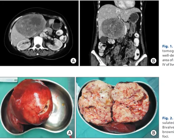
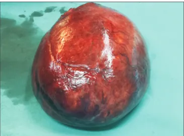
Primary hepatic liposarcoma is an extremely rare mesenchymal tumor that accounts for only 0.1% to 2% of primary malignant liver tumors. Due to its rarity, there is a lack of knowledge about its clinical course, management, and prognosis. Only 15 cases of primary liposarcoma of the liver have been reported since 1973. Among these 15 cases, only two involved primary liver liposarcoma with a pleomorphic subtype. Here we report the third and fourth cases of primary pleomorphic liver liposarcoma. A 57-year-old female pre- sented with abdominal discomfort and progressive abdominal distension for two weeks. Computed tomography (CT) of her abdomen revealed a large well-defined solid nodule mass with an area of necrosis and hemorrhage occupying segment IV-B of the liver. Wide local excision was performed. She had an uneventful recovery and remained well at six months post-treatment. A 65-year-old male presented with an abdominal mass for two-month. CT demonstrated a mass in the left lobe of the liver with mixed soft tissues and fat attenuation. He underwent wide local excision. He was discharged on day three postoperatively. Histological analysis for both cases revealed liposarcoma of the liver with a pleomorphic subtype.

Key Words: Liver neoplasm; Liposarcoma

INTRODUCTION

Primary hepatic liposarcoma is extremely rare, accounting for only 0.1% to 2% of primary malignant liver tumors [1]. Li- posarcoma originating from the mesenchymal tissue is typical- ly found in the shoulder, extremities, and the retroperitoneum space [2]. Primary hepatic liposarcoma can be divided into five subtypes (myxoid, well-differentiated, dedifferentiated, pleo- morphic, and myxoid pleomorphic), with pleomorphic lipo- sarcoma being the rarest subtype [3]. The clinical presentation of primary hepatic liposarcoma is highly variable. Early diag-

nosis of liver liposarcoma is challenging because most patients remain asymptomatic until mass effects such as abdominal pain, abdominal distention, or obstructive jaundice symp- toms emerge. Some might experience fever, nausea, vomiting, and weight loss. Physical examination might reveal a palpable abdominal mass. A computed tomography (CT) scan may provide certain typical characteristics of liver liposarcoma, making it the best tool to evaluate the resectability of liver sar- coma. Complete resection with clear margin is likely a curative therapy. To the best of our knowledge, only two cases of pri- mary pleomorphic liver liposarcoma have been reported in the literature. Here, we present two more cases of primary pleo- morphic liver liposarcoma with successful curative resection at our center. Written informed consent was obtained from both patients for publication of this case report and accompanying images.

CASES

Case 1

A 57-year-old female presented with abdominal discomfort and progressive abdominal distension for two weeks. She de-

Received: February 22, 2021, Revised: March 8, 2021, Accepted: March 10, 2021

Corresponding author: Chuah Jun Sen

Department of General Surgery, Hospital Sultanah Aminah, Ministry of Health Malaysia, Jalan Persiaran Abu Bakar Sultan, Johor Bahru 80100, Johor, Malaysia Tel: +60-16-7727965, Fax: +60-7-2231666, E-mail: vincentchuahjs@gmail.com ORCID: https://orcid.org/0000-0002-1439-8027

Copyright Ⓒ The Korean Association of Hepato-Biliary-Pancreatic Surgery

This is an Open Access article distributed under the terms of the Creative Commons Attri- bution Non-Commercial License (http://creativecommons.org/licenses/by-nc/4.0) which permits unrestricted non-commercial use, distribution, and reproduction in any medium, provided the original work is properly cited.

(2)

nied nausea, vomiting, jaundice, reduced effort intolerance, and constitutional symptoms. She had no risk factors such as drug abuse, alcoholism, or viral infection. She had no signifi- cant medical or surgical history. Physical examination revealed a palpable large mass that occupied the right upper quadrant of the abdomen. There was no stigmata of a chronic liver disease such as edema, ascites, splenomegaly, or spider naevi. All blood investigation results were normal. Viral markers of hepatitis B and hepatitis C were negative. Both serum tumor markers alpha-fetoprotein (AFP) and carcinoembryonic antigen (CEA) were normal. A CT scan of the abdomen revealed a large well-defined predominantly solid nodule mass (11 cm × 7 cm

× 9.8 cm) with an area of necrosis and hemorrhage occupying segment IV-B of the liver (Fig. 1). The mass displaced the sec- ond part of the duodenum, the head of the pancreas, and the pylorus. No other abnormality was detected in the abdominal viscera. Further investigation and workup showed no other primary lesions.

An elective laparotomy and wide local excision of the tumor was scheduled. Intraoperatively, we found a huge lipomatic liver mass (12 cm × 11 cm) arising from segment IV-B, extend- ing laterally into segments 5 and 6. The liver was smooth and non-cirrhotic. A macroscopic examination revealed a well-cir- cumscribed and an encapsulated fleshy, pale yellowish tumor.

A bi-valved section of the specimen appeared brownish and gelatinous with haemorrhage foci (Fig. 2).

Microscopically, the mass showed heterogeneous histology composed of neoplastic spindled to markedly pleomorphic cells with a variable loose myxoid background. Furthermore, the tumor comprised large cells with markedly pleomorphic vesicular nuclei and moderate-to-ample amounts of variably eosinophilic cytoplasm. There were numerous bizarre tumor giant cells. Several areas with spindled cells arranged in short fascicles were found. Numerous interspersed uni-vacuolated and multi-vacuolated variable pleomorphic lipoblasts were identified. Mitosis was easily seen. Intermittent large foci of coagulative necrosis were noted. Lymphovascular permeation was also observed. However, well-differentiated liposarcoma- tous areas were not seen. Immunohistochemical staining was performed. The tumor exhibited diffuse Vimentin positively.

The resection margin was clear. The histological diagnosis was pleomorphic hepatic liposarcoma (Fig. 3). There were no intra- or postoperative complications. She made an uneventful recovery. She was discharged on postoperative day three. A CT scan of the abdomen at six months postoperatively showed no disease recurrence.

Case 2

A 65-year-old male without significant past medical or surgi- cal history presented with an abdominal mass for two-month with losses of weight and appetite. He had no risk factors such as drug abuse, alcoholism, or viral infection. A physical exam-

A B

Fig. 1. Axial (A) and coronal (B) computed tomographyof the abdomen showing a well-defined predominantly solid mass with area of necrosis and hemorrhage at segment IV of liver.

Fig. 2. (A) Well circumscribed and encap- sulated fleshy pale yellowish tumour. (B) Bivalved section of specimen appearing brownish gelatinous with haemorrhagic foci.

A B

(3)

ination showed a palpable huge mass in the epigastric region. A CT scan demonstrated a mass in the left lobe of the liver with mixed soft tissues and fat attenuation (Fig. 4). He then under- went a laparotomy and tumor excision. Intraoperatively, there was a huge mass (approximately 15 cm × 15 cm) arising from the undersurface of segment III of the liver (Fig. 5). Otherwise, the rest of the liver was grossly normal. No ascites was noted.

Microscopically, the tumor showed a picture of pleomorphic liposarcoma with the presence of pleomorphic malignant cells, epitheloid cells, and central necrosis (about 25% of the tumor mass). The resection margin was clear. Postoperative recovery was uneventful. He was discharged on day three postoperative- ly. No evidence of primary liposarcoma was found in further history or subsequent investigation. He was lost to a scheduled three-month follow-up postoperatively.

Fig. 3. (A) Sheets of pleomorphic tumour cells (H&E, ×40). (B) Multiple bizarre tumour giant cells admixed with lipoblasts (blue arrows) are seen (H&E, ×200). (C) Tumour cells are diffusely positive for Vimentin (×200). (D) S100 stain highlighting lipoblasts (×400).

A B

C D

Fig. 4. Abdomen computed tomography (A, B) showing a large mass at the left lobe liver with mixed soft tissues and fat attenuation.

A B

Fig. 5. Gross specimen of the tumour.

(4)

DISCUSSION

Liposarcoma is one of common soft tissue malignancies that occur in the extremities and visceral areas, particularly in the retroperitoneum. Metastatic spread of soft tissue liposarcomas is relatively common, with liver involvement in 10% of cases [2].

However, primary liver liposarcoma is extremely rare, account- ing for only 0.1% to 2% of primary malignant liver tumors.

Classification of liposarcoma is based on a combination of two

basic histological aspects of the tumor: (1) the stage of lipoblast differentiation based on relative amounts of lipids in the cells and myxoid materials in extracellular spaces; and (2) the over- all degree of cellularity and cellular pleomorphism. There are five recognized subtypes: myxoid, well differentiated, dediffer- entiated, pleomorphic, and myxoid pleomorphic [3]. To date, only 15 cases of primary liver liposarcoma have been reported in the literature (Table 1). Hence, our knowledge towards this disease remains scarce. Apparently, its long-term prognosis Table 1. Characteristics of previously reported cases of primary liposarcoma of the liver

No Case source Age (yr)

/sex Location WHO classification Presentation Management Outcome

1 Kuo et al. [4] 61/F Right lobe Myxoid Fever, nausea, vomiting, jaundice, tea coloured urine and loss of weight

Right

hemihepatectomy

5 mon survival with recurrence 2 Naik et al. [5] 42/M Left lobe Pleomorphic Abdominal pain, loss of weight

and palpable mass

Extended left lateral hepatectomy

-

3 Wolloch et al. [8] 22/F Right lobe - - Right lobectomy Survival 46 days

4 Kim and Reyes [9]

86/M Right lobe Myxoid Right sided abdominal pain, dyspnea, cough and right shoulder pain for 1 yr

Supportive treatment Died rapidly

5 Kim et al. [10] 30/F Left lobe Dedifferentiated Abdominal pain and dyspnea for 4 mon

Left lateral

subsegmentectomy

10 mon disease free

6 Soares et al. [11] 2/M Hilum - Fever, jaundice, choluria, abdominal mass and anorexia for 1 mon

Laparotomy biopsy, and chemotherapy

5 mon survival

7 Wright et al. [12] 3/M Hilum Myxoid Right upper abdominal mass Wide local excision Disease free 12 yr.

Recurrence.

8 Aribal and Berberoglu [13]

48/F Hilum Myxoid Right abdominal pain Laparotomy biopsy,

and chemotherapy

- 9 Nelson et al. [14] 54/F Right and

left lobe

Myxoid Abdominal distension, pain, weight loss, nausea and vomiting for 1 yr

Laparotomy and biopsy

Post surgery bleeding and death 10 Kim et al. [15] 63/F Left lobe Well differentiated Abnormal serum liver enzyme Left

hemihepatectomy

- 11 Nakhai and

Motabar [16]

21/F Right lobe - Fever, dyspnea and abdominal pain for 3 mon

Right

hemihepatectomy and chemotherapy

9 mon survival

12 Lin et al. [17] 64/M Left lobe Myxoid Leg pain for 1 mon Left

hemihepatectomy

4 yr disease free 13 Binesh et al. [18] 83/F Left lobe Myxoid Nausea, vomiting, palpable

painful epigastric mass, loss of weight for 6 mon

Tumour resection and chemotherapy

19 mon disease free. Recurrence 14 Chen et al. [19] 43/M Left lobe Pleomorphic Abdominal distention, pain,

nausea, vomiting, weight loss for 1 yr

Left

hemihepatectomy

9 mon disease free. Recurrence 15 Rayya [20] 59/M Right lobe Myxoid Nonspecific right hypochondrium

pain, weight loss for 9 mon

Lateral partial right hepatectomy

More than 3 mon survival 16 Our 1st case 57/F Left lobe Pleomorphic Abdominal discomfort and

distention for 2 wk

Wide local excision More than 6 mon survival (remains disease free)

17 Our 2nd case 65/M Left lobe Pleomorphic Abdominal mass, weight loss for 2 mon

Wide local excision More than 3 mon

survival

WHO, World Health Organization; F, female; M, male; -, not available.

(5)

and survival are poor. Among reported cases (Table 1), there was no sex (9 females: 7 males) predilection. It appearred to be more common at the left lobe of the liver. The age range was between 2 and 86 years old, with a median age of 54 years old.

The myxoid subtype appeared to be the commonest.

Early diagnosis of primary liver liposarcoma is difficult because a tumor in the liver is usually undetected unless it is large. In addition, its symptoms are non-specific (Table 1).

Most symptoms are due to the mass effect of the tumor on nerves, vessels, the biliary tract, and intestines [4]. Common symptoms reported are fever, nausea, vomiting, loss of weight, jaundice, abdominal distention, abdominal pain, and palpable abdominal mass. In our case series, both patients presented with abdominal distention and a palpable abdominal mass.

However, there was no obstructive jaundice symptom because the tumor was confined to the liver lobe without having any compressive effect on the biliary duct.

A CT scan of the abdomen is the imaging modality of choice for identifying certain typical characteristics of liposarcoma [4-6] and for evaluating the resectablitiy of liver liposarcoma prior to surgery. It remains a challenge to differentiate liposar- coma from other liver diseases due to their varied histological compositions including areas of necrosis and hemorrhage.

For well-differentiated liposarcoma, it has a predominantly adipose mass containing non-lipomatous components. The fat component is usually > 75%, while non-lipomatous compo- nents are most often seen as prominent thick septa (> 2 mm) with or without nodularity. There might be a presence of focal nodular or globular non-adipose area [6]. For dedifferentiating liposarcoma, it has features similar to those of well-differenti- ated liposarcoma. However, it has a focal, nodular non-lipoma- tous region over 1 cm in size. Myxoid liposarcoma may appear normal or as a non-specific soft tissue mass. It has less calcifi- cation and radiolucent fat. The fat component is usually < 10%

of the lesion. It is often seen in the septa (lacy or linear pattern) or as subtle small nodules in the lesion [6].

Pleomorphic liposarcoma is typically presented as a non-spe- cific soft tissue mass. It is usually associated with an area of necrosis and hemorrhage, accounting for the prominent het- erogenecity seen in these lesions (similar to the one in our 1st case). Furthermore, it seldom contains adipose tissues, reflect- ing a higher degree of anaplasia of the lesion [6]. Both our cases involved pleomorphic type of hepatic liposarcoma, although their CT features were entirely different. The 1st case demon- strated a predominantly solid nodule mass with an area of ne- crosis and hemorrhage, while the 2nd case showed a mixed soft tissue and fat mass. These non-specific features of pleomorphic liposarcoma can make imaging diagnosis difficult. Tumor markers (such as AFP, CEA, or cancer antigen 19-9) were not increased. Liposarcoma should be one of our differential diag- noses when dealing with a liver mass.

Based on the literature review, the mainstay of treatment is curative surgical resection with clear margins [7]. However, the

disease is accompanied by a high rate of tumor recurrence with poor prognosis [4,5,8-20]. Palliative treatment should be of- fered for those with advanced liposarcoma and metastases. The role of chemotherapy and radiotherapy in hepatic liposarcoma remains less defined with a lack of strong evidence. Among the 15 cases reported, only four of them [11,13,16,18] underwent chemotherapy post-surgical resection, with survival duration ranging from 5 to 19 months. Our patient in the 1st case is disease free currently at six months post-surgical resection of the hepatic pleomorphic liposarcoma. She is still on a regular follow up schedule. However, the patient in the 2nd case had lost his follow up at three months post-surgery. Table 1 shows outcomes of all reported primary hepatic liposarcoma cases in the literature.

In conclusion, our two cases highlight our experience on the clinical course of primary hepatic liposarcoma which is extremely rare. Although there is no clear consensus, surgi- cal resection with clear margin remains to be the mainstay of treatment. Radiotherapy and chemotherapy in hepatic liposar- coma remain uncertain. They might play a role in potentially achieving a long-term survival. Additional strong evidence is required. Based on cases reported in the literature, prognosis of this disease is poor, showing a high recurrence rate.

ACKNOWLEDGEMENTS

The authors would like to thank Dr. Tan Jih Huei for contrib- uting his ideas to this study.

CONFLICT OF INTEREST

No potential conflict of interest relevant to this article was reported.

ORCID

Chuah Jun Sen, https://orcid.org/0000-0002-1439-8027 Siaw Jia Yng, https://orcid.org/0000-0001-7153-6986 Soon Koon Choon, https://orcid.org/0000-0003-3653-5667 Fatin Izni Zamri, https://orcid.org/0000-0003-4464-445X

AUTHOR CONTRIBUTIONS

Conceptualization: CJS, SJY, SKC. Data curation: All authors.

Methodology: CJS, SJY. Visualization: CJS, SJY, FIZ. Writing - original draft: CJS. Writing - review & editing: All authors.

REFERENCES

1. Liver Cancer Study Group of Japan. Primary liver cancer in Japan.

Clinicopathologic features and results of surgical treatment. Ann Surg 1990;211:277-287.

2. Teas S, Ronan SG, Ghosh L. Solitary metastatic liposarcoma of the

(6)

liver. Arch Pathol Lab Med 1978;102:605.

3. WHO Classification of Tumours Editorial Board. Soft tissue and bone tumours. 5th ed. Lyon: International Agency for Research on Cancer, 2020.

4. Kuo LM, Chou HS, Chan KM, Yu MC, Lee WC. A case of huge pri- mary liposarcoma in the liver. World J Gastroenterol 2006;12:1157- 1159.

5. Naik PR, Kumar P, Kumar PV. Primary pleomorphic liposarcoma of liver: a case report and review of the literature. Case Reports Hepatol 2013;2013:398910.

6. Murphey MD, Arcara LK, Fanburg-Smith J. From the archives of the AFIP: imaging of musculoskeletal liposarcoma with radiolog- ic-pathologic correlation. Radiographics 2005;25:1371-1395.

7. Nassif NA, Tseng W, Borges C, Chen P, Eisenberg B. Recent advances in the management of liposarcoma. F1000Res 2016;5:2907.

8. Wolloch Y, Dintsman M, Garti I. Primary malignant tumors of the liver. Isr J Med Sci 1973;9:6-11.

9. Kim TW, Reyes CV. Myxoid liposarcoma mimicking fluid density. J Surg Oncol 1985;30:80-82.

10. Kim YI, Yu ES, Lee KW, Park EU, Song HG. Dedifferentiated liposar- coma of the liver. Cancer 1987;60:2785-2790.

11. Soares FA, Landell GA, Peres LC, Oliveira MA, Vicente YA, Tone LG.

Liposarcoma of hepatic hilum in childhood: report of a case and re- view of the literature. Med Pediatr Oncol 1989;17:239-243.

12. Wright NB, Skinner R, Lee RE, Craft AW. Myxoid liposarcoma of the porta hepatis in childhood. Pediatr Radiol 1993;23:620-621.

13. Aribal E, Berberoglu L. Primary liposarcoma of the liver. AJR Am J Roentgenol 1993;161:1331-1332.

14. Nelson V, Fernandes NF, Woolf GM, Geller SA, Petrovic LM. Pri- mary liposarcoma of the liver: a case report and review of literature.

Arch Pathol Lab Med 2001;125:410-412.

15. Kim JL, Woo JY, Lee MJ, Kim KR, Jung JP, Lee NJ, et al. Imaging findings of primary well-differentiated liposarcoma of the liver: a case report. Acta Radiol 2007;48:1061-1065.

16. Nakhai B, Motabar AR. Primary liposarcoma of the liver: a case re- port and review of the literature. Med J Islam Repub Iran 2007;21:167- 172.

17. Lin YC, Tsai WC, Liu YC, Yu CP. Primary liposarcoma of liver: a case report and literature review. J Med Sci 2011;31:81-84.

18. Binesh F, Akhavan A, Kargar S, Navabii H. Primary liposar- coma of liver: a rare case and literature review. BMJ Case Rep 2012;2012:bcr0120125711.

19. Chen L, Luo J, Wen Q, Chu S, Wang W, Fan S. Primary pleomorphic liposarcoma of liver: a case report and review of the literature. Int J Clin Exp Pathol 2016;9:7629-7633.

20. Rayya F. Resection of a rare hepatic myxoid liposarcoma: case report.

Clin Surg 2017;2:1310.

수치

Fig. 1. Axial (A) and coronal (B) computed  tomographyof the abdomen showing a  well-defined predominantly solid mass with  area of necrosis and hemorrhage at segment  IV of liver.
Fig. 4. Abdomen computed tomography (A,  B) showing a large mass at the left lobe liver  with mixed soft tissues and fat attenuation.

참조

관련 문서

- leaves the abdominal cavity lateral to the inferior epigastric vessels and enters the deep inguinal ring. - traverses the entire inguinal canal - exits through

3D 모델 작성 도면 시공 계획 사업비 확인. 해석

Purpose:To evaluate stability and bone loss and histomorphometric bone reaction of dental implants in case of immediate nonfunctional loading(INFL) and

Time series of vertical cross section of potential vorticity and wind vector calculated by Case 1 along the A-A' line indicated at Fig.. Same

15.3 Functions Given by Power Series 15.4 Taylor and Maclaurin Series.

15.3 Functions Given by Power Series 15.4 Taylor and Maclaurin Series..

Substitute the series with undetermined coefficients and its derivatives.. [Reference] 5.4 Bessel’s Equation.. 11.6 Orthogonal Series.. 11.6 Orthogonal Series..

Ø Fourier series: Infinite series designed to represent general periodic functions in terms of simple ones (e.g., sines and cosines).. Ø Fourier series is more general