논문접수일 :2016년 08월 22일 논문수정일 :2016년 09월 30일 심사완료일 :2016년 10월 21일
교신저자 :이재훈, 54538 전북 익산시 무왕로 895 원광대학교 의과대학 이비인후과학교실
전화 :(063) 859-1441・전송:(063) 841-6556 E-mail:[email protected]
Introduction
Schwannomas are benign slow-growing tumors that can arise from the nerve sheath of any myelinated nerve. Although schwannomas are common in the head and neck, only 4% of head and neck schwanno- mas occur in the sinonasal tract.1) Various origins of nasal schwannomas have been reported in the litera- ture, including the inferior turbinate,2,3) middle turbi- nate,4,5) nasal septum,6,7) and nasal vestibule.8,9) Schwan- noma originating in the superior turbinate is extremely rare.
Case Report
A 40-year-old man presented to the clinic with a 5- month history of left-sided nasal obstruction. He de-
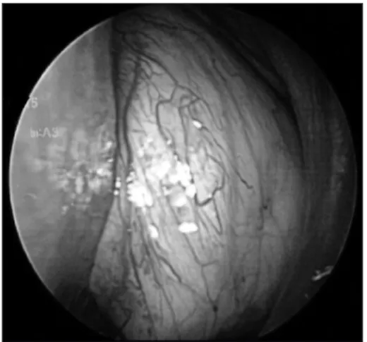
nied any history of underlying systemic disease, trau- ma, or nasal surgery. Nasal endoscopy showed a large mass obstructing the entire anterior and posteri- or nasal cavity. The mass appeared well-demarcated, smooth-margined, soft, and yellowish in color (Fig. 1).
A computed tomography (CT) scan of the sinus showed soft-tissue densities occupying the frontal si- nus, anterior ethmoid sinus, maxillary sinus, spheno- ethmoidal recess, and posterior choanae on the left side (Fig. 2A, B). CT revealed the presence of an ex- pansile mass without invasion or destruction of sur- rounding bony structures. Magnetic resonance imag- ing (MRI) showed that the mass was isointense on coronal T1-weighed images, with a hyperintense but heterogenous pattern on coronal T2-weighed images (Fig. 3A, B). Contrast enhanced fat-suppressed coro- nal T1-weighted images revealed the cystic portion of the mass and well-enhancement in the remaining por- tion (Fig. 3C). Preoperative biopsy was not performed because of showing the benign nature on the radiolog- ic findings.
Endoscopic excision of the mass was performed under general anesthesia. When the mass was opened, its tissue appeared fragile and yellowish in color, and there was no discernible capsule. Since the mass was
Schwannoma Originating from the Superior Turbinate
Dam Ho Lee, MD, Dong Hwan Oh, MD, Jin Yoon, MD and Jae Hoon Lee, MD, PhD
Department of Otolaryngology, Institute of Wonkwang Medical Science, Wonkwang University School of Medicine, Iksan, Korea
- ABSTRACT -
Schwannoma in the nasal cavity is rare. Only 4% of head and neck schwannomas occur in the sinonasal cavity.
Sinonasal schwannomas are postulated to arise from the ophthalmic and maxillary branches of the trigeminal nerve, or from autonomic nerves to the septal vessels and mucosa. Various origins of nasal schwannomas have been reported in the literature, including the inferior turbinate, middle turbinate, nasal septum, and nasal vesti- bule. We report the findings for a 40-year-old man who presented with nasal obstruction due to a schwannoma originating in the superior turbinate. (J Clinical Otolaryngol 2016;27:357-361)
KEY WORDS:SchwannomaㆍNasal cavityㆍNasal neoplasmㆍNose.
J Clinical Otolaryngol 2016;27:357-361 증 례
so fragile, it was removed in pieces instead of en bloc.
Freer elevator and a 0° endoscope was used in an at- tempt to trace the mass toward its origin. The mass occupied the sphenoethmoidal recess and pushed the middle turbinate in a lateral direction. After removal of the mass in the sphenoethmoidal recess, it was found to originate from the superior turbinate (Fig.
4A, B). A microdebrider was used to remove the por- tion of the superior turbinate to which the mass was attached. The bleeding was minimal during the mass resection.
The removed mass was sent for optimal histopath- ological analysis, which was consistent with those of a schwannoma: it comprised hypercellular Antoni type A and hypocellular Antoni type B areas (Fig.
Fig. 2. A : Coronal CT scan shows soft-tissue densities occupying the left frontal, anterior ethmoid, and maxillary sinus.
B : Coronal CT scan shows soft-tissue densities extending the sphenoethmoidal recess and posterior choanae on the left side. The CT shows the appearance of an expansile mass (arrowheads) without invasion or destruction of sur- rounding bony structures. Two arrows indicate the superior turbinate ; the origin site of the mass.
A B
Fig. 1. Endoscopy shows a large mass obstructing the entire anterior and posterior portions of the left nasal cavity. The mass appears well-demarcated, smooth- margined, soft, and yellowish in color.
Fig. 3. A : Coronal T1-weighed image shows that the mass is isointense. B : Coronal T2-weighed image shows that the mass has a hyperintense but heterogenous pattern. C : Contrast enhanced fat-suppressed coronal T1-weighted im- age shows the cystic portion (asterisk) of the mass and well-enhancement in the remaining portion.
A B C
5A). The tumor cells tested positive for S-100 protein on immunostaining (Fig. 5B), confirming the diagno- sis of schwannoma. The patient’s condition was good post-operatively, with resolution of the nasal obstruc- tion (Fig. 6). There was no evidence of recurrence at the 15-month follow-up.
Discussion
Schwannomas are rare in the nasal cavity. Sinona- sal schwannomas (SNSs) are postulated to arise from the ophthalmic and maxillary branches of the trigemi- nal nerve, or from autonomic nerves to the septal ves- sels and mucosa.10) All turbinate structures of the later-
Fig. 4. A : The mass occupies the sphenoethmoidal recess. B : After removal of the mass in the sphenoethmoidal re- cess, it is found to originate from the superior turbinate. # : mass, MT : middle turbinate, ST : superior turbinate.
Fig. 5. A : The tumor is composed of uniformly spindled Schwann cells with Antoni A (cellular fascicular, arrow), Anto- ni B (myxoid : vacuolated, arrowhead) and anuclear zone (Verocay body, asterisk) (H-E stain, ×200). B : These tumor cells are immunoreactive for S-100 protein (S-100 protein, ×400).
Fig. 6. There is no evidence of recurrence at the 3-month follow-up.
A B
al nasal wall are innervated by the maxillary branches of the trigeminal nerve.11) However, identification of the originating nerve may be difficult.
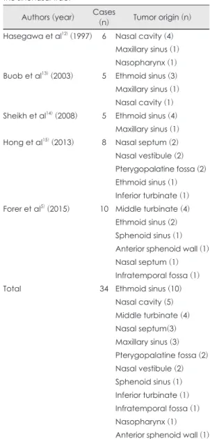
Most of SNSs in previous case series reports origi- nated from the ethmoid (10 cases), followed by the nasal cavity (5 cases), middle turbinate (4 cases), na- sal septum (3 cases), maxillary (3 cases), pterygopal- atine forssa (2 cases), and nasal vestibule (2 cases)
(Table 1).5,12-15) The exact origin site of the nasal cavi- ty was not described in two previous case series re- ports.12,13) The most common origin site was ethmoid, but this result is pretty different between previous case series reports. This result might be drawn the data from small case series (5-10 cases). The cases originated from the nasal turbinate were middle tur- binate (4 cases) and inferior turbinate (1 case), but the case originating from the superior turbinate has not been reported.
SNSs usually have a benign course. They are slow- growing and can reach a considerable size without obvious symptoms. The common signs and symp- toms of SNSs are similar to those of other benign tu- mors in the sinonasal tract, and include nasal obstruc- tion, epistaxis, and secondary sinus infection due to compromised drainage.5) In the present case, the pre- senting symptom of this case was only nasal obstruc- tion due to the large-sized tumor.
Since SNSs commonly have a polypoid appearance and lack distinctive features, they are usually mistak- en for nasal polyps, that are later histologically prov- en to be schwannomas.9) The differential diagnosis should include melanomas and olfactory neuroblasto- mas.7)
Radiological examinations such as CT and MRI are very helpful for localizing and defining the extent of SNSs. On MRI scans, schwannomas frequently appear as an isointense lesion on Tl-weighted imag- es, and as an intermediate to hyperintense lesion on T2-weighted images.16) Cystic or hemorrhagic degen- eration is a characteristic feature of schwannomas, particularly in large tumors, and MRI better demon- strates the signal intensities of cystic or hemorrhagic degeneration than CT due to higher soft-tissue contrast resolution.17) In the present case, contrast enhanced fat-suppressed coronal T1-weighted image showed the cystic degeneration within the large tumor.
Histologically, schwannomas can exhibit both An- toni type A (palisading cells surrounding an acellular central area) and Antoni type B (less cellular non-pal- isading cells without any distinct arrangement) pat-
Table 1. Previous schwannoma case series reports of the sinonasal tract
Authors (year) Cases
(n) Tumor origin (n) Hasegawa et al12) (1997) 6 Nasal cavity (4)
Maxillary sinus (1) Nasopharynx (1) Buob et al13) (2003) 5 Ethmoid sinus (3) Maxillary sinus (1) Nasal cavity (1) Sheikh et al14) (2008) 5 Ethmoid sinus (4)
Maxillary sinus (1) Hong et al15) (2013) 8 Nasal septum (2) Nasal vestibule (2) Pterygopalatine fossa (2) Ethmoid sinus (1) Inferior turbinate (1) Forer et al5) (2015) 10 Middle turbinate (4)
Ethmoid sinus (2) Sphenoid sinus (1) Anterior sphenoid wall (1) Nasal septum (1) Infratemporal fossa (1)
Total 34 Ethmoid sinus (10)
Nasal cavity (5) Middle turbinate (4) Nasal septum(3) Maxillary sinus (3) Pterygopalatine fossa (2) Nasal vestibule (2) Sphenoid sinus (1) Inferior turbinate (1) Infratemporal fossa (1) Nasopharynx (1) Anterior sphenoid wall (1)
terns in the same specimen.13) Immunohistochemical staining is important to the pathologist in the identifi- cation of these lesions. Schwannomas usually show positive immunoreactivity for S-100 protein (particular- ly in Antoni A areas), that may help to distinguish pe- ripheral nerve sheath neoplasms from other tumors.18)
The treatment of choice for SNSs is complete sur- gical excision.19,20) A resection approach that allows adequate exposure, depending on the location and extent of the tumor, must be chosen. This can involve various combinations of techniques, including endo- nasal endoscopic resection and external approaches such as lateral rhinotomy, external ethmoidectomy, Caldwell-Luc approach, and midfacial degloving.7) Functional and cosmetic aspects have to be consid- ered when selecting the surgical approach due to the noninvasive and slow-growing nature of this tumor.
SNSs prognosis is excellent because the tumor is be- nign and the postoperative recurrence is rare.9) In the present case, endonasal endoscopic resection of SNS was performed, there was no evidence of recurrence at the 15-month follow-up. Although the postoperative re- currence is rare, long-term follow-up should be need- ed. The authors present a case of schwannoma originat- ing from the superior turbinate with literature review.
REFERENCES
1) Yu E, Mikulis D, Nag S. CT and MR imaging findings in sinonasal schwannoma. Am J Neuroradiol 2006;27(4):929-30.
2) Kaufman SM, Conrad LP. Schwannoma presenting as a nasal polyp. Laryngoscope 1976;86(4):595-7.
3) Khnifies R, Fradis M, Brodsky A, Bajar J, Luntz M. Inferi- or turbinate schwannoma: report of a case. Ear Nose Throat J 2006;85(6):384-5.
4) Yang TL, Hsu MC, Liu CM. Nasal schwannoma: a case report and clinicopathologic analysis. Rhinology 2001;39(3):
169-72.
5) Forer B, Lin LJ, Sethi DS, Landsberg R. Endoscopic re- section of sinonasal tract schwannoma: presentation, treat-
ment, and outcome in 10 cases. Ann Otol Rhinol Laryngol 2015;124(8):603-8.
6) Pagella F, Giourgos G, Matti E, Colombo A. An asymptom- atic schwannoma of the nasal septum: report of a unique case. Ear Nose Throat J 2009;88(12):1264-5.
7) Dhingra S, Bakshi J, Mohindra S. Schwannoma of the nasal septum: an unusual finding. Ear Nose Throat J 2014;93(3):
E4-6.
8) Ling L, Chen HH, Zhou SH, Teng XD, Lu YY. Neurilem- momas of the nasal vestibule: report of 2 cases. Chin Med J (Engl) 2006;119(12):1053-5.
9) Fadzilah I, Salina H, Khairuzzana B, Rahmat O, Primu- harsa Putra SH. Nasal vestibule schwannoma: report of a rare case. Ear Nose Throat J 2014;93(6):E33-5.
10) Hegazy HM, Snyderman CH, Fan CY, Kassam AB. Neuri- lemmomas of the paranasal sinuses. Am J Otolaryngol 2001;
22(3):215-8.
11) Levin HL, Clemente MP. Sinus surgery: Endoscopic and Microscopic Approaches. New York: Thieme Medical Pub- lisher;2004. p.24-9.
12) Hasegawa SL, Mentzel T, Fletcher CD. Schwannomas of the sinonasal tract and nasopharynx. Mod Pathol 1997;
10(8):777-84.
13) Buob D, Wacrenier A, Chevalier D, Aubert S, Quinchon JF, Gosselin B. Schwannoma of the sinonasal tract: a clin- icopathologic and immunohistochemical study of 5 cases.
Arch Pathol Lab Med 2003;127(9):1196-9.
14) Sheikh HY, Chakravarthy RP, Slevin NJ, Sykes AJ, Ba- nerjee SS. Benign schwannoma in paranasal sinuses: a clinicopathological study of five cases, empahsising diag- nostic difficulties. J Laryngol Otol 2008;122(6):598-602.
15) Hong SM, Shin JM, Park IH, Lee SH, Kim YD, Lee HM.
Clinical experience of sinonasal schwannomas. J Rhinol 2013;20(1):26-30.
16) Som PN, Shapiro MD, Biller HF, Sasaki C, Lawson W.
Sinonasal tumors and inflammatory tissues: differentiation with MR imaging. Radiology 1988;167(3):803-8.
17) Beaman FD, Kransdorf MJ, Menke DM. Schwannoma: ra- diologic-pathologic correlation. Radiographics 2004;24(5):
1477-81.
18) Donnelly J, al-Sader MH, Blayney AW. Benign nasal schwannoma. J Laryngol Otol 1992;106(11):1011-5.
19) Shugar MA, Montgomery WW, Reardon EJ. Management of paranasal sinus schwannomas. Ann Otol Rhinol Lar- yngol 1982;91(1 Pt 1):65-9.
20) Nam GY, Jung MJ, Kwon JH, Kim JY. A Case of Schwan- noma in the nasal vestibule. J Clinical Otolaryngol 2014;
25:195-8.