Journal of Korean Arthroscopy Soc.
V o l u m e9 , N u m b e r1 , June, 2005
급성 전방십자인대 손상의 진단에 있어 관절경 소견과의 비교분석을 통한 자기공명영상의 유용성
연세대학교 의과대학 정형외과학교실, 연세대학교 의과대학 진단방사선과학교실*
최종혁・윤한국・김보람・윤춘식*
A Comparison of Accuracy between MRI and Arthroscopic Finding in the Diagnosis of Acute ACL Tear
Chong-Hyuk Choi, M.D., Han-Kook Yoon, M.D., Bo-Ram Kim, M.M., Choon-Sik Yoon, M.D.*
Department of Orthopedic Surgery, Yonsei University College of Medicine ,Seoul, Korea Department of Radiology, Yonsei University College of Medicine ,Seoul, Korea*
Purpose: The purpose of this study is to evaluate the accuracy of magnetic resonance imaging (MRI) in the diagnosis of acute anterior cruciate ligament (ACL) injury and its tear pattern in comparison with arthroscopic finding.
Materials and Methods: Sixty consecutive patients with acute ACL injury were taken MRI followed by arthroscopic examination between January 2002 and June 2004. MRI findings were reviewed according to the presence of ACL discontinuity, diffuse swelling or thickening, focal edema, collapse on distal end, and any combined tear. The pathologic findings were then confirmed arthroscopi- cally. The diagnostic accuracy of MRI on ACL tear pattern was analyzed by obtaining its positive predictive value.
Results: All fifty two cases with presence of discontinuity on MRI showed ACL rupture arthroscopically. The location of ACL tear, diffuse swelling and focal edema on MRI also corresponded with arthroscopic findings respectively. However, the diagnostic accuracy of MRI was relatively lower in the presence of other ACL patterns such as collapse and combined tear.
Conclusion: Preoperative MRI findings seem to be in accordance with arthroscopic findings and is significantly accurate in detec- tion of location and diffuse swelling and focal edema of ACL tear.
KEY WORDS: ACL, Acute rupture, MRI, Arthroscopy
서 론
최근 스포츠 손상과 교통사고 및 산업재해의 증가로 슬관 절의 전방십자인대 손상은 증가하는 추세에 있다. 급성 전 방십자인대 손상시 치료는 크게 보존적 치료와 수술적 치료
로 나눌수 있고 치료 방법을 결정하는 데는 환자의 연령 및 파열의 양상, 동반손상여부, 활동도 등이 고려되고 있다.
자기공명영상( M R I )의 진단적 유용성은 관절경 검사소견 과의 비교연구를 통해 이미 가치가 인정되고 있고 여러 저 자들에 의해 진단의 정확도가 7 8 %에서 1 0 0 %까지 보고 되고 있다7 , 8 , 1 2 ). 아울러 자기공명영상을 통해 정확한 진단 뿐 아니라 동반손상 및 파열부위, 파열정도 등을 알 수 있 다면 치료방법을 결정하는데 많은 도움을 받을 수 있을 것 이다.
본 연구에서는 급성 전방십자인대의 손상에 있어 자기공 명영상의 진단적 정확성을 관절경 검사소견과의 비교연구를 통해 알아 보았다.
*Adress correspondence and reprint requests to Chong-Hyuk Choi, M.D.
Department of Orthopedic Surgery, Yonsei University College of Medicine, Youngdong P.O. Box 1217, Seoul, Korea
Tel: 82-2-3497-3415, Fax: 82-2-573-5393 E-mail: [email protected]
연구 대상 및 방법
2 0 0 2년 1월부터 2 0 0 4년 6월까지 급성 슬관절 손상을 받고 임상적으로 전방십자인대 손상이 의심되어 자기공명영 상검사를 시행한 후 이어서 관절경검사를 시행받은 환자 6 0례를 대상으로 하였다. 환자들의 연령분포는 1 6세에서 4 7세의 분포를 보였으며 평균연령은 3 1세 였으며 성별분 포는 남자가 3 5명, 여자가 2 5명으로 남자가 더 많았다. 수 상후 MRI 검사까지의 기간은 최단 1일에서 최장 3주로 평 균 1 2일 이었고 수상후 관절경검사까지의 기간은 최단 2주 에서 최장 6주로 평균 2 5일 이었다.
MRI 판독은 본원 진단방사선과 전문의 1인이 시행하였 으며 전방십자인대 손상의 MRI 소견중 Robertson 등1 5 )의 기준중에서 직접 소견중 불연속성(discontinuity), 전반 적인 부종(diffuse swelling or thickening), 국소부종 (focal edema), 원위부 허탈( c o l l a p s e )과 파열부위 및 복합파열(combined tear)등에 대하여 관절경소견과 비교 하였다.
위의 파열양상에 대하여 양성예측도(positive predic- tive value, PPV)를 사용하였다.
본 연구에 사용된 자기공명영상기는 GE Sigma Knee C o i l과 3.0 Tesla초전도 영상장치를 이용한 M a g n e t i c Resonnace Scanner로 슬관절 촬영시 환자는 앙와위에 서 슬관절을 신전시키고 1 5도 외회전시킨 상태에서 촬영하 였다.
결 과
자기공명영상에서 불연속성은 5 2례에서 보였고 모두에서 관절경검사상 전방십자인대 손상이 확인되었다. 이 중 파열 부위는 근위부가 4 6례(Fig. 1), 중간부가 3례(Fig. 2), 원위부가 3례였으며 관절경검사소견과 자기공명영상소견이 모두 일치하여 1 0 0 %의 P P V를 나타냈다. 자기공명영상에 서 전반적인 부종을 보인 5 7례 중 관절경검사상 5 7례
( 1 0 0 % )에서 실제 전반적인 부종이 확인되었다. 자기공명 영상상에서의 국소부종은 4례 중 4례( 1 0 0 % )에서, 원위부 허탈은 2 4례 중 1 4례( 5 8 % )에서, 복합손상은 2 6례 중 1 6례( 6 1 % )에서 관절경검사상 확인되었다(Table 1).
고 찰
급성전방십자인대 손상시 진단에 있어 자기공명영상의 장 점으로는 비침습적으로 통증없이 검사를 시행할 수 있고 다 평면 영상을 얻을 수 있고 연부조직간 대조도를 조절 할 수 있는 등 많은 장점이 있으나 단점으로는 검사 비용이 비싸고 영상이 환자의 움직임에 영향을 받을 수 있다는 것이다3 , 5 , 1 3 ). 한편 전방십자인대 손상시 자기공명영상의 정확도에 대하 여 다양한 의견이 보고되고 있다. 이학적 검사가 자기공명 영상보다 진단율이 더 높다고 보고하는 저자도 있으며6 ) 특 히 Lee 등7 )은 손상받은 부위의 확인에 있어서 자기공명영 상의 정확도가 떨어진다고 보고하고 있으나 최근에는 기기 의 발달로 인하여 정확도가 크게 향상되었다.
급성 전방십자인대 손상시 전방전위검사와 L a c h m a n검 사는 동통과 근육경련, 관절강내 출혈에 의해 가음성이 생 길 수 있어 진단을 어렵게 하는 요인이 될 수 있으며 이러 한 이유로 B o m b e r g와 M c G i n t y2 ) 그리고 Noyes 등1 1 )은 부분파열시 관절경 검사가 더 효과적이라고 하고 있다.
이처럼 급성전방십자인대 손상에 대한 자기공명영상의 진 단적 유용성에 회의적인 의견도 있지만 이미 여러 저자들에 의해 그 가치가 인정되고 있다.
Ruwe 등1 6 )은 전방십자인대 파열의 진단에 있어 자기공 명영상 검사가 관절경검사를 5 1 %에서 대체 할 수 있었고 이로써 비용을 절감할 수 있는 효과를 얻을 수 있었다고 보 고하였다.
Mink 등9 )은 급성전방십자인대 파열의 진단시 자기공명 영상의 정확도가 9 5 %까지 된다고 보고 하였고 Vahey 등
1 9 )
은 자기공명영상검사를 통한 부종의 유무 확인을 통해서 전방십자인대 손상을 급성과 만성으로 구분이 가능하다고
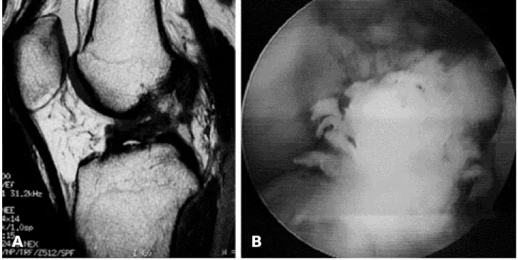
Fig. 1. (A) Sagittal magnetic resonace image shows proximal tear and discontinuity of substance. (B) Arthroscopic finding shows that ACL was grossly intact with synovial coverage. (C) Proximal tear was found with retraction.
A B C
보고하였다.
자기공명영상에서 전방십자인대 손상의 진단에 있어 직접 적인 소견으로 전방십자인대의 불규칙한 모양과 방향, 불연 속성 그리고 신호강도의 증가 등이 보고되고 있으며4 , 1 4 , 1 8 )
간접소견으로는 후방십자인대의 형태변화, 경골과 후방의 골멍, 경골의 전방전위 등이 있다1 , 1 0 , 1 7 ).
본 연구에서는 급성전방십자인대 손상의 진단에 있어 관 절경 검사를 기준으로 하여 비교하였고 자기공명영상의 정 확도가 1 0 0 %를 보이고 있었고 거의 대부분에서 부종을 확 인 할 수 있었다. 특히 파열양상에 있어서는 파열부위와 전 반적인 부종과 국소부종의 확인에 있어서 자기공명영상이 정확한 것을 알 수 있었으며 그 외 복합파열, 원위부허탈의 확인에 있어서는 유용성이 적은 것을 알 수 있었다.
결 론
결론적으로 급성전방십자인대 손상의 자기공명영상의 소
견은 관절경적 소견과 대체로 일치하였으며 특히 파열부위 의 위치와 전반적인 부종, 국소부종의 확인에 있어서는 매 우 정확한 것을 알 수 있었다. 그러나 동반손상여부, 원위 부 허탈등의 평가에 있어서는 유용성이 적은 것을 알 수 있 었다.
REFERENCES
01) Boerre NR and Ackroyd CE: Magnetic resonance imag- ing of anterior cruciate ligament rupture: A new diagnostic sign: J Bone Joint Surg, 74B:614-616, 1992.
02) Bomberg BC and McGinty JB: Acute hemarthrosis of the knee: indication for diagnosis arthroscopy. A r t h r o s c o p y, 6 : 2 2 1 - 2 2 5 . 1 9 9 0 .
03) Edelman RR and Hesselink JR: Clinical magnetic reso- nance imaging. 1st ed, Philadelphia, WB Saunders Co:996,1990.
04) Falchook FS, Tigges S, Carpenter WA, Branch TP and
Table 1. Positive predictive value of MRI as determined at arthroscopy
True positive (No.) False positive (No.) PPV* (%)
Tear site 52 00 100
Diffuse swelling 57 00 100
Focal edema 04 00 100
Collapse 14 10 058
Combined tear 16 10 061
*PPV: positive predictive value= true-positive results/(true-positive results+false positive results)*100
Fig. 2. (A) Sagittal magnetic resonace image shows midsubstance tear and discontinuity of substance. (B) Arthroscopic finding shows that midsubstance tear was found.
A B
Stiles RG: Accuracy of direct signs of tears of the anterior cruciate ligament. Canadian Association of Radiologists Journal, 47(2):114-120, 1996.
05) Glashow J, Katz R, Schneider M and Scott W: Double- blind assessment of the value of magnetic resonace imag- ing in the diagnosis of anterior cruciate and meniscal lesions. J Bone Joint Surg,71-A:113-119,1989.
06) Katz JW and Fingeroth RJ: The diagnostic accuracy of rupture of the anterior cruciate ligament comparing the Lachman test, the anterior drawer sign, and the pivot shift test in acute and chronic knee injuries. Am J Sports Med,14:88-91,1986.
07) Lee JK, Yao L, Phelps CT, Wirth CR, Czajka J and Lozman J: Anterior cruciate ligament tears: MR imaging compared with arthroscopy and clinical test. R a d i o l o g y, 166:861-864,1988.
08) Mink JH, Levy T and Crues JV: Tears of the anterior cruciate ligament and menisci of the knee: MR imaging evaluation. Radiology,167:769-774,1998.
09) Mink JH, Reicher MA, Crues JV and Deutsch AL:
MRI of the knee. 2nd ed, New York, Raven Press:141- 162,1993.
10) Murphy BJ, Smith RL, Uribe JW, Janecki CJ, Hechtman KS and Mangasarian RA : Bone signal abnormalities in the posterolateral tibia and lateral femoral condyle in complete tears of the anterior cruciate ligament:
A specific sign? Radiology, 182:221-224, 1992.
11) Noyes FR, Bassett RW, Grood ES and Butler DL:
Arthroscopy in acute traumatic hemarthrosis of the knee incidence: Incidence of anterior cruciate tears and others injuries. J Bone Joint Surg,62-A:687-695,1980.
12) Polly DW, Callaghan JJ, Sikes RA, McCabe JM, McMahon K and Savory CG: The accuracy of selective magnetic resonance imaging as compared with the find- ings of arthroscopy of the knee. J Bone Joint Surg. 70- A:192-198,1988.
13) Remer EM, Fizgerald SW, Friedman H, Rogers LF, Hendrix RW and Schafer MF: Anterior cruciate liga- ment injury: MR imaging diagnosis and pattern of injury.
Radiology,181:251-253,1991.
14) Remer EM, Fitzgerald SW, Friedman H, Rogers LF, Hendrix RW and Schafer MF: Anterior cruciate liga- ment injury: MR imaging diagnosis and patterns of injury.
Radiographics, 12:901-915, 1992.
15) Robertson PL, Schweitzer ME, Bartolozzi AR and Ugoni A : Anterior cruciate ligament tears : evaluation of multiple signs with MR imaging. R a d i o l o g y 1 9 3 : 8 2 9 - 834,1994.
16) Ruwe PA, Wright J, Randall RL, Lynch JK, Jokl P and McCarthy S: Can MRI imaging effectively replace diagnostic arthroscopy? Radiology,183:335-339,1992.
17) Schweitzer ME, Cervilla V, Kursunoglu-Brahme S and Resnick D: The posterior cruciate legament line: An indi- rect sign of anterior cruciate ligament injury. C l i n Imaging, 16:43-48, 1992.
18) Tung GA, Davis LM and Wiggins ME: Tears of the anterior crucate ligament: Primary and secondary signs at MR imaging. Radiology, 188:661-667, 1993.
19) Vahey TN, Broome DR, Kayes KJ and Shelbourne KD:
Acute and chronic tears of the anterior cruciate ligament : differential features at MR imaging. Radiology, 187:251- 253,1991.
목적: 급성 전방십자인대의 손상에 있어 자기공명영상의 진단적 정확성 및 파열양상에 대한 유용성을 관절경 검 사소견과의 비교연구를 통해 알아 보았다.
대상 및 방법: 2002년 1월부터 2 0 0 4년 6월까지 급성 슬관절 손상을 받고 임상적으로 전방십자인대 손상이 의심 되어 자기공명영상검사를 시행한 후 이어서 관절경검사를 시행받은 환자 6 0례를 대상으로 하였다. 급성 전방십자인 대의 손상에 있어 자기공명영상의 진단의 정확도, 파열양상에 대한 유용성에 대하여 관절경검사소견을 기준으로 양 성예측도를 이용하여 분석하였다.
결과: 자기공명영상에서 불연속성을 보인 5 2례 모두에서 관절경검사상 전방십자인대 손상이 확인되었다. 파열양 상에 있어서는 파열부위, 전반적인 부종, 국소 부종의 확인에 있어서는 관절경검사소견과 자기공명영상소견이 모두 일치한 결과를 보였고 원위부 허탈, 복합파열의 확인에 있어서는 정확도가 떨어지는 결과를 보였다.
결론: 급성 전방십자인대 손상의 자기공명영상의 소견은 관절경적 소견과 대체로 일치하였으며 특히 파열 양상에 있어서는 파열부위, 전반적인 부종, 국소부종의 확인에 있어서 매우 정확한 것을 알 수 있었다.
색인단어: 전방십자인대, 급성전방십자인대손상, 자기공명영상, 관절경검사 초 록