특발성 간질성 폐렴은 침윤성 병변으로 나타나는, 염증과 섬 유화 병소로 이루어진 다양한 질환군이다 (1, 2). Liebow (3) 는 조직학적 소견을 근거로 이를 통상성 간질성 폐렴(usual interstitial pneumonia, UIP), 박리성 간질성 폐렴(de-squa- mative interstitial pneumonia, DIP), 림프양 간질성 폐렴(lym- phocytic interstitial pneumonia, LIP), 거대세포 간질성 폐렴 (giant cell interstitial pneumonia, GIP), 그리고 세기관지염 및 간질성 폐렴(bronchiolitis with interstitial pneumonia, BIP) 등 다섯 가지로 나누었다. 이 후 GIP는 거의 예외 없이 텅스텐과 코발트를 포함하는 합금을 다루는 금속제련업 종사자에게서 나 타나는 진폐증의 일종으로 분류되었으며, LIP는 소림프구성 림 프종(small lymphocytic lymphomas), 만성 림프구성 백혈병 (chronic lymphocytic leukemiais), 그리고 림프구양 증식증 (lymphoid hyperplasia) 등 병리적으로 다양한 질환군을 포함 하는 림프 증식성 질환으로 분류되었다 (1, 4). BIP는 UIP, 일 부 부위의 폐쇄성 세기관지염을 동반한 미만성 폐포 손상, 그 리고 폐쇄성 세기관기염 및 기질화 폐렴(bronchiolitis obliter- ans organizing pneumonia, BOOP) 등을 포함하는 용어로 지 금은 쓰이지 않는다 (5). BOOP는 주로 폐포 강(alveolar space) 을 침범하는 질환이므로 병리학적인 간질성 폐렴의 분류에서 제외하는 경우가 더 많다. 최근, 급성 간질성 폐렴(acute inter- stitial pneumonia, AIP)과 비특이적 또는 비분류 간질성 폐렴 (nonspecific or nonclassifiable interstitial pneumonia, NSIP
or NCIP)이 특발성 간질성 폐렴의 분류에 포함되어 (1, 4), 현 재는 UIP, DIP, AIP, 그리고 NSIP 등 네 개의 질환군으로 분 류하는 것이 가장 보편적이며, 최근에는 LIP와 특발성 폐쇄성 세기관기염 및 기질화 폐렴(idiopathic BOOP)를 포함하는 여 섯 개의 질환군으로 분류하는 다른 분류도 있다 (6). 이 외에 호흡 세기관지렴-간질성 폐렴(respiratory br-onchiolitis interstitial lung disease, RB-ILD)도 특발성 간질성 폐질환의 일종이나 이는 DIP와 유사한 조직소견을 보이며 이의 초기소 견으로 생각하는 견해가 있고 DIP와 같은 분류에 포함시킨다.
본 론
통상성 간질성 폐렴 (Usual Interstitial Pneumonia)
통상성 간질성 폐렴과 특발성 폐 섬유화증(idiopathic pul- monary fibrosis, IPF)이 때로는 동의어처럼 혼용되기도 하나, 전자는 조직병리학적 진단명이며 후자는 특발성 간질성 폐렴 중 UIP의 조직학적 소견을 보이고 폐에 병변이 국한된 경우를 지칭한다 (6). 즉, 교원성 혈관질환의 폐 침범 시, 또는 약물반 응에 의해서도 IPF와 동일한 병리소견을 보일 수 있으므로 (7), UIP로 조직병리학적 진단이 내려지면, 교원성 혈관질환의 유 무 등 원인을 찾아보아야 한다.
임상소견
통상성 간질성 폐렴은 중년(40-70세)에 발생하며 남자에서 여자보다 두 배 가량 흔히 발생한다. 임상증상은 서서히 진행 하는 운동시 호흡곤란(exertional dyspnea)과 마른 기침이며 (8), 고열, 전신쇠약감, 관절통, 그리고 체중감소 등 전신증상
특발성 간질성 폐렴: 병리소견과 방사선소견의 연관1
윤영철・서지영2・한정호3・이경수
특발성 간질성 폐렴은 통상성 간질성 폐렴, 비특이적 간질성 폐렴, 급성 간질성 폐렴, 그리고 박리성 간질성 폐렴 등 네 군으로 분류한다. 급성 간질성 폐렴은 예후가 좋지 않아 높은 사망 률을 보이며 박리성 간질성 폐렴이 가장 예후가 좋다. 통상성 간질성 폐렴은 가장 흔하며 고해 상도 CT에서 늑막하 부위의 반점상 간유리음영, 불규칙한 선상음영, 그리고 벌집모양을 보인 다. 비특이적 간질성 폐렴은 두 번 째로 흔한 빈도를 보이며, 고해상도 CT 소견은 늑막하 부 위의 반점상 간유리음영과 불규칙한 선상음영이다. 급성 간질성 폐렴의 고해상도 CT 소견은 광범위한 기강경화와 반점상, 또는 미만성의 간유리음영이다. 박리성 간질성 폐렴의 흔한 고해 상도 CT 소견은 양측 하부 폐 늑막하 부위의 대칭성 간유리음영이다.
1성균관대학교 의과대학 삼성서울병원 진단방사선과학교실
2성균관대학교 의과대학 삼성서울병원 내과학교실
3성균관대학교 의과대학 삼성서울병원 진단병리과학교실
이 논문은 2001년 9월 20일 접수하여 2001년 12월 11일에 채택되었음.
이 환자의 약 반 수에서 보이고, 곤봉지는 반 수 이상에서 관 찰된다 (7, 9). 폐기능 검사에서는 제한성 장애와 폐 확산능 감 소가 두드러진다 (10). 혈액검사에서는 교원성 혈관질환이 없 는 환자에서도 류마티스양 인자(rheumatoid factor)와 항핵 항 원 (antinuclear antibody)이 양성반응을 보일 수 있다 (7).
통상성 간질성 폐렴의 예후는 매우 불량하여 진단 후 5-10 년 이내에 대부분의 환자가 사망하며, 때로는 급성악화의 임상 적 경과를 보이기도 한다 (11-13). 사망원인은 대부분 호흡 부전이며 일부는 폐암 등의 합병증에 의한다. 남자, 방사선 검 사 상 이상소견의 정도가 심한 경우, 그리고 진단 시 생리적 기능손상의 정도가 심할수록 예후가 좋지않다 (14, 15). 스테 로이드 등의 약물치료는 별 도움이 되지 않으며 (15, 16), 일 부 환자에서는 폐 이식이 치료방법으로 쓰이기도 한다 (17).
병리소견
통상성 간질성 폐렴은 폐 조직의 반복적인 손상과 염증반응, 그리고 이의 복구의 결과로 생각된다. 따라서 UIP의 조직학적 소견은 여러 부위에 염증성 병변, 증식성 병변, 그리고 섬유화 병변이 동시에 보이며, 이들의 사이 사이에 정상 폐 실질이 섞 여 있는 것이 전형적이다 (Fig. 1). 이를 시간적 다양성(tem-
poral variegation)이라 하며, 이는 시간적 차이를 두고 발생한 수 차례의 진행성 폐 손상의 결과이다 (18, 19). 대부분의 섬 유화는 호산구성 교원질과 약간의 염증세포로 이루어져 있으 며, 이러한 교원질의 침착은 폐포벽의 비후와 반흔형성, 그리 고 벌집모양 낭포(honeycombing cyst)를 형성한다.
폐 손상은 폐포상피세포나 내피세포, 또는 이 두 군데 모두 에서 시작하는 것으로 보인다 (1, 20, 21). 이로 인하여 폐포 내에 단백양 액체(proteinaceous fluid)와 세포 잔해물(cellu- lar debris)이 고이게 되며, 대식세포(macrophages)와 호중구 (neurtophils)가 손상 받은 폐포 내로 이동하게 된다. 기관지 폐포 세척액에서 이 대식세포와 호중구가 발견되는 것은 폐포 염(alveolitis)이 진행되는 중임을 의미한다 (22). 세포손상의 복구기전에는 세포매개 면역(cell mediated immunity)에 의한 제 1 형 사이토킨 반응(type-I cytokine response)과 체액성 면역 (humoral immunity)에 의한 제 2 형 사이토킨 반응의 균 형이 중요한 역할을 한다. 제 1 형 사이토킨 반응은 손상된 상 피를 구조왜곡(architectural distortion) 없이 복구시키나, 제 2 형 사이토킨 반응이 우세하게 되면, 섬유모세포(fibroblast)가 활성화되고 증식하게 되며, 세포 외 기질 단백(extracellular matrix protein)의 침착과 섬유화가 진행된다 (23). 대부분의
A
C
B
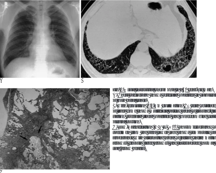
Fig. 1. Usual interstitial pneumonia in a 61 year-old man.
A. Chest radiograph shows reticular opacities in subpleural areas of both basal lungs.
B. High-resolution (1.0-mm collimation) CT scan obtained at level of liver dome shows multifocal patchy areas of ground-glass atten- uation, irregular linear opacities, and honeycombing cysts at sub- pleural portion.
C. Low-magnification (H & E, ×12) photomicrography of biopsy specimen from the right lower lobe shows temporally variegated appearance with alternating areas of active inflammation (small arrows), fibrosis (large arrows), and interspersed normal lung tis- sue (arrow heads).
섬유화는 이러한 교원질 다발로 이루어지나, 적은 양의 증식성 섬유모세포 군집이 거의 항상 발견되며 (24), 이러한 섬유모 세포 병소(fibroblastic foci)는 비특이적인 소견이나, UIP의 진 단에 꼭 필요한 요소이자 증식기 폐 손상의 증거이다 (20, 21).
폐포 내에 침착된 교원질의 표면에 제 2 형 폐포상피세포(type II pneumocyte)가 증식하게 되면, 섬유조직이 폐포벽의 일부 를 구성하게 되며 폐포벽 비후를 일으키게 된다 (20). 지속적 인 섬유화, 폐포허탈(alveolar collapse), 그리고 폐포관의 과팽 창(overdistention of the alveolar ducts) 등은 벌집모양 낭포 와 견인성 기관지 확장(traction bronchiectasis) 등을 초래한 다 (22). 염증은 비교적 미약하며 대부분 림프구로 이루어져 있다. 이러한 염증 반응은 대개 교원질 침착부위나, 벌집모양 낭포 부위에서 보이며 정상 폐포벽에서는 거의 관찰되지 않는 다. 심한 염증이 있다면 UIP보다는 다른 질환을 생각해야 한
다 (1). 벌집모양 낭포를 보이는 부위는 기관지상피 세포로 덮 여 있고 낭성 섬유화를 보이는 기강(air space)으로 구성되며, 내부에 점액을 포함 하기도 한다.
방사선소견
가장 흔한 흉부 방사선 소견은 폐 변연부와 양측 폐 기저부 의 불규칙한 선상음영, 간유리음영, 벌집모양 낭포, 그리고 폐 용적 감소이다 (1). 종격동 림프절 종대는 드물며, 미만성으로 분포하는 1.5-3 mm 크기의 폐결절(15-29%)이 있을 수 있 고 , 흉막삼출은 6% 정도에서 보인다 (1, 4). 약 5%에서는 정 상 흉부 방사선 소견을 보일 수도 있다 (18, 19). 불규칙한 선 상음영의 정도(profusion), 벌집모양 낭포 및 폐 용적 감소의 정도는 질병의 심각도(severity), 생존기간 그리고 전체적인 예 후와 대략적인 관계가 있다. 그러나 방사선학적 소견과 임상양
A B
C D
Fig. 2. Usual interstitial pneumonia (end-stage fibrosis) in a 57 year-old man.
A. Chest radiograph shows coarse, irregular, linear opacities and honeycombing in subpleural areas of both lower lung zones.
B, C. High-resolution (1.0-mm collimation) CT scans obtained at level of bottom of left atrium and liver dome show extensive hon- eycombing cysts, irregular linear opacities, and traction bronchiectasis (white arrows) at both subpleural and central lung zones.
D. Low-magnification (H & E, ×12) photomicrography of biopsy specimen from the right lower lobe shows marked interstitial thickening with fibrosis and bronchiectasia (arrow heads).
상 및 생리적 지표(physiologic index)의 상관관계는 미약하다 (25). 초기의 방사선 소견은 폐 변연부와 기저부의 대칭성의 작거나 중간 크기의 불규칙한 선상음영 또는 간유리음영이다 (Fig. 1). 진행성 병변은 거친 망상음영, 혹은 망상결절 음영이 특징이며, 말기 섬유화가 동반되면 직경 1cm 정도의 뚜렷한 벌집모양 낭포들과 진행성 폐 용적 감소 소견이 보인다 (Fig.
2) (1).
고해상도 CT 소견은 불규칙한 선상음영(82%), 간유리음영 (65-76%), 벌집모양 낭포(96%), 그리고 견인성 기관지 확장 (50%)이며 이러한 소견들은 특징적으로 폐 기저부(68-90%) 와 늑막하 부위(79%)에 분포한다 (Fig. 1, 2). 중심부 폐는 질 병의 경과가 상당히 진행되기 전에는 잘 침범하지 않는다 (4).
그 외에 명확한 결절(4-15%), 소엽간격막 비후(10%), 경한 종격동 림프절 종대 등이 보일 수 있으며, 2-3 cm 크기의 림 프절 종대는 드물다 (26).
Nishmura 등 (27)은 UIP의 고해상도 CT소견과 병리조직 소견의 상관관계를 기술하였는데, 고해상도 CT상의 벌집모양 낭포는 섬유화를 동반한 확장된 세기관지에 해당하였으며, 간 유리음영은 폐포벽의 섬유화나 염증에 해당된다고 하였다.
Akira 등 (28)은 추적검사 상 간유리음영이 있던 부위에 벌집 모양 낭포가 발생하며, 치료여부에 관계없이 벌집모양 낭포는 진행된다고 보고하였다. 간유리음영만으로 이루어진 병소는 진 행 중인 염증성 병변에 해당한다 (29, 30). 하지만 불규칙한 선상음영 또는 견인성 기관지 확장이 있는 부위에 섞여 있는 간유리음영은 섬유화만으로도 나타날 수 있다 (31). 박 등 (32) 은 치료 후 추적검사 상 간유리음영 단독으로 보인 경우, 또는 불규칙 선상음영만을 동반한 경우는 병변의 범위가 감소하였 으며, 불규칙한 선상음영과 기관지 확장 모두와 동반된 간유리 음영은 범위가 변화 없거나 증가하였다고 보고하였다.
감별해야 할 질환으로는 교원성 혈관질환, 만성 과민성 폐 렴, 그리고 폐실질을 침범한 유육종증(sarcoidosis) 등이 있다.
UIP 중 교원성 혈관질환이나 석면증을 동반한 경우와 특발성 인 경우는 CT소견으로 구분할 수 없으나, 이들 질환을 동반한 경우는 동반하지 않은 경우에 비해 병변의 진행속도가 느린 경 향이 있다. 만성 과민성 폐렴이나 유육종증이 드물게 UIP의 CT소견과 유사하게 보일 수 있다. 경계가 불분명한 소결절이 보이거나, 폐 기저부가 보존되어 있는 경우 만성 과민성 폐렴 의 가능성을 염두에 두어야 하며, 낭포의 크기가 크거나 기관
A
C
B
Fig. 3. Nonspecific interstitial pneumonia (group I) in a 54 year- old woman.
A. Chest radiograph shows areas of ground-glass opacities at both middle and lower lung zones.
B. High-resolution (1.0-mm collimation) CT scans obtained at level of liver dome shows geographic areas of grond-glass atten- uation in subpleural portion of both lower lobes.
C. Low-magnification (H & E, ×12) photomicrography of biop- sy specimen from the right lower lobe shows diffuse areas of in- flammation with lymphocytic infiltration (arrows).
지혈관속에 연한 결절이 있을 때는 유육종증을 의심해야 한다.
비특이적 간질성 폐렴 (Nonspecific Interstitial Pneumonia) 비특이적 간질성 폐렴은 1994년 Katzenstein과 Fiorelli가 처 음 기술하였는데 (7), 만성 간질성 폐렴 중 UIP, DIP, AIP 등 다른 분류에 포함되지 않는 모든 경우를 포함하는 일종의 쓰 레기통 용어(wastebasket term)로 출발하였다. Katzen-stein 의 첫 보고 중에는 교원성 혈관 질환(16%), 유해물질 흡입 (17%), 그리고 최근의 수술이나 심한 폐렴, 또는 급성 호흡곤 란 증후군 등의 병력(8%) 등을 가진 환자들이 포함되었다 (7).
그러나Naghai 등(32)이 특발성 NSIP는 UIP와 비교하여 매우 좋은 예후를 갖고 임상적으로 다른 특발성 간질성 폐렴과 구 별이 가능하다고 보고한 이후 특발성 간질성 폐렴 중 하나의 고유질환으로 받아들여지고 있다.
임상소견
평균 발병연령은 50대로 UIP보다 젊고 (7), 특징적으로 여 성에서 많이 발생한다 (4). 수 개월 정도의 아급성 증상 발현 을 보이며, 호흡곤란, 마른 기침, 그리고 미열을 호소한다. 곤 봉지는 비교적 드물며(9.7%), 이러한 임상소견의 차이와 기관 지 폐포 세척(Bronchoalveolar lavage, BAL) 세포검사 상 림 프구 증가와 T-림프구의 CD4:CD8 비율 감소 등이 UIP와의 감별에 도움이 된다 (32). NSIP의 예후는 비교적 양호하며 대 부분의 환자가 스테로이드 치료에 반응을 보여, 약 50%의 환 자는 완전히 회복되며, 사망률은 11%이다(8).
병리소견
비특이적 간질성 폐렴의 진단은 특발성 간질성 폐렴 중 다 른 질환의 가능성을 모두 배제한 후 내릴 수 있다. 조직학적으 로는 다양한 정도의 염증과 섬유화를 보이는데, 각 예에서 시
A B
C D
Fig. 4. Nonspecific interstitial pneumonia (group II) in a 46 year-old woman.
A. Chest radiograph shows areas of ground-glass opacities at subpleural portion of both lower lobes.
B, C. High-resolution (1.0-mm collimation) CT scans obtained at level of left atrium and liver dome show irregular linear opacities and patchy areas of ground-glass attenuation at subpleural portion of both lower lungs, and areas of air-space consolidation with bronchiectasis at the right middle lobe.
D. Low-magnification (H & E, ×12) photomicrography of biopsy specimen from the left lower lobe shows temporally uniform mixed areas of interstitial inflammation and fibrosis. Also noted interlobular septal thickening (arrows).
간적 균일성(temporal uniformity)을 보이는 것이 특징이다. 환 자의 약 반 수에서 폐쇄성 세기관지염 및 기질화 폐렴(BOOP) 의 특징인 기강 내 기질화 병소를 볼 수 있으나, 전체 병소의 약 10% 이하의 적은 부위를 차지한다 (1).
Katzenstein (7)은 NSIP를 세 군으로 나누었다. 제 1 군은 세포성 간질성 폐렴(cellular interstitial pneumonia)으로 섬유 화는 거의 없는 경우이며 약 50%를 차지한다 (Fig. 3). 제 2 군은 세포성 간질성 폐렴의 소견에 상당한 정도의 섬유화가 동 반된 경우이며 약 40%를 차지한다 (Fig. 4). 제 3 군은 섬유 화가 주된 소견이며 나머지 약 10%에 해당한다 (Fig. 5). 제 3 군의 경우 섬유화가 주된 소견이므로 UIP와 유사하나, 시간 적 균일성을 보이며, 진행 중인 섬유화는 거의 없다는 점이 차 이점이다.
Travis 등 (34)은 NSIP를 Katzenstein에 의한 분류 제 1 군 에 해당하는 세포성 형태(cellular pattern)와 제 2 군과 제 3 군을 모두 포함하는 섬유화 형태(fibrosing pattern)로 분류하 여야 한다고 주장하였다. 세포성 형태 NSIP는 DIP와 유사한 정도의 5년, 그리고 10년 생존율을 보였다. 하지만, 성유화 형 태 NSIP는 UIP와 비교하여 10년 생존율은 높지만 5년 생존 율은 거의 비슷하였다. 따라서 NSIP를 예후가 다른 이 두 형
태로 분류하는 것이 임상적으로 중요한 의미가 있다고 하였다.
방사선소견
비특이적 간질성 폐렴의 가장 흔한 방사선 소견은 양측 폐 기저부와 변연부의 간유리음영이며 불규칙한 선상음영이 동반 될 수 있다 (Fig. 3-5). 폐문부 림프절 종대(6%) 늑막 삼출 (5%) 등이 동반 될 수 있으며. 6-14%에서는 정상 소견을 보 인다 (1).
고해상도 CT에서는 늑막하 부위에 간유리음영과 불규칙한 선상음영이 혼재된 소견이 가장 흔하며, 기관지혈관속의 비후 와 기관지 확장 등도 흔히 보인다 (Fig. 3-5) (35, 36). 기강 경화는 약 35%에서 주로 하부 폐에서 보이며, 벌집모양 낭포 는 잘 동반되지 않는다 (36). 고해상도 CT에서 간유리음영으 로 보이는 부위는 조직학적으로는 시간적 균일성을 갖는 다양 한 정도의 염증과 섬유화로 인한 간질의 비후에 해당한다. 기 강경화 부위는 폐쇄성 세기관지렴 및 기질화 폐렴, 폐포 내 포 말세포(foamy cell) 침착, 또는 점액정체를 동반한 현미경적 벌 집모양 낭포 형성에 해당한다. NSIP환자에서 고해상도 CT에 서 보이는 간유리음영은 추적 검사에서 면적이 감소할 수 있 으며, 그 정도는 폐활량과 폐확산능의 변화정도와 상관관계가
A
C
B
Fig. 5. Nonspecific interstitial pneumonia (group III) in a 50 year-old woman.
A. Chest radiograph shows reticular opacities and patchy areas of ground-glass opacities in subpleural portion of both lower lung zones.
B. High-resolution (1.0-mm collimation) CT scan obtained at lev- el of liver dome shows irregular linear opacities, ground-glass at- tenuation, and traction bronchiectasis with temporal homogene- ity.
C. Low-magnification (H & E, ×12) photomicrography of biop- sy specimen from the right lower lobe shows relatively homoge- neous areas of diffuse interstitial fibrosis (arrows) with mild chronic inflammation (arrow heads).
있다 (1).
비특이적 간질성 폐렴의 CT소견은 매우 다양하므로 UIP, BOOP, 과민성 폐렴, 그리고 특히 DIP등과 감별이 어렵다.
급성 간질성 폐렴 (Acute Interstitial Pneumonia)
급성 간질성 폐렴은 이전에 건강했던 환자에서 발생한, 원인 을 알 수 없고, 수 일에서 수 주 간에 걸쳐 급속히 진행하는 호 흡부전과, 병리조직학적으로는 미만성 폐포손상(diffuse alve- olar damage, DAD)으로 특징 지워지는 증후군이며 (37-39), 1944년 Hamman과 Rich가 처음으로 기술하였다 (40). 이전 에는 UIP의 가속형(accelerated form)으로 생각하였으나, 현 재는 UIP와 관련이 없는 독립된 임상 및 병리적 질환군으로 분류한다 (15, 40).
임상소견
급성 간질성 폐렴은 특발성 급성 호흡곤란 증후군(idiopathic acute respiratory distress syndrome)이라 할 수 있다 (38).
평균연령은 7-77세로 광범위하며 남녀 비의 차이는 없다 (1).
수 주 이내의 기침, 발열, 호흡곤란 등의 병력이 있으며, 심한 호흡곤란과 저산소증, 그리고 인공호흡기 치료를 요하는 호흡 부전으로 급속히 진행한다 (4, 41). AIP는 박테리아, 바이러 스, 그리고 진균 배양에서 모두 음성 소견을 보이고, 독성물질 의 흡입이나 폐 약물독성, 그리고 급성 호흡곤란 증후군의 다 른 원인의 가능성이 모두 배제되어야 진단 할 수 있다 (4). 스 테로이드가 치료제로 사용되고 있으나, 그 효과는 입증되지 않 고 있다 (42). 사망률은 60-90%에 이르며 이는 원인이 밝혀 진 급성 호흡곤란 증후군과 유사하다. 생존한 환자에서는 병의 재발은 없다고 보고되어 왔으나 최근에는 재발하여 만성 진행 성 간질성 폐 질환으로 발전하는 경우도 보고되었다 (38, 42).
나. 병리소견
급성 간질성 폐렴의 병리소견은 폐포벽의 미만성 비후와 부 종, 유리질막(hyaline membrane) 형성, 간질의 심한 섬유모세 포 증식에 의한 미만성 폐포 손상이며 (38), 약간의 성숙 교원 질 침착을 보인다 (4, 39). 염증세포와 혈관 색전 등도 보일 수 있다 (38). 용어 상 앞에 붙은 미만성이란 단어 때문에 모
A
C
B
Fig. 6. Acute interstitial pneumonia (exudative phase) in a 63 year- old woman.
A. Chest radiograph shows bilateral asymmetric air-space consoli- dation and areas of ground-glass opacities.
B. High-resolution (1.0-mm collimation) CT scan obtained at level of right main bronchus shows bilateral diffuse areas of air-space consolidation, ground-glass attenuations with crazy-paving ap- pearance, and bilateral pleural effusions.
C. Low-magnification (H & E, ×12) photomicrography of biopsy specimen from another 58 year-old woman shows scattered hya- line membrane lining alveolar septa (arrows) and mononuclear cell infiltrations.
든 폐포가 다 연루된 것으로 이해하기 쉬우나 폐포를 구성하 는 상피세포, 내피세포 그리고 간질이 모두 연루되었다는 의미 로 쓰인 것이며 폐 전체적으로 볼 때 미만성 또는 국소성으로 침범 된다. 미만성 폐포 손상은 간질과 폐포의 부종, 유리질막 형성을 특징으로 하는 급성 삼출기(acute exudative phase) (Fig. 6), 삼출액의 기질화(organization)와 폐포상피세포의 재 생이 이루어지는 아급성 증식기(subacute proliferative phase) (Fig. 7), 그리고 폐포벽 내에서 증식한 섬유모세포가 손상된 폐포 기저막(basement membrane)을 통해 폐포내 공간으로 이동하게 되는 만성 섬유화기(chronic fibrotic phase)로 구분 할 수 있다 (43, 44).
급성 간질성 폐렴의 섬유화가 UIP에서 보이는 섬유화와 다
른 점은 시간적 균일성이 있으며, 성숙 교원질 침착과 구조 왜 곡(architectural distortion)이 적고, 활성상태의 섬유모세포 증 식이 대부분을 차지한다는 점 등이다. 또한 병의 진행이 한 달 이상 경과하면 UIP와 유사한 현미경적 벌집모양 낭포 형성을 보일 수 있지만 AIP에서의 벌집모양 낭포는 기강 벽이 주로 섬유모세포 침착으로 이루어져 있으며, 기관상피세포 보다는 폐포상피로 덮여있다. 그리고 짧은 시간 내에 폐 실질에 심한 반흔형성을 일으킬 수 있으며, 회복 직후에는 안정화되는 점도 UIP와의 차이점이다 (38).
방사선 소견
급성 간질성 폐렴의 방사선 소견은 급성 호흡곤란 증후군과
A B
C D
Fig. 7. Acute interstitial pneumonia (proliferative phase) in a 43 year-old woman.
A. Chest radiograph shows mixed areas of air-space consolidations and ground-glass opacities at bilateral lungs.
B. High-resolution (1.0-mm collimation) CT scan obtained at level of aortic root shows bilateral geographic areas of ground-glass at- tenuations, air-space consolidation, and mild bronchiectasis.
C. Follow up High-resolution (1.0-mm collimation) CT scan 22 months after initial examination shows irregular linear opacities, ground-glass attenuations, and honeycombing cysts at subpleural portion of both lower lobes.
D. Low-magnification (H & E, ×12) photomicrography of biopsy specimen from the right lower lobe shows extensive fibroblast proliferation (arrows), mainly within the interstitium, with a few residual intra-alveolar exudates.
매우 유사하여, 반점상 또는 미만성의 진행하는 기강경화이며 주로 하부 폐를 침범한다 (Fig. 6, 7) (43).
고해상도 CT 소견은 광범위한 양측성 기강경화가 가장 흔 한 소견(67%)이며 반점형 분포(67%)나 미만성 분포(33%)를 보이는 간유리음영도 흔히 관찰된다. 림프절 종대, 흉막삼출, 격막 비후, 구조 왜곡, 그리고 벌집모양 낭포 등은 드물다 (1, 4). 벌집모양 낭포가 좀 더 자주 보이고, 하부 폐에 대칭성 병 변을 보이는 것이 급성 호흡곤란 증후군과의 차이라는 보고도 있다 (44). CT에서 소엽간 격막 비후로 보이는 것은 병리조직 에서 격막주위 폐포(juxta-septal alveoli)의 허탈(collapse)과 기질화(organization)에 해당한다. Ichikado 등 (43)은 견인성 기관지 확장을 동반하지 않은 증가 음영 부위는 삼출기나 증 식기에 해당하였으며 (Fig. 6), 견인성 기관지 확장을 동반한 증가 음영 부위는 증식기나 섬유화기에 해당한다고 하였다 (Fig. 7). 벌집모양 낭포는 원위부 기도 재구성과 심한 간질 섬
유화에 의하며, 증가 음영 주위의 정상으로 보이는 부위도 조 직학적으로는 삼출기에 해당한다고 하였다. 생존한 경우, 간유 리음영이나 기강경화는 점차 호전된다. 회복 후 가장 흔한 CT 소견은 저음영 부위, 폐 낭종, 그리고 주로 비중력 부위(non- dependent portion)의 망상음영과 동반된 실질왜곡 등이다.
박리성 간질성 폐렴 (Desquamative Interstitial Pneumonia) 호흡성 세기관지렴(respiratory bronchiolitis, RB)과 호흡성 세기관지렴-간질성 폐렴(repiratory bronchiolitis interstitial lung disease, RB-ILD), 그리고 DIP는 모두 흡연과 깊은 관 계가 있다. 호흡성 세기관지렴은 1974년 흡연자에서 소기도 (small airway) 기능장애를 일으키는 독립된 질환으로 정의되 었으며, 1987년 호흡성 세기관지렴과 연관된 미만성 간질성 폐렴이 처음 보고되었다. RB-ILD와 DIP는 임상소견과 병리 소견이 유사하여 같은 질환의 다른 단계로 여겨진다 (45, 46).
A B
C D
Fig. 8. Desquamative interstitial pneumonia in a 49 year-old man.
A. Chest radiograph shows diffuse areas of ground-glass opaciteis in subpleural portions of both middle and lower lung zones.
B, C. High-resolution (1.0-mm collimation) CT scans obtained at level of right main bronchus and liver dome show diffuse areas of ground-glass attenuations and focal air-space consolidations in subpleural portions of both lungs.
D. Low-magnification (H & E, ×12) photomicrography of biopsy specimen from the right middle lobe shows diffuse intra-alveolar accumulation of macrophages (arrows).
하지만 RB-ILD에서 DIP로 진행한 예의 보고가 없으며, DIP 의 경우 흡연 경력이 전혀 없는 소아나 폐이식 후 발병한 예 가 있어, 임상적으로 다른 질환이라는 견해도 있다 (45).
임상소견
박리성 간질성 폐렴의 평균 발병연령은 30-50세로 UIP보 다 열 살 정도 적다. 남자에서 여자보다 두 배 자주 발생한다.
약 90%의 DIP 환자는 흡연자이다 (41). UIP보다 증상은 비 교적 급성경과를 보이며, 진행하는 호흡곤란증과 마른 기침을 호소한다. 곤봉지는 약 반 수 에서 보인다 (1, 4). 폐기능 검사 에서 제한성 장애를 보이나, 그 정도는 UIP에 비해 미약하다.
평균 생존기간 12년, 사망률 27%로 UIP에 비해 예후가 좋으 며 환자의 60%가 스테로이드 치료 후 완치된다 (3, 10). 추적 검사 상 모든 환자가 병변이 진행되는 UIP와 달리 DIP 환자 의 대부분은 호전되거나 별 변화를 보이지 않아 DIP가 UIP의 전구질환이 아닌 별개의 질환으로 분류된다 (47).
호흡성 세기관지렴(RB)은 증상이 없으며, RB-ILD의 임상
증상은 DIP와 동일하나 약간 경미하다. 기침과 운동시 호흡곤 란이 주 증상이며 폐기능 검사 상 제한성 장애를 보인다. 대부 분의 경우 금연, 또는 스테로이드 치료에 잘 반응하여 호전된 다 (45, 48).
병리소견
많은 양의 폐포 내 대식세포가 발견되고 간질 섬유화는 미 약하며, 부위마다 유사한 소견을 보이는 것이 DIP의 특징적인 병리소견이다 (Fig. 8) (49). 대부분의 대식세포는 단핵세포이 다. 미약한 폐포벽 비후는 약간의 염증세포를 동반한 교원질 침착에 의하며, 대개 폐포 내 병소를 동반한다. 벌집모양 낭포 도 보일 수는 있으나 그 정도는 미약하다. 조직학적으로 UIP 와 다른 점은 시간적 균일성이 있고, UIP에서 보이는 폐포 내 대식세포 축적은 항상 부분적이라는 점인데 반하여 DIP에서는 광범위하며, 섬유모세포 병소가 DIP에서는 없다는 점 등이다 (1, 4).
폐포 내 대식세포 축적이 세기관지 주변에만 있고 그 원위
A B
C D
Fig. 9. Respiratory bronchiolitis-interstitial pneumonia in a 48 year-old man.
A. Chest radiograph shows bilateral subpleural areas of subtle opacities and linear opacities in the middle lung zones.
B, C. High-resolution (1.0-mm collimation) CT scans obtained at level of aortic arch and inferior pulmonary vein show bilateral areas of ground-glass attenuation in subpleural and peribronchial portions. The findings of centrilobular and panlobular emphyse- ma are also seen.
D. Low-magnification (H & E, ×12) photomicrography of biopsy specimen from the left lower lobe shows accumulation of intra- alveolar macrophages which are patchy in distribution and localized to peribronchiolar parenchyma (arrows).
부는 정상일 경우에는 호흡성 세기관지렴이라 한다 (Fig. 50) (50). 병변 부위의 세기관지 및 폐포벽에 만성 염증세포 침윤, 미약한 섬유화, 그리고 제 2 형 폐포상피세포 증식 등이 동반 될 수 있다. DIP에서는 RB-ILD와 달리 폐포 중심성(bron- chocentricity)을 보이지 않고 미만성 침범을 보이며, 미약하지 만 섬유화 병변이 RB-ILD 보다는 점 더 자주, 뚜렷이 보이는 것이 차이점이다 (46, 48).
방사선 소견
양측 하부 폐의 망상음영이 가장 흔한 단순 흉부 방사선 소 견이다(10). 이전에 DIP의 특징적 소견으로 생각되었던 간유 리음영이나 기강경화는 25-33%에서만 보인다 (1). 그 이외 의 소견으로는 폐결절(15%), 벌집모양 낭포(12.5%) 등이 보 일 수 있으며, 림프절 종대나 흉막삼출은 드물다 (Fig. 9). 3- 22%에서는 정상 흉부 방사선 소견을 보인다 (4, 10).
가장 흔한 고해상도 CT소견은 간유리음영이며 거의 모든 환 자에서 볼 수 있다. 이는 양측 대칭적으로 보이는 경우가 86%
이며 어느 부위라도 침범할 수 있지만 하부 폐에서 가장 잘 보 인다 (73%). 또한 59%가 변연부에 위치한다 (Fig. 8). 그 외 에 불규칙한 선상음영과 구조왜곡이 반 수에서 있을 수 있으 며, 벌집모양 낭포와 견인성 기관지 확장도 32%에서 보인다.
이러한 섬유화를 의미하는 소견들도 주로 하부 폐의 변연부에 서 잘 보인다 (4). 이와 같이 DIP의 CT 소견은 UIP와 유사한 점이 많다. 하지만 폐 기저부에서 섬유화를 의미하는 소견이 없거나 적으면서 간유리음영이 주된 소견일 때 DIP로 진단할 수 있다 (4).
가장 흔한 RB-ILD의 고해상도 CT 소견은 주로 상부 폐에 보이는 경계가 불분명한 소엽중심성 결절(centrilobular nod- ule), 간유리음영 등이며, 무기폐(atelectasis), 소엽간간질 비 후, 폐기종, 그리고 기포(bleb) 등이 동반될 수 있다 (Fig. 9) (46, 48).
감별해야 할 질환으로는 급성, 또는 아급성 과민성 폐렴, 유 육종증, 그리고 뉴모시스트 카리니 폐렴 등을 들 수 있다.
결 론
특발성 간질성 폐렴에 속하는 질환들은 매우 유사한 방사선 학적 소견을 보일 수 있으며, 따라서 각 질환들의 비교적 특징 적인 소견을 잘 관찰하여 진단에 도움을 줄 수 있어야 하겠다.
또한 현재 활발히 연구되고 있는 이 질환군의 병인과 발생기 전 등이 보다 명확히 밝혀져 좀 더 효과적인 치료방법이 개발 되기를 기대한다.
부록
2001년 6월 채택된 American Thoracic Society/ European Respiratory Society International Multidisciplinary Consensus Classification of the Idiopathic Interstitial Pneumonias에서 Idiopathic pulmonary fibrosis/cryptogenic fibrosing alveolitis, Nonspecific interstitial pneumonia,
Cryptogenic organizing pneumonia, Acute interstitial pneu- monia, Respiratory bronchiolitis interstitial lung disease, Desquamative interstitial pneumonia, Lymphoid interstitial pneumonia로 분류하였음(Am J Respir Crit Care Med 2002;165:277-304).
참 고 문 헌
1. Lee KS, Chung MP. Idiopathic interstitial pneumonias: clinical findings, pathogenesis, pathology and radiologic findings. J Korean Med Sci 1999;14:113-127
2. Muller ML, Colby TV. Idiopathic interstitial pneumonias: high-res- olution CT and histologic findings. RadioGraphics 1997;17:1016- 1022
3. Liebow AA. New concepts and entities in pulmonary disease. Mo- nogr Pathol 1960;8:332-365
4. McAdams HP, Rosade-de-Christenson ML, Wehunt WD, Fishback MF. The alphabet soup revisited: the chronic interstitial pneumo- nias in the 1990s. RadioGraphics 1996;16:1009-1033
5. Katzenstein ALA, Myers JL. Nonspecific interstitial pneuonia and the other idiopathic interstitial pneumonias: classification and diag- nostic criteria. Am J Surg Pathol 2000;24:1-3
6. American Thoracic Society. Idiopathic pulomonary fibrosis: diag- nosis and treatment: international consensus statement. Am J Respir Crit Care Med 2000;161:646-664
7. Katzenstein ALA. Fiorelli RF. Nonspecific interstitial pneumo- nia/fibrosis: histologic features and clinical significance. Am J Surg Pathol 1994; 18:136-147
8. Crystal RG, Fulmer JD, Roberts WC, Moss ML, Line BR, Reynolds HY. Idiopathic pulmonary fibrosis: clinical, histologic, radiograph- ic, physiologic, scintigraphic, cytologic and biochemical aspects.
Ann Intern Med 1976;85:769-788
9. Guerry-Force ML, Muller ML, Wright JL, Wiggs B, Coppin C, Pare P, Hogg JC. A comparison of bronchiolitis obliterans with organiz- ing pneumonia, usual interstitial pneumonia, and small airway dis- ease. Am Rev Respir Dis 1987;135:705-712
10. Carrington CB, Gaensler EA, Coutu RE, FitsGerald MX, Gupta RG. Natural history and treated course of usual and desquamative interstitial pneumonia. N Engl J Med 1978;298:801-809
11. Pratt DS, Schwarz MI, May JJ, Dreisin RS. Rapidly fatal pul- monary fibrosis: the accelerated variant of interstitial pneumonitis.
Thorax 1979;34:587-593
12. Colby TV, Lombard CL, Yousem SA, Kitaichi M, Interstitial disease.
In: Bordin GM(Ed.) Atlas of pulmonary surgical pathology.
Philadelphia, WB Saunders. 1991;228-306
13. Akira M, Hamada H, Sakatani M, Kobayashi C, Nishioka M, Yamamoto S. CT findings during phase of accelerated deteriora- tion in patients with idiopathic pulmonary fibrosis. AJR Am J Roentgenol 1997; 168:79-83
14. Schwartz DA, Van Fossen DS, Davis CS, et al. Determinants of progression in idiopathic pulmonary fibrosis. Am J Respir Crit Care Med 1994; 149:444-449
15. Rudd RM, Haslam PL, Turner-Warwick M. Cryptogenic fibrosing alveolitis. Am Rev Respir Dis 1981;124:1-8
16. Johnson MA, Kwan J, Snell NJ, Nunn AJ, Darbyshire JH, Turener- Warwick M. Randomized controlled trial comparing prednisolone alone with cyclophosphamide and low dose prednisolone in com- bination in cryptogenic fibrosing alveolitis. Thorax 1989;44:280- 288
17. Hunninghake G, Kalica AR. Approaches to the treatment of pul-
monary fibrosis. Am J Respir Crit Care Med 1995;131:915-918 18. Muller NL, Guerry-Force ML, Staples CA, Wright JL, Wiggs B,
Coppin C, Pare P, Hogg JC. Differential diagnosis of bronchiolitis obliterans with organizing pneumonia and usual interstitial pneu- monia: clinical, functional, and radiologic findings. Radiology 1987;
162:151-156
19. Chandler PW, Shin MS, Friedman SE, Myers JL, Katzenstein A.
Radiographic manifestations of bronchiolitis obliterans with orga- nizing pneumonia vs usual interstitial pneumonia. AJR Am J Roent- genol 1986;147:899-906
20. Kuhn C, Boldt H, King TE, Crouch E, Vartio T, McDonald JA. An immunohistochemical study of architectural remodeling and con- nective tissue synthesis in pulmonary fibrosis. Am Rev Respir Dis 1989;140:1693-1703
21. Myers JA, Katzenstein AL. Epithelial necrosis and alveolar collapse in the pathogenesis of usual interstitial pneumonia. Chest 1988;
94:1309-1311
22. Hogg JC. Chronic interstitial lung disease of unknown cause: a new classification based on pathogenesis. AJR Am J Roentgenol 1991;156:225-233
23. Sime PJ, O’Reilly KM, Fibrosis of the lung and other tissues: new concepts in pathogenesis and treatment. Clin Immunol 2001;99:
308-319
24. Katzenstein ALA, Myers JL, Prophet WD, Corley LS, Shin MS.
Bronchiolitis obliterans and usual interstitial pneumonia: a com- parative clinopathologic study. Am J Surg Pathol 1986;106:373-381 25. Schwartz DA, Helmers RA, Galvin JR, Van Fossen DS, Frees KL,
Dayton CS, Burmeister LF, Hunninghake GW. Determination of survival in idiopathic pulmonary fibrosis. Am J Respir Crit Care Med 1994;149:450-454
26. Bergin C, Castellino RA. Mediastinal lymph node enlargement on CT scans in patients with usual interstitial pneumonitis. AJR Am J Roentgenol 1990;154:251-254
27. Nishimura K, Kitaichi M, Izumi T, Nagai S, Kanaoka M, Itoh H.
Usual interstitial pneumonia: histologic correlation with high-reso- lution CT. Radiology 1992;183:337-342
28. Akira M, Sakatani M, Ueda E. Idiopathic pulmonary fibrosis: pro- gression of honeycombing at thin-section CT. Radiology 1993;189:
687-691
29. Wells AU, Rubens MB, du Bois RM, Hansell DM. Serial CT in fi- brosing alveolitis: prognostic significance of the initial pattern. AJR Am J Roentgenol 1993;161:1159-1165
30. Muller NL, Staples CA, Miller RR, Vedal S, Thurlbeck WM, Ostrow DN. Disease activity in idiopathic pulmonary fibrosis: CT and pathologic correlation. Radiology 1987;165:731-734
31. Remy-Jardin M, Giraud F, Jemy J, Copin MC, Gosselin B, Duhamel A. Importance of ground-glass attenuation in chronic dif- fuse infiltrative lung disease: pathologic-CT correlation. Radiology 1993;189:693-698
32. Park JS, Lee KS, Choi DL, et al. Ground-glass attenuation in usual interstitial pneumonia: histologic correlation and serial changes on thin-section CT. J Korean Radiol Soc 1998;39:313-320
33. Nagai S, Kitachi M, Itoh H, Nishimura K, Izumi T, Colby TV.
Idiopathic nonspecific pneumonia/fibrosis: comparison with idio- pathic pulmonary fibrosis and BOOP. Eur Respir J 1998;12:1010-
1019
34. Travis WD, Matsui K, Moss J, Ferrans VJ. Idiopathic nonspecific interstitial pneumonia: prognostic significance of cellular and fi- brosing patterns; survival comparison with usual interstitial pneu- monia and desquamative interstitial pneumonia. Am J Surg Pathol 2000;24(1);19-33
35. Park JS, Lee KS, Kim JS, Park CS, Suh YL, Choi DL, Kim KJ.
Nonspecific interstitial pneumonia with fibrosis: radiographic and CT findings in seven patients. Radiology 1995;195:645-648 36. Kim TS, Lee KS, Chung MP, Han J, Park JS, Hwang JH, Kwon OJ,
Rhee CH. Nonspecific interstitial pneumonia with fibrosis: high- resolution CT and pathologic findings. AJR Am J Roentgenol 1998;
171:1645-1650
37. Katzenstein ALA, Myers JL, Mazur MT. Acute interstitial pneumo- nia: a clinicopathologic, ultrastructural, and cell kinetic study: Am J Surg Pathol 1986;10:136-147
38. Olson J, Colby TV, Eliott CG. Hamman-Rich syndrome revisited.
Mayo Clin Proc 1990;65:1538-1548
39. Primack SL, Hartman TE, Ikezoe J, Akira M, Sakatani M, Muller NL. Acute interstitial pneumonia; radiographic and CT findings in nine patients. Radiology 1993;188:817-820
40. Hamman L, Rich AR. Acute diffuse interstitial fibrosis of the lungs.
Bull Johns Hopkins Hosp 1944;74:177-212
41. Katzenstein ALA. Idiopathic interstitial pneumonia: classification and diagnosis. In: Churg A, katzenstein ALA, eds. The lung: current con- cepts. Baltimore, Md: Williams & Wilkins, 1994;1-31
42. Vourlekis JS, Broun KK, Cool CD, etc. Acute interstitial pneumoni- tis; case series and review of the literature. Medicine 2000;79:369- 378
43. Ichikado K, Johkoh T, Ikezoe J, et al. Acute interstitial pneumonia:
high-resolution CT findings correlated with pathology. AJR Am J Roentgenol 1997;168:333-338
44. Tomiyama N, Muller NL, Johkoh T, etc. Acute respiratory distress syndrome and acute interstitial pneumonia: comparison of thin- section CT findings. J Comput Assist Tomogr 2001;25:28-33 45. Nagai S, Hoshino Y, Hyasi M, Ito I. Smoking-related interstitial
lung disease. Curr Opin Pulm Med 2000;6:415-419
46. Heyneman LE, Ward S, Lynch DA, Remy-Jardin M, Johkoh T, Muller NL. Respiratory bronchiolitis, respiratory bronchiolitis-as- sociated interstitial lung disease, and desquamative interstitial pneumonia: different entities or part of the spectrum of the same disease process? AJR Am J Roentgenol 1999;173:1617-1622 47. Akira M, Yamamoto S, Hara H, Sakatani M, Ueda B. Serial com-
puted tomographic evaluation in desquamative interstitial pneu- monia. Thorax 1997;52:680-693
48. Holt RM, Schmidt RA, Godwin JD, Raghu G. High resolution CT in respiratory bronchiolitis-associated interstitial lung disease. J Comput Assist Tomogr 1993;17:46-50
49. Hartman TE, Primack SL, Swensen SJ, Hansell D, McGuinness G, Muller NL. Desquamative interstitial pneumonia: thin-section CT findings in 22 patients. Radiology 1993;187:789-790
50. Myers JL, Veal CF, Shin MS, katzenstein ALA. Respiratory bron- chiolitis causing interstitial lung disease: a clinopathologic study of six cases. Am Rev Respir Dis 1987;135:880-884
J Korean Radiol Soc 2002;46:403-415
Address reprint requests to : Kyung Soo Lee, M.D., Department of Radiology, Samsung Medical Center, Sungkyunkwan University School of Medicine, 50, Ilson-dong, Kangnam-gu, Seoul 135-710, Korea.
Tel. 82-2-3410-2511 Fax. 82-2-3410-2559 E-mail: [email protected]
Idiopathic Interstitial Pneumonias: Radiologic-Pathologic Correlation1
Young Cheol Yoon, M.D., Gee Young Suh, M.D.2, Joungho Han, M.D.3, Kyung Soo Lee, M.D.
1Department of Radiology, Samsung Medical Center, Sungkyunkwan University School of Medicine
2Department of Internal Medicine, Samsung Medical Center, Sungkyunkwan University School of Medicine
3Department of Pathology, Samsung Medical Center, Sungkyunkwan University School of Medicine
Idiopathic interstitial pneumonias are at present classified as one of four types: usual, nonspecific, acute, or desquamative. The acute form has the worst prognosis, followed by the usual and the nonspecific form; it is in desquamative cases that prognosis is best. At high-resolution CT, usual interstitial pneumonia, the most fre- quent type, manifests as patchy subpleural areas of ground-glass attenuation, irregular linear opacity, and hon- eycombing, which the nonspecific type, the second most frequent, appears as subpleural patchy areas of ground-glass attenuation with associated areas of irregular linear opacity. Acute interstitial pneumonia demon- strates extensive bilateral airspace consolidation and patchy or diffuse bilateral areas of ground-glass attenua- tion in middle and lower lung zones.
Index words :Lung fibrosis Lung diseases
Pneumonia, interstitial with fibrosis