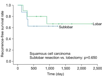
Lobectomy versus Sublobar Resection in Non-Lepidic Small-Sized Non-Small Cell Lung Cancer
9
0
0
전체 글
(2)
(3)
(4)
(5)
(6)
(7)
(8)
(9)
수치
+2
관련 문서
Background : The prognosis of patients with stage III non-small cell lung cancer (NSCLC) who achieve complete response or downstaging following
Kaplan-Meier survival curves according to plasma osteo- pontin (OPN) levels in patients with non-small cell lung cancer (NSCLC).. Overall survival curves of patients with plasma
Background: Epidermal growth factor receptor (EGFR) tyrosine kinase inhibitors, gefitinib and erlotinib, are effective therapies for non-small cell lung cancer (NSCLC)
To our knowledge, this is the first case report of histopatholgically confirmed ILD following erlotinib therapy in a live patient with non-small cell lung cancer
The genomic DNA from 9 subjects with a non-small cell lung cancer (squamous cell cancer 6, adenocarcinoma 2, non-small cell lung cancer1) and 9 age
The patients’ age, sex, disease types (adenocarcinoma, squamous cell carcinoma), tumor location (supratentorial, in- fratentorial), timing of metastasis (synchronous, metachro-
Forest plot showing a comparison of recurrence-free survival between segmentectomy and lobectomy in (A) stage I and (B) stage IA non-small cell lung cancer. All of them used
The results of this study showed that FXIII activity was higher in patients with NSCLC than in the healthy group. Patients with squamous cell carcinoma and stage III and IV
