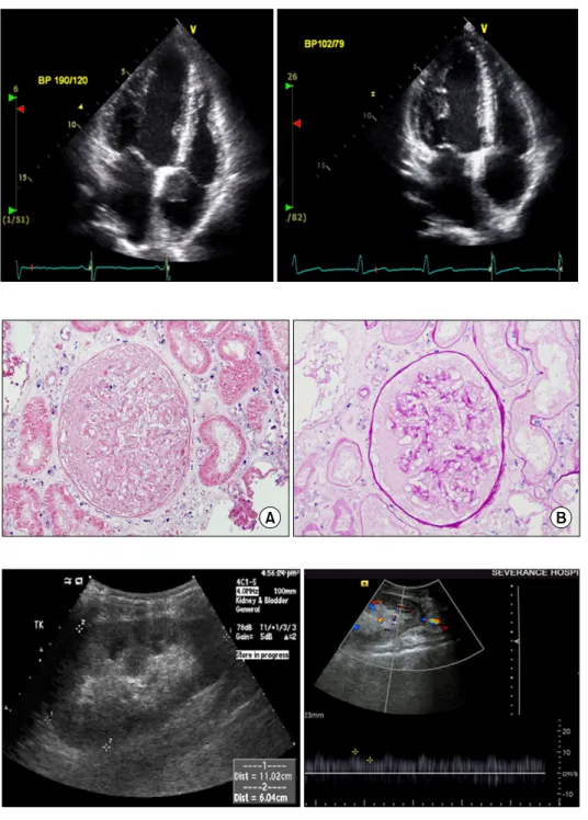
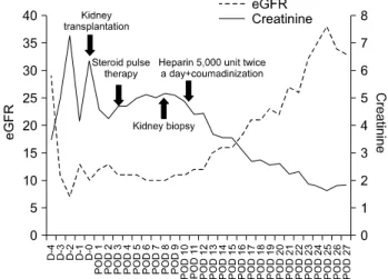
Successful Graft Recovery from Thrombotic Acute Kidney Injury in a Kidney Transplant Patient with Antiphospholipid Syndrome
4
0
0
전체 글
(2)
(3)
(4)
수치
관련 문서
Based the above study results, balance relaxation therapy is considered a therapy that can help the body recover its flexibility if there is an imbalance
We evaluated the general characteristics, mechanism of injury, Revised Trauma Score(RTS), Injury Severity Score(ISS), Trauma Injury Severity Score(TRISS). More men
Alternative splicing; 그림은 하나의 유전자가 다양한 splicing을 통하여 multiple mRNA를 만드는 것을 보여준다.. WT1 유전자 (돌연변이가 생기면 kidney tumor를
P.( Kidney
Long-term outcome after an early invasive versus selective invasive treatment strategy in patients with non.ST-elevation acute coronary syndrome and elevated
Chronic kidney disease (partial update): Early identification and management of chronic kidney disease in adults in primary and secondary care (Clinical
Adopting ISS 16+as severe injury criteria, the collision speed( ) making the reduced injury equal to induced injury is estimated as 32.5km/h. The airbag deployment rate
• In itself, this decline poses little danger however it renders the aging kidney more vulnerable to acquired insults (volume depletion, nephrotoxins, contrast dye). •
