Bisphosphonates are agents used to treat or prevent os- teoporosis and treat malignant bone lesion. These agents have been increasingly recommended for use in patients with osteoporosis, Paget’s disease of bone, osteolytic bone metastases, and so on.1-4Also, these agents reduce skele- tal complications such as pain, pathologic bony fracture, and size of osteolytic lesion.1-3Oral bisphosphonates such as alendronate (Fosamax) and risedronate (Actonel) are used for the treatment of postmenopausal osteoporosis.
These medications are usually prescribed once a week (aledronate 70 mg, risedronate 35 mg).1
Despite the benefits related to the use of these medica- tions, osteonecrosis of the jaws is a significant complica- tion in a subset of patients receiving these drugs.1,2,5,6 It appears that the pathogenesis of this process is most con- sistent with a defect in the jaw bone physiologic remodel- ing or wound healing. The negative impact of bisphospho- nate is the inhibition of osteoclast function.1-3,5Once depo- sited on the surface of bone, bisphosphonates are internal-
ized by osteoclast, causing disruption of osteoclast-medi- ated bone turnover and resorption.2-4
This complication usually presents after dento-alveolar trauma, elective dental surgery or tooth extraction.5-8How- ever, a subset of edentulous and dentate patients have de- veloped necrotic bone spontaneously.2There is an increas- ing concern that the oral bisphosphonates are implicated in osteonecrosis of the jaws.8
Recently there were patients showing the features of bis- phosphonate related osteonecrosis of the jaws (BRONJ).
This report demonstrates 2 cases of BRONJ examined using plain radiography and computed tomography (CT).
Case Report
Case 1
A 74-year-old woman had a complaint of bony exposure on the left posterior maxilla after spontaneous loss of the left maxillary first and second molars 3 months ago. She had been taking oral risedronate (Actonel), a kind of bis- phosphonates, 35 mg once weekly for 4 years because of osteoporosis. The intraoral examination revealed the bony exposure on the left maxillary alveolar region, pus dis- charge, and bleeding on the same site as well as sequestrum receiving these drugs. This complication occurs either spontaneously or after a simple dento-alveolar surgery. Recent- ly there were two patients who showed the features of bisphosphonate related osteonecrosis of the jaws (BRONJ) in Gangneung-Wonju National University Dental Hospital. The patients revealed the clinical and radiological features of classical osteomyelitis. This report presents two cases of BRONJ which were examined by plain radiography and computed tomography. (Imaging Sci Dent 2011; 41 : 129-34)
KEY WORDS : Bisphosphonate; Osteonecrosis; Tomography, X-ray Computed; Osteoporosis
Received January 22, 2011; Revised February 21, 2011; Accepted April 8, 2011 Correspondence to : Prof. Jin-Woo Han
Department of Oral and Maxillofacial Radiology, College of Dentistry, Gangneung- Wonju National University, 123 Jibyun-Dong, Gangneung, Gangwon-do 210-702, Korea
Tel) 82-33-640-3135, Fax) 82-33-640-3113, E-mail) [email protected]
Copyright ⓒ 2011 by Korean Academy of Oral and Maxillofacial Radiology
This is an Open Access article distributed under the terms of the Creative Commons Attribution Non-Commercial License (http://creativecommons.org/licenses/by-nc/3.0) which permits unrestricted non-commercial use, distribution, and reproduction in any medium, provided the original work is properly cited.
Imaging Science in Dentistry∙pISSN 2233-7822 eISSN 2233-7830
detached from the area (Fig. 1).
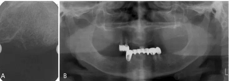
The patient took a panoramic radiograph and CT. On the panoramic radiograph, a large osseous crater and cor-
tical bone destruction were found on the left maxillary ede- ntulous area (Fig. 2). CT images demonstrated cortical bone destruction on the left maxillary edentulous area and
Fig. 1.Intraoral examination reveals alveolar bone exposure (A) and detached sequestrum (B) on left maxillary area of case 1.
A B
Fig. 2.Panoramic radiograph shows a large osseous crater on left maxil- lary edentulous area.
Fig. 3.A. Axial CT image reveals cortical bone destruction, sclerotic change of adjacent trabecular bone.
B. Axial CT image reveals numer- ous bony fragments that is consider- ed as sequestrum.
A B
infiltrated with chronic inflammatory cells. There was not vivid osteocyte in the lacunae (Fig. 5). Through the histo- pathologic feature, this case was confirmed as BRONJ.
During 6 months follow-up period, there was no sign of recurrence (Fig. 6).
Case 2
A 72-year-old woman had a chief complaint of swelling
dura from the extraction socket of the canine and mild mucositis on the left maxillary sinus (Fig. 8). The patient was diagnosed with osteomyelitis from the clinical and radiological examinations. The treatment was performed with saucerization, fistulorraphy, and sequestrectomy.
The patient revisited 1 year and 6 months later. The chief
Fig. 4.Saucerization and decortication are performed on that area with sequestrectomy.
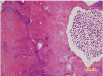
Fig. 5.Photomicrograph of specimen of biopsy shows infiltration of chronic inflammatory cell and no vivid osteocyte in the lacunae (H&E stain, ×200).
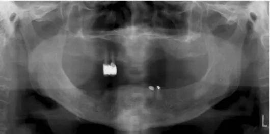
Fig. 6.Panoramic radiograph shows bone remodeling on the 6 months follow-up period in case 1. There is no sign of recurrence on left maxil- lary area.
complaint was bony exposure on the right mandibular eden- tulous area with pus discharge (Fig. 9). Plain radiographic
examination showed the decrease in bone density on the right mandibular alveolar process, resorptive area with irregular shape, and sequestrum on the crestal area (Fig. 10).
The patient was diagnosed with BRONJ from the clinical and radiological examinations. Treatment began with the cessation of bisphosphonates therapy. And then, sauceriza- tion and decortication were performed on that area with sequestrectomy. Biopsy from the bony lesion demonstrated sequestrum infiltrated with chronic inflammatory cell as well as bacterial flora (Fig. 11). Histopathologic findings confirmed the diagnosis of BRONJ. During 3 months follow-up period, there was no recurrence sign.
Discussion
Bisphosphonates inhibit osteoclast-mediated bone turn- over and endothelial cell function. Cells treated with bis- phosphonates demonstrate decreased proliferation, incre-
Fig. 8.A. Axial CT image reveals separated lamina dura on socket of left maxillary canine. B. Axial CT image shows mild buccal soft tissue swelling.
A B
Fig. 7.A. Intraoral radiograph of case 2 reveals bony fragment with trabeculation on socket of left maxillary canine. B. Panoramic radio- graph shows separated lamina dura on socket of left maxillary canine.
A B
Fig. 9.Intraoral examination of case 2 reveals recurrence sign of alveolar bone exposure and pus discharge on right mandibular edentulous area.
ased rate of apoptosis, and a decrease in capillary-tube for- mation.1,4Bisphosphonates also demonstrate antiangioge- nic properties owing to their ability to significantly decre- ase circulating levels of vascular endothelial growth factor (VEGF).1,8
BRONJ was first reported in 2003, recognized a few years after bisphosphonates approval for use.9There was a growing number of BRONJ in patients receiving oral bis- phosphonates therapy. Ortega et al reported that the inci- dence of BRONJ varied from 0.15% to 12% of patients receiving bisphosphonates for treatment of osteoporosis.10
There is an increasing concern that oral bisphosphonates are implicated in osteonecrosis of the jaws, albeit less com- monly than that observed with the more potent intravenous- ly administered bisphosphonates. Recent retrospective cli- nical studies have identified several potential risk factors associated with the development of BRONJ. These include
a history of dento-alveolar trauma, duration of bisphospho- nates exposure, and the type of bisphosphonates.3,7,9-12 In the majority of BRONJ cases reported to date, recent dento- alveolar trauma was the most prevalent and consistent risk factor.3
The lesion related with BRONJ is most frequently symp- tomatic when surrounding tissues become inflamed or there is a clinical evidence of infection. Signs and symp- toms include pain, tooth mobility, mucosal swelling, ery- thema, and ulceration.2-8Some patients may also present with complaints of altered sensation in the affected area.2 These cases showed the clinical features such as pain, bone exposure, pus discharge, bleeding, and swelling. The pati- ents had been taking oral risedronate (Actonel), a kind of bisphosphonates 35 mg, once weekly for treatment of osteo- porosis for 4 years and 3 years 5 months, respectively. In both cases, there were loss of teeth that could be regarded as the causative factor of dento-alveolar trauma and sub- sequent osteonecrosis of the jaws.
Radiographic changes are not evident until there is sig- nificant bone involvement. Therefore, panoramic and peri- apical radiographs may not reveal significant changes in the early stages of osteonecrosis.1-3Early or late radiogra- phic changes may mimic classic periapical pathology or osteomyelitis. In the cases presented, plain radiographs and CT images were taken, which showed signs of bone destruction with sequestrum and mucositis on the adjacent maxillary sinus. These features were not differed from clas- sic osteomyelitis.
Magopoulos et al3 reported that cessation of bisphos- phonates therapy combined with surgical debriment and long term antibiotic therapy was the treatment of choice in BRONJ patients. In case 1, the patient was treated with saucerization, decortications, and sequestrectomy on the
Fig. 11.Photomicrograph of sequestrum shows heavy infiltration of chronic inflammatory cell and bacterial flora (H&E stain, ×200).
problematic area with the cessation of bisphosphonates therapy. During the follow-up period of 6 months, there was no recurrence sign. In case 2, the patient was treated with saucerization, fistulorraphy, and sequestrectomy with- out cessation of medication. However, there was recurrence of osteonecrosis on the mandible after 1 year 6 months.
Treatment was performed again on the recurrent area. Sau- cerization, decortications, and sequestrectomy were per- formed with the cessation of bisphosphonates therapy.
During the follow-up period of 3 months, there was no sign of recurrence after the retreatment.
It is critical that the dentist should be aware of this sig- nificant complication which can occur spontaneously or after any dento-alveolar procedure in the population receiv- ing bisphosphonates. Because the oral examination and review of the patient’s medical history are currently the most effective and sensitive means of detecting BRONJ, dental professionals are in a unique position to identify and diagnose this disease process early in its course.
This report serves to alert dentists about the potential complication of bone necrosis in patients receiving bis- phosphonates therapy. For the treatment of BRONJ, den- tists need to consider ceasing the use of bisphosphonates.
References
1. Ruggiero SL, Fantasia J, Carlson E. Bisphosphonate-related osteonecrosis of the jaw: background and guidelines for diag- nosis, staging and management. Oral Surg Oral Med Oral Path- ol Oral Radiol Endod 2006; 102 : 433-41.
2. Kos M, Kuebler JF, Luczak K, Engelke W. Bisphosphonate- related osteonecrosis of the jaws: a review of 34 cases and eval-
uation of risk. J Craniomaxillofac Surg 2010; 38 : 255-9.
3. Magopoulos C, Karakinaris G, Telioudis Z, Vahtsevanos K, Dimitrakopoulos I, Antoniadis K, et al. Osteonecrosis of the jaws due to bisphosphonate use. A review of 60 cases and treat- ment proposals. Am J Otolaryngol 2007; 28 : 158-63.
4. Lehrer S, Montazem A, Ramanathan L, Pessin-Minsley M, Pfail J, Stock RG, et al. Normal serum bone markers in bis- phosphonate-induced osteonecrosis of the jaws. Oral Surg Oral Med Oral Pathol Oral Radiol Endod 2008; 106 : 389-91.
5. Campisi G, Di Fede O, Musciotto A, Lo Casto A, Lo Muzio L, Fulfaro F, et al. Bisphosphonate-related osteonecrosis of the jaw (BRONJ): run dental management designs and issues in diagnosis. Ann Oncol 2007; 18 Suppl 6 : vi168-72.
6. Morag Y, Morag-Hezroni M, Jamadar DA, Ward BB, Jacob- son JA, Zwetchkenbaum SR, et al. Bisphosphonate-related osteonecrosis of the jaw: a pictorial review. Radiographics 2009; 29 : 1971-84.
7. Ruggiero SL. Bisphosphonate-related osteonecrosis of the jaw:
an overview. Ann N Y Acad Sci 2011; 1218 : 38-46.
8. Ficarra G, Beninati F. Bisphosphonate-related osteonecrosis of the jaws: an update on clinical, pathological and management aspects. Head Neck Pathol 2007; 1 : 132-40.
9. Marx RE. Pamidronate (Aredia) and zoledronate (Zometa) in- duced avascular necrosis of the jaws: a growing epidemic. J Oral Maxillofac Surg 2003; 61 : 1115-7.
10. Ortega C, Montemurro F, Faggiuolo R, Vormola R, Nanni D, Goia F, et al. Osteonecrosis of the jaw in prostate cancer pati- ents with bone metastases treated with zoledronate: a retro- spective analysis. Acta Oncol 2007; 46 : 664-8.
11. Conte-Neto N, Bastos AS, Spolidorio LC, Marcantonio RA, Marcantonio E Jr. Oral bisphosphonate-related osteonecrosis of the jaws in rheumatoid arthritis patients: a critical discus- sion and two case reports. Head Face Med 2011; 7 : 7.
12. Abughazaleh K, Kawar N. Osteonecrosis of the jaws: what the physician needs to know: practical considerations. Dis Mon 2011; 57 : 231-41.