This is an Open Access article distributed under the terms of the Creative Commons Attribution Non-Commercial License (http://creativecommons.org/licenses/by-nc/3.0) which permits unrestricted non-commercial use, distribution, and reproduction in any medium, provided the original work is properly cited.
V O L U M E 4 5 ㆍ N U M B E R 2 ㆍ J u n e 2 0 1 0
THE KOREAN JOURNAL OF HEMATOLOGY C A S E R E P O R T
A variant acute promyelocytic leukemia with t(11;17) (q23;q12);
ZBTB16-RARA showing typical morphology of classical acute promyelocytic leukemia
Sang Bong Han
1, Jihyang Lim
1, Yonggoo Kim
1, Hee-Je Kim
2, Kyungja Han
1Departments of 1Laboratory Medicine and 2Division of Hematology, Internal Medicine, College of Medicine, The Catholic University of Korea, Seoul, Korea
p-ISSN 1738-7949 / e-ISSN 2092-9129 DOI: 10.5045/kjh.2010.45.2.133 Korean J Hematol 2010;45:133-5.
Received on April 26, 2010 Revised on May 23, 2010 Accepted on May 24, 2010
A subgroup of acute leukemia with morphology resembling acute promyelocytic leuke- mia (APL) shows variant translocations involving RARA and has a different morphology from that of classical APL. The variant APL with t(11;17)(q23;q12); ZBTB16-RARA sub- group has been reported to have leukemic cells with regular nuclei, many granules, ab- sence of Auer rods, an increased number of Pelgeroid neutrophils, strong myeloperox- idase (MPO) activity, and all-trans-retinoic-acid (ATRA) resistance. Here, we report a case of variant APL with t(11;17)(q23;q12); ZBTB16-RARA showing typical morphological fea- tures of classical APL, including numerous Auer rods and faggot cells. The leukemic cells expressed CD13, CD33, CD117, human leukocyte antigen (HLA)-DR, and cytoplas- mic-MPO on the immunophenotyping study. The diagnosis was confirmed by cytoge- netic and molecular studies. To distinguish variant APL cases from classical APL cases, regardless of whether morphologically the findings are consistent with those of classical APL, combining morphologic, immunophenotypic, cytogenetic, and molecular studies before chemotherapy is very important.
Key Words APL, t(11;17), ZBTB16-RARA, PLZF
Correspondence to Kyungja Han, M.D.
Department of Laboratory Medicine, College of Medicine, The Catholic University of Korea, 505, Banpo-dong, Seocho-gu, Seoul 137-701, Korea Tel: +82-2-2258-1644 Fax: +82-2-2258-1719 E-mail: hankja@catholic.ac.kr
Ⓒ 2010 Korean Society of Hematology
INTRODUCTION
Acute promyelocytic leukemia (APL) with t(15;17)(q22;q12);
PML-RARA is characterized by the presence of promyelo- cytes and multiple Auer rods, hemorrhagic tendency, and PML/RARA fusion mRNA [1-4]. A subgroup of acute leuke- mia that has a similar morphology as APL shows variant translocations involving RARA. These variant fusion partners include ZBTB16 at 11q23, NUMA1 at 11q13, NPM1 at 5q35, and STAT5B at 17q11.2 [5-8]. Some of the variant APL with t(11;17)(q23;q21) cases that are associated with the ZBTB16-RARA fusion gene have been reported as being resistant to all-trans-retinoic acid (ATRA) [9]. Therefore, differential diagnosis of variant APL with t(11;17)(q23;q12) from classical APL with t(15;17)(q22;q12); PML-RARA is very important. Variant APL cases usually show different morphological characteristics. The variant APL with t(11;17) (q23;q12); ZBTB16-RARA subgroup has been reported to have leukemic cells with regular nuclei, many granules, ab- sence of Auer rods, an increased number of Pelgeroid neu-
trophils, and strong myeloperoxidase (MPO) activity [10].
Therefore, whe n leukemic cells show typical morphology of classical APL with t(15;17)(q22;q12); PML-RARA, the possibility of variant APL is very low. We report a case of variant APL with the t(11;17)(q23;q12); ZBTB16-RARA showing typical morphology of classical APL.
CASE REPORT
In January 2010, a 52-year-old man was referred to our hospital because of pancytopenia. Complete blood count (CBC) showed pancytopenia with white blood cells, 1,620/μL (segment neutrophils, 12.3%; lymphocytes, 60.5%; mono- cytes, 27.2%; eosinophils, <1%; basophils, <1%; leukemic cells, 8%); hemoglobin, 8.8 g/dL; and platelets, 98,000/μL.
Bone marrow biopsy revealed about 100% cellularity, and that 80% of the nucleated elements were leukemic cells.
The leukemic cells showed medium to large size, irregular shape, finely chromatinized nuclei with distinct nucleoli, and moderate amount of blue cytoplasm with azurophilic
Korean J Hematol 2010;45:133-5.
134 Sang Bong Han, et al.
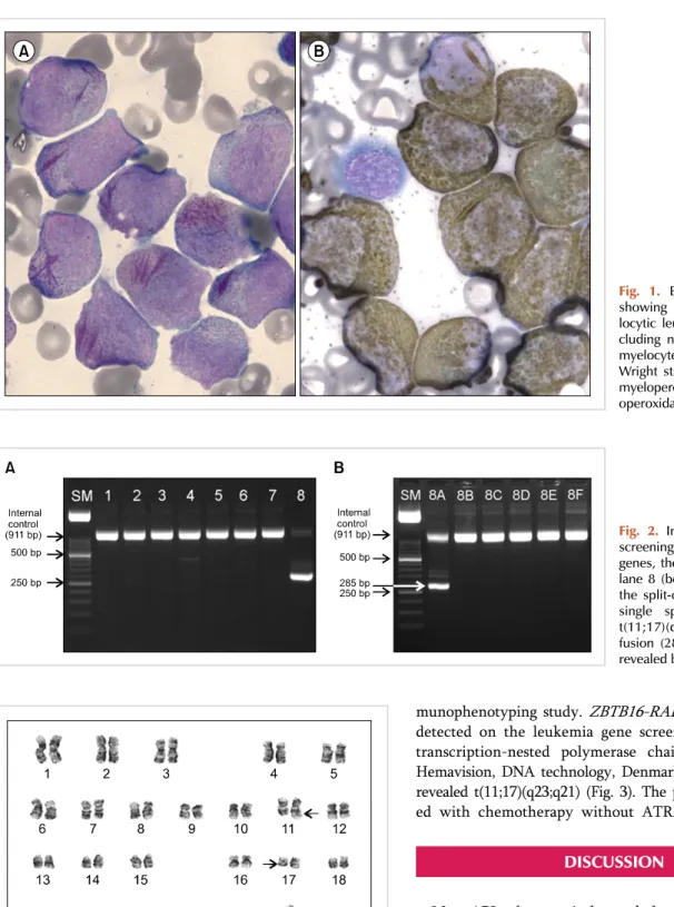
Fig. 1. Bone marrow aspirates showing typical acute promye- locytic leukemia morphology, in- cluding numerous leukemic pro- myelocytes with faggot cells (A) Wright stain; ×1,000 and strong myeloperoxidase activity (B) myel- operoxidase stain; ×1,000).
Fig. 2. In the first PCR (A) for screening of leukemia-associated genes, there is a positive band in lane 8 (between 250-300 bp). In the split-out reaction (B) using a single specific primer pair, a t(11;17)(q23;q21); ZBTB16-RARA fusion (285 bp) is confirmed, as revealed by the bands in lane 8A.
Fig. 3. The karyotype of the patient was 46,XY,t(11;17)(q23;q21)[17]/
46,XY[3].
granules. The leukemic cells showed multiple Auer rods and frequently showed faggot cells (Fig. 1A). They were strongly positive to MPO stain (Fig. 1B). The leukemic cells expressed CD13, CD33, CD117, human leukocyte antigen (HLA)-DR, and cytoplasmic-MPO, as revealed by the im-
munophenotyping study. ZBTB16-RARA translocation was detected on the leukemia gene screening test by reverse transcription-nested polymerase chain reaction (Fig. 2;
Hemavision, DNA technology, Denmark). Cytogenetic study revealed t(11;17)(q23;q21) (Fig. 3). The patient is being treat- ed with chemotherapy without ATRA.
DISCUSSION
Most APLs show typical morphology, including irregular and variable nuclei that are often kidney-shaped or bilobed;
cytoplasm containing densely packed or even coalescent large granules that stain bright pink, red, or purple in Romanowsky stains; and multiple Auer rods [1-4, 10]. Therefore, APL can be easily diagnosed at the time of morphologic evaluation.
However, variant APL t(11;17)(q23;q21) has been reported to have atypical APL morphology, and Auer rods are seldom observed [9, 10]. Absence of Auer rods or faggot cells is especially considered as a characteristic morphologic feature of this subtype [9, 10]. In variant APLs, including variant RARA translocation, the diagnosis can be made by chromo-
Korean J Hematol 2010;45:133-5.
A variant APL with t(11;17) (q23;q12) 135
some analysis or molecular study [11-13]. The results may show the typical karyotype t(15;17)(q22;q12); PML-RARA or the variant RARA translocation [11-13]. However, in our case, the typical morphology of classical APL, including multiple Auer rods in numerous leukemic cells and frequent faggot cells, was observed, and the karyotype and molecular studies showed t(11;17)(q23;q12) and ZBTB16-RARA fusion.
At the time of morphologic evaluation, our case showed findings consistent with those of APL with t(15;17)(q22;q12), before chromosome analysis and molecular study were performed. The typical immunophenotype of APL has been reported to be distinctive unlike those in other acute myeloid leukemia (AML) cases. The leukemic cells in the former express myeloid antigens such as CD13 and CD33 but show weak or negative reaction to CD34 and HLA-DR [14]. Some of the microgranular variant APL cases express CD34 and/or HLA-DR [15]. However, variant APL with t(11;17)(q23;q12);
ZBTB16-RARA subgroup has been reported to show the same immunophenotype as that in classical APL [10].
Interestingly, in our case, the leukemic cells expressed HLA-DR in addition to CD13, CD33, and CD117, unlike other cases that have previously been reported [10]. The variant APL with t(11;17)(q23;q12); ZBTB16-RARA sub- group usually shows resistance to ATRA therapy [9].
Therefore, our patient is being treated with alkylating agents rather than the classical APL regimen that includes ATRA.
The present study emphasizes the importance of combining morphologic, immunophenotypic, cytogenetic, and molec- ular studies to distinguish variant APL cases from classical APL cases before initiating chemotherapy, regardless of whether the morphological study reveals findings consistent with those of classical APL.
REFERENCES
1. Stone RM, Mayer RJ. The unique aspects of acute promyelocytic leukemia. J Clin Oncol 1990;8:1913-21.
2. Barbui T, Finazzi G, Falanga A. The impact of all-trans-retinoic acid on the coagulopathy of acute promyelocytic leukemia. Blood 1998;91:3093-102.
3. Menell JS, Cesarman GM, Jacovina AT, McLaughlin MA, Lev EA, Hajjar KA. Annexin II and bleeding in acute promyelocytic leukemia. N Engl J Med 1999;340:994-1004.
4. Bennett JM, Catovsky D, Daniel MT, et al. Proposals for the classi-
fication of the acute leukaemias. French-American-British (FAB) co-operative group. Br J Haematol 1976;33:451-8.
5. Chen Z, Brand NJ, Chen A, et al. Fusion between a novel Krüppel-like zinc finger gene and the retinoic acid receptor-alpha locus due to a variant t(11;17) translocation associated with acute promyelocytic leukaemia. EMBO J 1993;12:1161-7.
6. Corey SJ, Locker J, Oliveri DR, et al. A non-classical translocation involving 17q12 (retinoic acid receptor alpha) in acute promyelo- cytic leukemia (APML) with atypical features. Leukemia 1994;
8:1350-3.
7. Hummel JL, Wells RA, Dubé ID, Licht JD, Kamel-Reid S. Deregu- lation of NPM and PLZF in a variant t(5;17) case of acute promye- locytic leukemia. Oncogene 1999;18:633-41.
8. Wells RA, Hummel JL, De Koven A, et al. A new variant trans- location in acute promyelocytic leukaemia: molecular character- ization and clinical correlation. Leukemia 1996;10:735-40.
9. Licht JD, Chomienne C, Goy A, et al. Clinical and molecular char- acterization of a rare syndrome of acute promyelocytic leukemia associated with translocation (11;17). Blood 1995;85:1083-94.
10. Sainty D, Liso V, Cantù-Rajnoldi A, et al. A new morphologic clas- sification system for acute promyelocytic leukemia distinguishes cases with underlying PLZF/RARA gene rearrangements. group français de cytogénétique hématologique, UK cancer cytogenetics group and BIOMED 1 European coomunity-concerted acion
"molecular cytogenetic diagnosis in haematological malignancies.
Blood 2000;96:1287-96.
11. McKenna RW, Parkin J, Bloomfield CD, Sundberg RD, Brunning RD. Acute promyelocytic leukaemia: a study of 39 cases with iden- tification of a hyperbasophilic microgranular variant. Br J Haematol 1982;50:201-14.
12. Aventin A, Mateu R, Martino R, Colomer D, Bordes R. A case of cryptic acute promyelocytic leukemia. Leukemia 1998;12:1490- 506.
13. Neame PB, Soamboonsrup P, Leber B, et al. Morphology of acute promyelocytic leukemia with cytogenetic or molecular evidence for the diagnosis: characterization of additional microgranular variants. Am J Hematol 1997;56:131-42.
14. Paietta E, Andersen J, Gallagher R, et al. The immunophenotype of acute promyelocytic leukemia (APL): an ECOG study.
Leukemia 1994;8:1108-12.
15. Guglielmi C, Martelli MP, Diverio D, et al. Immunophenotype of adult and childhood acute promyelocytic leukaemia: correlation with morphology, type of PML gene breakpoint and clinical outcome. A cooperative Italian study on 196 cases. Br J Haematol 1998;102:1035-41.