https://doi.org/10.5624/isd.2018.48.4.261
Introduction
In paediatric dentistry, panoramic radiography is consid- ered an important diagnostic tool for monitoring the devel- opment of dentition and detecting caries, trauma, and oral anomalies.1 Panoramic imaging does not require placement of an intra-oral film; hence, better cooperation and toler- ance is expected from children.2
Modern advances in dental imaging have led to the de- velopment of various digital panoramic imaging techniques for minimizing the radiation dose and improving the qual- ity of radiographs.3 Compared to conventional radiograph- ic techniques, digital panoramic images have a number of advantages, including reduced radiation exposure, faster acquisition and processing time, the need for less storage space, and minimal environmental contamination.4 At the same time, the literature has suggested contradictory evi- dence related to image quality in conventional versus dig- itized radiographs.5
In digital panoramic radiography, the reduced radiation dose compromises the quality of the image, thereby affect- ing its diagnostic accuracy.6 For that reason, various tech-
Image quality assessment of pre-processed and post-processed digital panoramic radiographs in paediatric patients with mixed dentition
Isti Rahayu Suryani 1,*, Natalia Salvo Villegas 2, Sohaib Shujaat 3, Annelore De Grauwe 3, Azhari Azhari 4, Suhardjo Sitam 4, Reinhilde Jacobs 3,5
1Department of Dentomaxillofacial Radiology, Universitas Gadjah Mada, Yogyakarta, Indonesia
2Department of Oral and Maxillofacial Radiology, Universidad de Los Andes, Santiago, Chile
3OMFS IMPATH Research Group, Department of Imaging and Pathology, Faculty of Medicine, KU Leuven and Oral and Maxillofacial Surgery, University Hospitals Leuven, Leuven, Belgium
4Department of Dental Radiology, University of Padjajaran, Bandung, Indonesia
5Division of Oral Diagnostics and Rehabilitation, Department of Dental Medicine, Karolinska Institutet, Stockholm, Sweden
ABSTRACT
Purpose: To determine the impact of an image processing technique on diagnostic accuracy of digital panoramic radiographs for the assessment of anatomical structures in paediatric patients with mixed dentition.
Materials and Methods: The study consisted of 50 digital panoramic radiographs of children aged from 6 to 12 years, which were later on processed using a dedicated image processing method. A modified clinical image quality evaluation chart was used to evaluate the diagnostic accuracy of anatomical structures in maxillary and mandibular anterior and maxillary premolar region of processed images.
Results: A statistically significant difference was observed between pre and post-processed evaluation of anatomical structures(P<0.05) in the maxillary and mandibular anterior region. The anterior region was found to be more accu- rate in post-processed images. No significant difference was observed in the maxillary premolar region(P>0.05).
The Inter-observer and intra-observer reliability of both pre and post processed images were excellent(>0.82) for anterior region and good(>0.63) for premolar region.
Conclusion: The application of image processing technique in digital panoramic radiography can be considered a reliable method for improving the quality of anatomical structures in paediatric patients with mixed dentition.
(Imaging Sci Dent 2018; 48: 261-8)
KEY WORDS: Radiography; Dentition, Mixed; Diagnosis
Copyright ⓒ 2018 by Korean Academy of Oral and Maxillofacial Radiology
This is an Open Access article distributed under the terms of the Creative Commons Attribution Non-Commercial License(http://creativecommons.org/licenses/by-nc/3.0) which permits unrestricted non-commercial use, distribution, and reproduction in any medium, provided the original work is properly cited.
Imaging Science in Dentistry·pISSN 2233-7822 eISSN 2233-7830
*The research received funding from AREAS(Academic Relations between Europe and Asia)+Erasmus Mundus Action II.
Received July 13, 2018; Revised August 31, 2018; Accepted September 23, 2018
*Correspondence to : Dr. Isti Rahayu Suryani
Department of Dentomaxillofacial Radiology, Faculty of Dentistry, Universitas Gadjah Mada, Jln. Denta, Sekip Utara, Caturtunggal, Kec. Depok, Kabupaten Sleman, Daerah Istimewa, Yogyakarta 55281, Indonesia
Tel) 6281392709191, Fax) 62274515307, E-mail) [email protected]
niques have been developed for enhancing the image qual- ity without exposing the patient to additional radiation.7 A digital radiograph consists of anatomical structures with varying textures and intensity; as a result, these processing methods tend to enhance a single specific anatomical fea- ture, while obscuring the visibility of other structures.8
Only a few recent studies have investigated whether image enhancement and processing techniques improved the overall image quality of complete panoramic radiogra- phs, instead of specific anatomical structures.9-11 The aim of the present study was to determine whether the applica- tion of a dedicated image processing technique to digital panoramic radiographs improved the subjective accuracy of anatomical structures in children with mixed dentition.
Materials and Methods
The study was carried out in compliance with the World Medical Association Declaration of Helsinki on medical
research. Following approval from the local ethical com- mittee(Approval no.: S57587), 50 panoramic radiographs were retrospectively collected from the Dentomaxillofa- cial Imaging Centre. The inclusion criteria were paediatric patients(6-12 years) with mixed dentition, for whom pan- oramic radiographs with good image quality were avail- able and showed no craniofacial abnormalities. Patient- specific data were kept anonymous.
Image acquisition
Radiographic images were acquired using the VistaPano (Dürr Dental AG, Bietigheim-Bissingen, Germany) pano- ramic radiography device, operating at 70kVp, 8-12mA, and an exposure time of 13.5s. All pre-processed images were saved for further analysis without applying any tech- nique for image quality enhancement.
Image processing
Pre-processed panoramic radiographs were processed
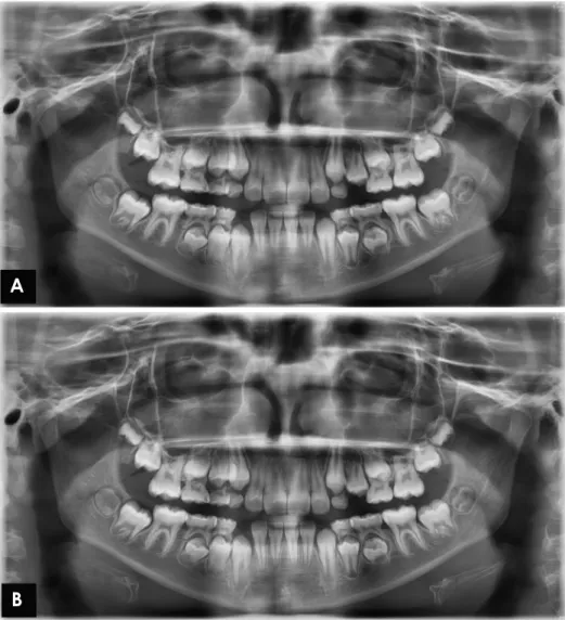
Fig. 1. Digital panoramic image. A.
Pre-processed image. B. Post-pro- cessed image.
A
B
using the Smart Panoramic(S-Pan) technology, which ac- companied the radiographic device. This technique enabled capturing multiple parallel layers of the radiograph from a single exposure. Each layer was then split into fragments automatically by the algorithm of the S-Pan technology, allowing it to reconstruct the image by selecting the most focussed and sharpest fragments. Thereafter, all the frag- ments that showed clear anatomical features were auto-re- compiled into a single panoramic image.
All pre- and post-processed data were saved as TIFF files for subsequent evaluation and comparison(Fig. 1).
Anatomical region selection
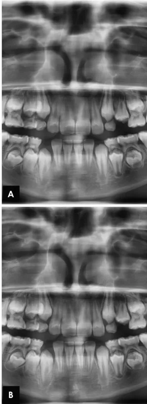
Similar anatomical structures were selected in both the pre- and post-processed images for assessment, including the dentinoenamel junction(DEJ), periodontal ligament (PDL) space, lamina dura, shape of the crown and roots of deciduous and permanent teeth, alveolar crest, and tra- becular pattern. All selected structures were identified for evaluation within the maxillary and mandibular anterior and maxillary premolar regions(Fig. 2).
Image quality assessment
A modified version of the clinical image quality evalua- tion chart proposed by Choi et al. was used to evaluate the subjective radiographic image quality(Table 1).12 All the anatomical structures were scored from 1 to 5(where 1=
not visible and 5=clearly visible in all regions). All pre and post-processed panoramic radiographs were assessed on a Dell computer monitor(Dell P2312H, Dell Inc., Texas, USA) in a room with dimmed light at a viewing distance of 60cm.
Reliability assessment
All images were assessed by 2 maxillofacial radiologists separately and blindly. Each image was evaluated twice by both observers(i.e., before and after processing) to deter- mine the inter-observer reliability. For intra-observer reli- ability, each pre-processed and post-processed image was assessed by both observers at an interval of 2 weeks.
Statistical methods
The data were analysed using SPSS version 22.0(IBM Corp., Armonk, NY, USA). The paired t-test was used to analyze differences in the observations of the anatomical structures between the pre- and post-processed radiographs, as well as the inter-and intra-observer variance. A P value of less than .05 was considered to indicate statistical sig- nificance. The intra and inter-observer agreement for ana-
tomical structures was estimated using the kappa test, with the results classified as follows: poor, kappa <0.20; fair, kappa =0:21-0:40; moderate, kappa=0.41-0.60; good, kappa=0.61-0.80; and excellent, kappa=0.81-1.00.13
Fig. 2. Subjective evaluation of the maxillary and mandibular an- terior maxillary premolar region. A. Pre-processed image. B. Post- processed image using the S-Pan technology.
A
B
Results
Table 2 and Figure 3 illustrate the subjective image qual- ity assessment scores of the pre- and post-processed images
in the maxillary and mandibular anterior region. The ante- rior region showed statistically significant differences for all anatomical structures between the pre- and post-pro- cessed images(P<.05). The score obtained from the pro-
Table 1. A modified clinical image quality evaluation scoring chart
Assessment Description Image score
DEJ in primary teeth
Clearly visible in all regions 5
Clearly visible for at least 1 tooth in one region and all teeth in the other region 4
Clearly visible for all teeth in one region 3
Clearly visible for 1 tooth in 1 region 2
Not visible for teeth in all regions 1
DEJ in permanent teeth
Clearly visible in all regions 5
Clearly visible for at least 1 tooth in one region and all teeth in the other region 4
Clearly visible for all teeth in one region 3
Clearly visible for 1 tooth in 1 region 2
Not visible for teeth in all regions 1
PDL space and lamina dura in primary teeth
Clearly visible in all regions 5
Clearly visible for at least 1 tooth in one region and all teeth in the other region 4
Clearly visible for all teeth in one region 3
Clearly visible for 1 tooth in 1 region 2
Not visible for teeth in all regions 1
PDL space and lamina dura in permanent teeth
Clearly visible in all regions 5
Clearly visible for at least 1 tooth in one region and all teeth in the other region 4
Clearly visible for all teeth in one region 3
Clearly visible for 1 tooth in 1 region 2
Not visible for teeth in all regions 1
Shape of roots in primary teeth
Clearly visible in all regions 5
Clearly visible for at least 1 tooth in one region and all teeth in the other region 4
Clearly visible for all teeth in one region 3
Clearly visible for 1 tooth in 1 region 2
Not visible for teeth in all regions 1
Shape of roots in permanent teeth
Clearly visible in all regions 5
Clearly visible for at least 1 tooth in one region and all teeth in the other region 4
Clearly visible for all teeth in one region 3
Clearly visible for 1 tooth in 1 region 2
Not visible for teeth in all regions 1
Shape of the crown in permanent teeth
Clearly visible in all regions 5
Clearly visible for at least 1 tooth in one region and all teeth in the other region 4
Clearly visible for all teeth in one region 3
Clearly visible for 1 tooth in 1 region 2
Not visible for teeth in all region 1
Definition of the alveolar crest in alveolar bone
Clearly visible in all regions 5
Clearly visible for at least 1 tooth in one region and all teeth in the other region 4
Clearly visible for all teeth in one region 3
Clearly visible for 1 tooth in 1 region 2
Not visible for teeth in all regions 1
Definition of trabecular pattern in alveolar bone
Clearly visible in all regions 5
Clearly visible for at least 1 tooth in one region and all teeth in the other region 4
Clearly visible for all teeth in one region 3
Clearly visible for 1 tooth in 1 region 2
Not visible for teeth in all regions 1
DEJ: dentinoenamel junction, PDL: periodontal ligament
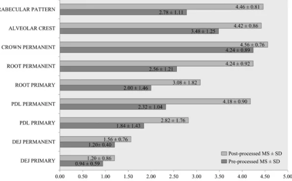
cessed images showed improved image quality compared to the original unprocessed data. The highest mean score difference was observed for the PDL space and lamina dura in permanent teeth(-1.86±1.01) and the lowest for the DEJ of the primary teeth(-0.26±0.60). The perma- nent tooth crown showed the highest post-processed score (4.56±0.76), whereas the DEJ of the primary teeth had the lowest score(1.20±0.86) based on the modified eval- uation chart.
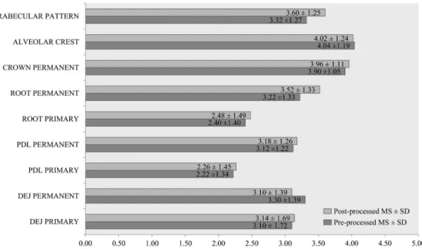
As shown in Table 3 and Figure 4, no significant differ- ences were observed between the pre- and post-processed anatomical structures in the maxillary premolar region(P>
.05), except for the root shape of permanent premolars (P=.01) and the trabecular pattern in alveolar bone(P=
.001). The root shapes of permanent teeth showed the high- est mean score difference(0.30±0.79), whereas the least difference was observed for the alveolar crest(0.02±0.92).
The DEJ of the permanent teeth had the lowest score(3.10
±1.39), whereas the alveolar crest showed the highest score(4.02±1.24) after being processed.
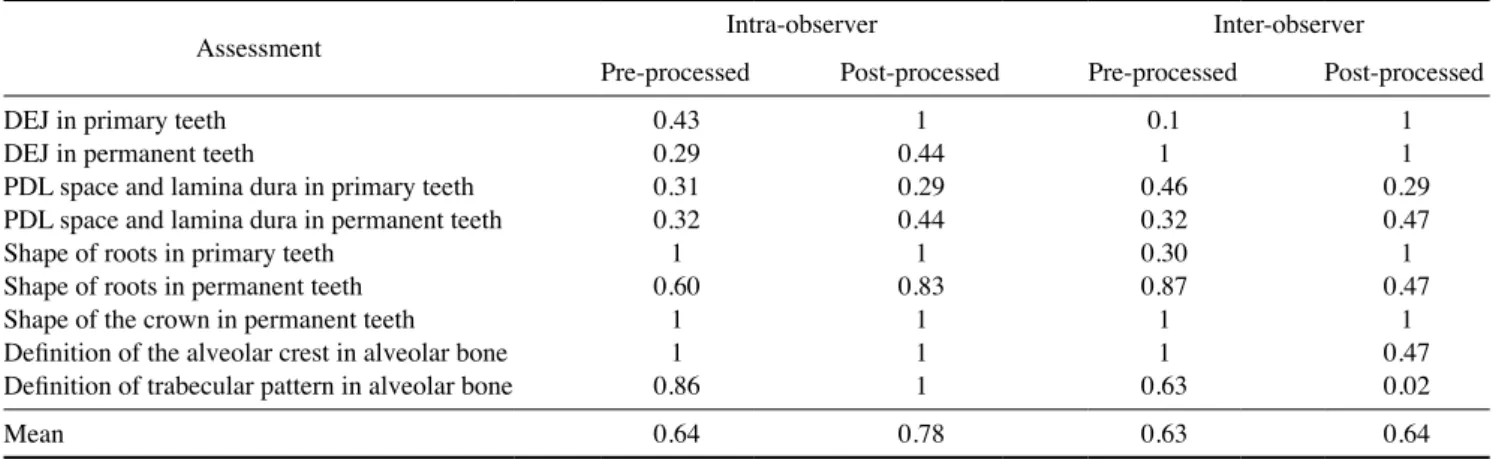
Table 4 and Table 5 present the Cohen kappa index for inter-observer and intra-observer reliability related to the pre- and post-processed images. The maxillary and mandib- ular anterior region showed excellent inter-observer(kap- pa≥0.82) and intra-observer≥0.86) agreement, whereas the premolar region showed good inter-observer(kappa≥ 0.63) and intra-observer agreement(kappa≥0.64) for both the pre- and post-processed images.
Table 2. Subjective image quality assessment of the maxillary and mandibular anterior regions
Assessment Score Significance
DEJ in primary teeth -0.26±0.60 P<0.05
DEJ in permanent teeth -0.36±0.60 P<0.05
PDL space and lamina dura in primary teeth -0.98±1.20 P<0.05
PDL space and lamina dura in permanent teeth -1.86±1.01 P<0.05
Shape of roots in primary teeth -1.08±1.31 P<0.05
Shape of roots in permanent teeth -1.68±1.12 P<0.05
Shape of the crown in permanent teeth -0.32±0.77 P<0.05
Definition of the alveolar crest in alveolar bone -0.94±1.04 P<0.05
Definition of trabecular pattern in alveolar bone -1.68±1.00 P<0.05
DEJ: dentinoenamel junction, PDL: periodontal ligament
Fig. 3. Mean score(MS) and standard deviation(SD) of image quality in the maxillary and mandibular anterior region. PDL: periodontal ligament, DEJ: dentinoenamel junction.
The post-processed images showed improved reliability when compared to the pre-processed data, but no signifi- cant differences were observed for inter-observer(anterior region, P=.93; premolar region, P=.97) or intra-observer reliability(anterior region, P=.86; premolar region, P=
.06).
Discussion
In modern dentistry, regular conventional panoramic de- vices are being replaced by digital technology in order to obtain high-quality images, while minimizing patients’
radiation exposure. At the same time, dose reduction direct- ly affects image quality, which in turn can lead to the inac- curate identification of anatomical structures.14 Other rea-
sons for diminished quality are related to noise and patient positioning during image acquisition.15 Image quality in paediatric patients with mixed dentition is of vital impor- tance for diagnosis and treatment planning.14 To the best of our knowledge, no evidence is available in the literature on the effects of processing techniques on image quality in paediatric patients. Therefore, this study was conducted to analyse the impact of a state-of-the-art image process- ing technique on image quality in paediatric patients with mixed dentition.
In our study, post-processed digital panoramic radiogra- phs allowed significantly better visualization of anatomi- cal structures than standard non-processed digital images.
This finding is in accordance with other studies that used processing techniques to improve image quality.16-18 In
Table 3. Subjective image quality assessment of maxillary premolar region
Assessment Score Significance
DEJ in primary teeth -0.04±0.64 .66
DEJ in permanent teeth 0.20±1.03 .18
PDL space and lamina dura in primary teeth -0.04±0.67 .67
PDL space and lamina dura in permanent teeth -0.06±0.89 .64
Shape of roots in primary teeth -0.08±0.85 .51
Shape of roots in permanent teeth -0.30±0.79 P<0.05
Shape of the crown in permanent teeth -0.06±0.68 .54
Definition of the alveolar crest in alveolar bone 0.02±0.92 .88
Definition of trabecular pattern in alveolar bone -0.28±0.54 P<0.05
DEJ: dentinoenamel junction, PDL: periodontal ligament
Fig. 4. Mean score(MS) and standard deviation(SD) of image quality in the maxillary premolar region. PDL: periodontal ligament, DEJ:
dentinoenamel junction.
contrast, according to Sabarudin and Tiau,4 no significant mean quality and scoring difference was observed between pre- and post-processed images related to the visualization of anatomical structures.
In comparison to conventional image processing techni- ques, which rely on filters, contrast, and brightness adjust- ments,19 our study involved a dedicated processing method.
The most focussed segments were automatically identified and processed in this technique to generate a sharp pano- ramic image. This technique allowed better visualization of the maxillary and mandibular anterior region without any blurring by ghost image artefacts from the spinal column, as shown by the significantly higher evaluation scores ob- tained using modified clinical image quality evaluation charts for the processed images in the anterior region. At the same time, the technique provided overall good image quality, allowing proper visualization in the premolar re- gion, in contrast to the pre-processed images. Improve- ments in inter-observer and intra-observer reliability were
also observed after image processing.
In this study, only the anatomical structures in the maxil- lary and mandibular anterior and maxillary premolar region in patients with mixed dentition were selected for evalua- tion. This choice was made because structures in those re- gions are more prone to overlap and blurriness, which can compromise the diagnosis and treatment planning.20-22
A blurred, shortened and narrowed appearance of the front teeth is a common error that occurs when the patient’s head is positioned in front of the focus.23 As a result, stud- ies performed in the mandibular premolar and posterior region showed minimal to non-significant distortion.24,25
To compensate for these limitations, clinicians sometimes take additional intra-oral radiographs, which leads to more radiation exposure and is time-consuming. Therefore, we recommend the application of an image processing tech- nique to improve image quality, instead of relying on sup- plementary radiographic investigations, especially in pae- diatric patients who are more vulnerable to radiation expo-
Table 4. Intra-observer and inter-observer reliability for the maxillary and mandibular anterior regions
Assessment Intra-observer Inter-observer
Pre-processed Post-processed Pre-processed Post-processed
DEJ in primary teeth 1 0.39 1 0.08
DEJ in permanent teeth 1 1 1 1
PDL space and lamina dura in primary teeth 0.71 1 0.40 1
PDL space and lamina dura in permanent teeth 0.74 1 0.63 1
Shape of roots in primary teeth 1 1 1 0.50
Shape of roots in permanent teeth 0.82 0.83 0.83 1
Shape of the crown in permanent teeth 1 1 1 1
Definition of the alveolar crest in alveolar bone 0.54 0.74 0.59 1
Definition of trabecular pattern in alveolar bone 1 1 1 1
Mean 0.87 0.89 0.83 0.84
DEJ: dentinoenamel junction, PDL: periodontal ligament
Table 5. Intra-observer and inter-observer reliability for the maxillary premolar region
Assessment Intra-observer Inter-observer
Pre-processed Post-processed Pre-processed Post-processed
DEJ in primary teeth 0.43 1 0.1 1
DEJ in permanent teeth 0.29 0.44 1 1
PDL space and lamina dura in primary teeth 0.31 0.29 0.46 0.29
PDL space and lamina dura in permanent teeth 0.32 0.44 0.32 0.47
Shape of roots in primary teeth 1 1 0.30 1
Shape of roots in permanent teeth 0.60 0.83 0.87 0.47
Shape of the crown in permanent teeth 1 1 1 1
Definition of the alveolar crest in alveolar bone 1 1 1 0.47
Definition of trabecular pattern in alveolar bone 0.86 1 0.63 0.02
Mean 0.64 0.78 0.63 0.64
DEJ: dentinoenamel junction, PDL: periodontal ligament
sure.
In conclusion, the presented image processing technique can be regarded as a useful tool for improving the image quality of panoramic radiographs in paediatric patients with mixed dentition. Further investigations are required to study the application of the proposed technique for image quali- ty in patients with dentofacial abnormalities.
References
1. Clark HC, Curzon ME. A prospective comparison between find- ings from a clinical examination and results of bitewing and panoramic radiographs for dental caries diagnosis in children.
Eur J Paediatr Dent 2004; 5: 203-9.
2. Abdinian M, Razavi SM, Faghihian R, Samety AA, Faghihian E. Accuracy of digital bitewing radiography versus different views of digital panoramic radiography for detection of proxi- mal caries. J Dent(Tehran) 2015; 12: 290-7.
3. Barot AA, Chaturvedi MK, Butala PB, Rao VV, Patel PS, Barot AA. A study on changes in image quality with dose reduction in digital panoramic radiographs. J Int Oral Health 2017; 9: 174-9.
4. Sabarudin A, Tiau YJ. Image quality assessment in panoramic dental radiography: a comparative study between conventional and digital systems. Quant Imaging Med Surg 2013; 3: 43-8.
5. Peker I, Toraman AM, Usalan G, Altunkaynak B. The compar- ison of subjective image quality in conventional and digital panoramic radiography. Indian J Dent Res 2009; 20: 21-5.
6. Parissis N, Angelopoulos C, Mantegari S, Karamanis S, Masood F, Tsirlis A. A comparison of panoramic image quality between a digital radiography storage phosphor system and a film-based system. J Contemp Dent Pract 2010; 11: E009-16.
7. Amiri SA, Moudi E. Image quality enhancement in digital pan- oramic radiograph. J AI Data Min 2014; 2: 1-6.
8. Kandan RS, John A, Kumar S. An improved contrast enhance- ment approach for panoramic dental x-ray images. ARPN J Eng App Sci 2015; 10: 1897-901.
9. Svenson B, Larsson L, Båth M. Optimization of exposure in panoramic radiography while maintaining image quality using adaptive filtering. Acta Odontol Scand 2016; 74: 229-35.
10. Baksi BG, Alpöz E, Sogur E, Mert A. Perception of anatomical structures in digitally filtered and conventional panoramic radio- graphs: a clinical evaluation. Dentomaxillofac Radiol 2010; 39:
424-30.
11. Gijbels F, De Meyer AM, Bou Serhal C, Van den Bossche C, Declerck J, Persoons M, et al. The subjective image quality of direct digital and conventional panoramic radiography. Clin Oral Investig 2000; 4: 162-7.
12. Choi BR, Choi DH, Huh KH, Yi WJ, Heo MS, Choi SC, et al.
Clinical image quality evaluation for panoramic radiography in Korean dental clinics. Imaging Sci Dent 2012; 42: 183-90.
13. Altman DG, Machin D, Bryant TN, Gardner MJ. Statistics with confidence. 2nd ed. London: BMJ Books; 2003.
14. Angelopoulos C, Bedard A, Katz JO, Karamanis S, Parissis N.
Digital panoramic radiography: an overview. Semin Orthod 2004; 10: 194-203.
15. Ahmad SA, Taib MN, Khalid NE, Taib H. Correlation between quantitative and qualitative analysis on image quality of digi- tal dental X-ray images. J Comput Sci Comput Math 2012; 2:
43-51.
16. Yalcinkaya S, Künzel A, Willers R, Thoms M, Becker J. Sub- jective image quality of digitally filtered radiographs acquired by the Dürr Vistascan system compared with conventional ra- diographs. Oral Surg Oral Med Oral Pathol Oral Radiol Endod 2006; 101: 643-51.
17. Kaeppler G, Axmann-Krcmar D, Reuter I, Meyle J, Gómez- Román G. A clinical evaluation of some factors affecting image quality in panoramic radiography. Dentomaxillofac Radiol 2000; 29: 81-4.
18. Lehmann TM, Troeltsch E, Spitzer K. Image processing and en- hancement provided by commercial dental software programs.
Dentomaxillofac Radiol 2002; 31: 264-72.
19. Batista SS, Panzarella FK, Tavano O, Filho AM, Junqueira JL.
Image adjustments on digital panoramic radiographs using Adobe Photoshop CS3 software. Rev Sul-Bras Odontol 2013;
10: 394-401.
20. Gijbels F, Sanderink G, Pauwels H, Jacobs R. Subjective image quality of digital panoramic radiographs displayed on monitor and printed on various hardcopy media. Clin Oral Investig 2004; 8: 25-9.
21. Bekiroglu N, Mete S, Ozbay G, Yalcinkaya S, Kargul B. Evalu- ation of panoramic radiographs taken from 1,056 Turkish chil- dren. Niger J Clin Pract 2015; 18: 8-12.
22. Temmerman A, Hertelé S, Teughels W, Dekeyser C, Jacobs R, Quirynen M. Are panoramic images reliable in planning sinus augmentation procedures? Clin Oral Implants Res 2011; 22:
189-94.
23. Rushton VE, Horner K, Worthington HV. The quality of pano- ramic radiographs in a sample of general dental practices. Br Dent J 1999; 186: 630-3.
24. Kayal RA. Distortion of digital panoramic radiographs used for implant site assessment. J Orthod Sci 2016; 5: 117-20.
25. Vazquez L, Nizamaldin Y, Combescure C, Nedir R, Bischof M, Dohan Ehrenfest DM, et al. Accuracy of vertical height mea- surements on direct digital panoramic radiographs using poste- rior mandibular implants and metal balls as reference objects.
Dentomaxillofac Radiol 2013; 42: 20110429.