172 Copyright © 2015 Korean Dementia Association
INTRODUCTION
Hashimoto’s encephalopathy (HE) is a rare neurological disorder with a heterogeneous group of neurological symp- toms.1 Sporadic Creutzfeldt-Jakob disease (CJD) is the most common human prion disease and usually manifests as a rap- idly progressive dementia, with ataxia and myoclonus lead- ing to death in less than 1 year.2 CJD shares common clinical features with HE, and dementia, myoclonus, extrapyramidal signs, ataxia, psychotic phenomena and personality changes characterize both diseases.3 The presence of 14-3-3 protein in
the cerebrospinal fluid (CSF) is a useful marker for in vivo di- agnosis of CJD. 14-3-3 protein is a relatively sensitive and spe- cific marker of CJD and is not commonly detected in HE. We report the case of a patient with HE with unusual features, in- cluding positive 14-3-3 protein in the CSF, and an atypical course mimicking that of CJD.
CASE REPORT
A 64-year-old male visited the hospital with progressively deteriorating cognitive function and abnormal behavior for 3 days. There were no specific findings in disease, drug, social, or family history. The patient complained of systemic fatigue and showed a dull demeanor and more-sluggish activity than usual. The patient was lucid, but showed impaired orienta-
Hashimoto’s Encephalopathy with Positive 14-3-3 Protein in the Cerebrospinal Fluid and Atypical Course
Mimicking Creutzfeldt-Jakob Disease
Jee-Eun Yoon, Ji Sun Kim, Kyung-Bok Lee, Hakjae Roh, Moo-young Ahn
Department of Neurology, Soonchunhyang University Seoul Hospital, Soonchunhyang University School of Medicine, Seoul, Korea
Background Creutzfeldt-Jakob disease (CJD) shares common clinical features with Hashimoto’s encephalopathy (HE). The 14-3-3 protein is a relatively sensitive and specific marker of CJD but is not commonly detected in HE. We report the case of a patient with HE with unusual features including positive 14-3-3 protein in the cerebrospinal fluid (CSF) and an atypical course mimicking that of CJD.
Case Report A 64-year-old male was admitted due to acute-onset cognitive dysfunction. HE was suspected based on increased titers of anti-thyroid microsomal antibody and an excellent response to steroid. However, 14-3-3 protein was detected in the CSF and a recurrent at- tack with progressive cognitive decline, pyramidal symptoms and myoclonus mimicking CJD occurred. Cognitive dysfunction showed pro- gressive worsening and the response to steroid treatment was decreased.
Conclusions 14-3-3 protein could be considered a general marker of neuronal destruction and not specific to CJD. The clinical manifesta- tions of HE are variable and its diagnosis is difficult due to the lack of a specific phenotype and reliable diagnostic criteria. We recommend that patients with clinical features of CJD and antithyroid antibodies should be considered for empirical steroid treatment for HE, despite a positive result for 14-3-3 protein.
Key Words Creutzfeldt-Jakob disease, Hashimoto’s encephalopathy, 14-3-3 protein.
Received: October 21, 2015 Revised: November 29, 2015 Accepted: November 29, 2015
Correspondence: Ji Sun Kim, MD, Department of Neurology, Soonchunhyang University Seoul Hospital, Soonchunhyang University School of Medicine, 59 Daesagwan-ro, Yongsan-gu, Seoul 04401, Korea
Tel: +82-2-709-9224, Fax: +82-2-795-3687, E-mail: [email protected]
cc This is an Open Access article distributed under the terms of the Cre- ative Commons Attribution Non-Commercial License (http://creative- commons.org/licenses/by-nc/3.0) which permits unrestricted non-com- mercial use, distribution, and reproduction in any medium, provided the ori- ginal work is properly cited.
DND
Print ISSN 1738-1495 / On-line ISSN 2384-0757
Dement Neurocogn Disord 2015;14(4):172-175 / http://dx.doi.org/10.12779/dnd.2015.14.4.172
CASE REPORT
www.dnd.or.kr 173
DND
tion regarding time, place, and person. He gave inappropriate answers to most questions and was able to perform tasks only after several repetitions of simple instructions. There was no apparent decrease in muscle power, and all sensory functions and the deep tendon reflex were normal, with no pathological reflexes. The cerebellar function test was normal, but frontal re- leasing signs, such as the glabellar reflex and palmomental reflex, were positive, and ideomotor apraxia of both hands was ob- served. The Korean Mini-Mental Status Examination (MMSE) score was decreased to 16 points, and all domains of the Seoul Neuropsychological Screening Battery showed deterioration.
The results of complete blood count, blood chemistry, syphi- lis reaction test, and vitamin B12 test were within normal limits, and levels of viral markers and autoimmune antibod- ies (RF, ANA, ANCA, C3, C4, anti-mitochondrial Ab, anti- dsDNA Ab, anti-Ro Ab, anti-La Ab, etc.) were within normal limits or negative. Paraneoplastic syndrome selection tests, including serum anti-Hu, anti-Yo, and anti-Ri, were normal.
In the thyroid function test, TSH was 1.00 μIU/m and free T4 was 1.04 ng/dL, both normal values, but T3 was decreased at 55.66 ng/dL. Anti-thyroid microsomal Ab and anti-thyroglob- ulin Ab levels were increased at 0.33 U/mL and 0.70 U/mL, re- spectively. The CSF test showed a normal intracranial pres- sure of 11 cm H2O, and a normal red blood cell (RBC) count of 2/μL, but the white blood cell (WBC) count was increased
to 54/μL (85% lymphocytes), and the protein level was in- creased at 64.5 mg/dL. Antibodies for anti-Hu, Yo, Ri, MA2, CV2/CRMP5, amphiphysin, recoverin, SOX1, titin, NMSAR, AMPA1, AMPA2, LGI1, CASPR2, and GABA-B in the CSF were negative, and 14-3-3 protein was weakly positive in the Western blot. High signal intensities were observed in the bi- lateral basal ganglia, the thalamus, and multifocal areas in the cerebral hemispheres in T2-weighted image (T2WI) and flu- id-attenuated inversion recovery (FLAIR) image on brain magnetic resonance imagining (MRI) (Fig. 1A). The brain electroencephalogram (EEG) showed mostly slow waves with no epileptiform spikes (Fig. 2A). After admission, the patient showed progressive worsening of abnormal behavior, and al- though viral antibodies were not found in the CSF, because the patient showed an increase in WBC count with lympho- cyte predominance, he was treated with acyclovir 1980 mg/
day intravenous due to a diagnosis of idiopathic viral en- cephalitis, but showed no significant clinical improvement.
Although TSH was normal, T3 was slightly decreased and an- ti-thyroid microsomal Ab and anti-thyroglobulin Ab levels were increased in the TFT. Under the suspicion of HE caused by Hashimoto’s thyroiditis, which is the most common cause of asymptomatic hypothyroidism, the patient was given high- dose steroids (methylprednisolone 1000 mg/day) for 5 days and was maintained with oral prednisolone. He showed a
Fig. 1. T2-weighted MRI on the first admission day showed high-signal-intense lesions in the basal ganglia, the thalamus and bilateral cere- bral white matter (A). Follow-up MR images 8 months after the initial study revealed an increased extent of high-signal-intense lesions in the basal ganglia, the thalamus and multifocal cerebral hemisphere, and progression of diffuse cerebral atrophy (B).
A
B
Jee-Eun Yoon et al.
Hashimoto Encephalopathy with 14-3-3 Mimicking CJD
174 Dement Neurocogn Disord 2015;14(4):172-175
clear improvement in cognitive function after initiation of treatment. The patient received a score of 22 in the MMSE performed 10 days after initiating steroid treatment, which is an improvement compared to the previous score, and partic- ular improvement in the areas of orientation and memory retrieval was evident. The patient was discharged from the hospital after stopping oral steroid treatment, but the disori- entation and abnormal behavior symptoms slowly began to recur after 2 months, and he was readmitted. The CSF test results showed an intracranial pressure of 14 cm H2O and RBC count of 2/μL, which is normal, but the WBC count was 48/μL (72% lymphocytes) and the protein level was in- creased at 54 mg/dL, and 14-3-3 protein was positive in West- ern blot. TSH was 1.23 μIU/m and free T4 was normal at 1.08 ng/dL, but T3 was decreased at 38.91 ng/dL, and although the anti-thyroid microsomal Ab level was normal at 0.18 U/mL, the anti-thyroglobulin Ab level was increased to 0.47 U/mL.
With the symptom recurring, high-dose steroids (methyl- prednisolone 1000 mg/day) were given intravenously for 5 days. Symptoms improved accordingly, and the patient was discharged. However, 2 weeks later the patient revisited the hospital due to generalized seizures, and the neurological ex- amination showed left upper-limb weakness, bilateral upper limb myoclonus, opsoclonus, visual hallucinations and optic ataxia. He also showed a tendency to lean to both sides due to trunk impairment. The patient was unable to perform simple tasks due to severe cognitive impairment and apraxia and it was difficult to understand what the patient was saying due to dysarthria. He also could not form proper sentences. The CSF test showed an intracranial pressure of 12 cm H2O, a WBC count of 3/μL, and a protein concentration of 26 mg/
dL, which is normal. 14-3-3 protein was positive in the West- ern blot, but the tau protein level decreased from 2704.3 pg/
mL to 205.7 pg/mL, which is a finding inconsistent with typi- cal CJD. Bilateral frontoparietal cortical atrophy was observed
in the brain MRI, and the high signal intensity in the bilateral cerebral cortex, subcortical white matter, right thalamus, and midbrain in the FLAIR and T2WI was shown (Fig. 1B). Angi- ography performed to differentiate primary central nervous system (CNS) angiitis showed no significant stenosis or occlu- sion of the cranial arteries. Whole-body positron emission to- mography (PET) was performed to rule out paraneoplastic syndrome, but no abnormalities were observed. The brain EEG showed periodic lateralized epileptiform discharges across the entire right cranial hemisphere (Fig. 2B). Loraze- pam and phenytoin were injected intravenously to control the seizures and were maintained orally thereafter, but 7 days later the patient showed status epilepticus, which is a state in which multiple seizures occur with no recovery of consciousness, and was given levetiracetam intravenously, upon which no further seizures occurred. Slowly progressing dementia, myoclonal seizures, generalized seizures, optic ataxia, cerebellar symp- toms, visual hallucinations, and dysarthria suggested a diagno- sis of recurrent HE, and high-dose steroid (methylpredniso- lone 1000 mg/day) were readministered for 5 days, but symptoms did not improve. Disorientation, memory loss, and optic ataxia are still apparent at the time of writing, after 5 months.
DISCUSSION
Our case of HE showed progressive cognitive dysfunction with relapsing and remitting course and positive 14-3-3 pro- tein in CSF. The clinical manifestations of HE may mirror that of CJD, and the two diseases can be confused.4 HE can be dis- tinguished from CJD based on several features. First, a posi- tive response to steroid treatment is an important identifying characteristic of HE.5 Another distinguishing point is the pres- ence of seizures, which is common in HE but rare in CJD.6 Moreover, the presence of periodic spike wave discharges on
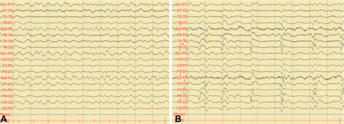
Fig. 2. Initial EEG showed generalized continuous slow activity (A). Follow-up EEG recorded 8 months later shows periodic lateralized epi- leptiform discharges over the right hemisphere (B). EEG: electroencephalogram.
A B
www.dnd.or.kr 175
DND
EEG2 and characteristic MRI findings of T2/FLAIR and dif- fusion weighted imaging (DWI) pattern in the basal ganglia7 are also helpful in distinguishing the two conditions. Our pa- tient showed a definite response to steroid pulse therapy, fre- quent seizure attacks, absence of periodic spike wave dis- charges on EEG and nonspecific diffuse white matter changes on MRI. However, 14-3-3 protein was positive in CSF. Al- though the presence of 14-3-3 protein is commonly associat- ed with CJD, there are two reports of HE cases with positive 14-3-3 protein.4,8 Elevated 14-3-3 protein levels have been also reported in patients with stroke, CNS infections, intracranial metastasis and metabolic encephalopathy.9 In view of these findings, 14-3-3 protein is considered to be a general marker of neuronal destruction and not a specific tool for the identi- fication of CJD.
Clinical subtypes of HE are proposed as a vasculitic type with relapsing and remitting course and another subtype with a progressive course of significant decline in cognitive func- tion. Our patient showed a relapsing and remitting course of cognitive function and an excellent initial response to steroid treatment. Over time, his cognitive dysfunction worsened, and new psychiatric symptoms developed. Furthermore, opsoclo- nus-myoclonus syndrome, which is a common feature of para- neoplastic limbic encephalitis (PLE), was observed and was not improved by steroid pulse therapy. MRI findings did not re- veal temporal lobe abnormalities and no primary malignancy was detected by whole-body PET. The clinical manifestations of HE are variable and its diagnosis is difficult due to the ab- sence of a specific phenotype pattern. A decreased response to steroid or opsoclonus-myoclonus resembling PLE is rarely re- ported in HE. The differential diagnosis is broad and includes CNS infection, autoimmune-related encephalopathy, neuro- degenerative disease and PLE. Likewise, other possible causes
should be ruled out before making a diagnosis of HE.
In conclusion, we recommend that patients with clinical features of CJD and antithyroid antibodies should be consid- ered for empirical steroid treatment for HE, despite being positive for 14-3-3 protein.
Conflicts of Interest
The authors have no financial conflicts of interest.
REFERENCES
1. Chong JY, Rowland LP, Utiger RD. Hashimoto encephalopathy: syn- drome or myth? Arch Neurol 2003;60:164-171.
2. Steinhoff BJ, Zerr I, Glatting M, Schulz-Schaeffer W, Poser S, Kretzschmar HA. Diagnostic value of periodic complexes in Creutzfeldt- Jakob disease. Ann Neurol 2004;56:702-708.
3. Utsumiya K, Arakawa R, Fujimoto S, Ueyama H, Kumamoto T. [A case of steroid-responsive encephalopathy with positive 14-3-3 pro- tein of the cerebrospinal fluid clinically resembling Creutzfelt-Jakob disease]. Rinsho Shinkeigaku 2004;44:618-622.
4. Vander T, Hallevy C, Alsaed I, Valdman S, Ifergane G, Wirguin I. 14- 3-3 protein in the CSF of a patient with Hashimoto’s encephalopathy.
J Neurol 2004;251:1273-1274.
5. Doherty CP, Schlossmacher M, Torres N, Bromfield E, Samuels MA, Folkerth R. Hashimoto’s encephalopathy mimicking Creutzfeldt-Ja- kob disease: brain biopsy findings. J Neurol Neurosurg Psychiatry 2002;73:601-602.
6. Seipelt M, Zerr I, Nau R, Mollenhauer B, Kropp S, Steinhoff BJ, et al. Hashimoto’s encephalitis as a differential diagnosis of Creutzfeldt- Jakob disease. J Neurol Neurosurg Psychiatry 1999;66:172-176.
7. Schröter A, Zerr I, Henkel K, Tschampa HJ, Finkenstaedt M, Poser S.
Magnetic resonance imaging in the clinical diagnosis of Creutzfeldt- Jakob disease. Arch Neurol 2000;57:1751-1757.
8. Hernández Echebarría LE, Saiz A, Graus F, Tejada J, García JM, Clavera B, et al. Detection of 14-3-3 protein in the CSF of a patient with Hashimoto’s encephalopathy. Neurology 2000;54:1539-1540.
9. Zerr I, Bodemer M, Gefeller O, Otto M, Poser S, Wiltfang J, et al.
Detection of 14-3-3 protein in the cerebrospinal fluid supports the di- agnosis of Creutzfeldt-Jakob disease. Ann Neurol 1998;43:32-40.