원위 대퇴골 관절내 골절에서 최소 침습적 잠김 금속판 고정술의 결과
김성현ㆍ윤성현ㆍ박희곤 ㆍ정재욱
단국대학교 의과대학 정형외과학교실
Results after Less Invasive Locking Plating in Intra-Articular Fractures of the Distal Femur
Sung Hyun Kim, M.D., Sung Hyun Yoon, M.D., Hee Gon Park, M.D., Ph.D. , Jae Uk Jung, M.D.
Department of Orthopedic Surgery, Dankook University College of Medicine, Cheonan, Korea
Received April 29, 2018 Revised (1st) June 15, 2018
(2nd) September 26, 2018 Accepted October 16, 2018 Correspondence to:
Hee Gon Park, M.D., Ph.D.
Department of Orthopedic Surgery, Dankook University Hospital, 201 Manghyang-ro, Dongnam-gu, Cheonan 31116, Korea Tel: +82-41-550-6290 Fax: +82-41-556-0551 E-mail: [email protected] Financial support: None.
Conflict of interests: None.
Purpose: The purpose of this study was to determine the clinical outcomes after a less invasive locking plating technique in intra-articular fractures of the distal femur.
Materials and Methods: This was a retrospective 19 case series of patients with distal femoral intra- articular fractures treated with a less invasive locking plating technique in a single center (Dankook University Hospital) from June 2010 to April 2016. Nineteen patients (11 males and 8 females) with a mean age of 55.9 years were enrolled. The functional outcomes were evaluated using the visual analogue scale (VAS), range of knee joint motion (flexion & extension), and Knee Society score. The radiology outcomes were evaluated with parameters measured in a plain radiograph (deviation angle of alignment axis on coronal and sagittal plane, mechanical lateral distal femur angle).
Results: The mean follow-up period was 26.4 months (range, 12-72 months) and the mean duration to union was 15.94 weeks (range, 11-28 weeks). The mean VAS was 1.36 (range, 0-8) and the range of motion of the knee joint was extension 4.73° (range, 0°-30°) and flexion 107.36° (range, 60°-135°).
The mean Knee Society score was 85.47 (range, 47-100). The mean deviation angle of the coronal alignment axis was 4.07° (range, 1.3°-8.8°), the mean deviation angle of the sagittal alignment axis was 3.23° (range, 0.7°-7.0°), and the mechanical lateral femoral angle was 87.75° (range, 82.8°-95.5°). Six patients had traumatic osteoarthritis at the final follow-up.
Conclusion: The purpose of this study was to evaluate the clinical and radiologic outcomes of intraar- ticular fractures of the distal femur in patients who underwent an anatomical reduction through an open reduction, and converted to an extra-articular fracture with rigid internal fixation. The results were relatively satisfactory.
Key Words: Distal femur, Complete articular fracture, Less invasive locking plating
Copyright © 2019 The Korean Fracture Society. All rights reserved.
This is an Open Access article distributed under the terms of the Creative Commons Attribution Non-Commercial License (http://creativecommons.org/licenses/by-nc/4.0) which permits unrestricted non-commercial use, distribution, and reproduction in any medium, provided the original work is properly cited.
서 론
원위 대퇴골 골절은 젊은 나이 환자에서 고 에너지의 외상 과 골다공증이 있는 고령 환자에서 저 에너지 외상으로 종종
발생한다. 원위 대퇴골 완전 관절내 골절의 치료는 어렵고 여 러 연구에서 부정유합, 이상회전, 슬관절 강직, 외상 후 골관 절염 등과 같은 합병증 및 후유증이 보고되었다.1-11) 또한 여 러 연구들1-6)에서 복잡한 AO/OTA 분류상 C3 유형의 원위
대퇴골 골절의 수술적 치료 이후 비교적 불만족스러운 결과 를 보고하였다.
원위 대퇴골의 관절내 골절의 수술적 치료로서 전통적으 로 관혈적 정복술 및 금속판, 나사 내고정술이 흔히 사용되었 고 슬관절 통증 및 관절 운동 범위 등 슬관절의 임상적 결과 를 개선시켜 주었지만 부정유합, 이상회전, 골결손, 감염과 같 은 합병증이 나타날 수 있다. 이러한 문제들 때문에 ‘간접적 정복술’, ‘최소 침습적 금속판 골유합술(minimally invasive plate osteosynthesis)’과 같은 술기들이 생물학적 보존 및 골 절의 개선을 위해 정립되었다.12) 생물학적 우수성 때문에 최 소 침습 금속판 골유합술은 최근에 자주 사용되며, 관혈적 정복술과 비교하여 감염 발생률이 적고, 불유합의 비율 또 한 적어 골유합이 되는 시간을 줄여줄 수 있다고 보고되고 있
다.13-17) 따라서 본 연구의 목적은 원위 대퇴골의 관절내 골절,
특히 분쇄골절에서 최소 침습적 잠김 금속판 고정술 후 임상 적, 영상학적 결과를 보고하고자 하였다.
대상 및 방법
1. 연구대상
의무기록을 분석하여 2010년 6월부터 2016년 4월까지 원 위 대퇴골 관절내 골절로 최소 침습적 잠김 금속판 고정술을 이용한 수술적 치료를 시행한 환자들을 후향적으로 조사하 였다. (1) 양측 원위 대퇴 골절, (2) 분절 대퇴골 골절, (3) 골절 된 하지의 동측에 경골 또는 족관절 부위 골절이 동반된 환 자, (4) 인공 고관절 또는 슬관절 술 후 상태인 환자, 또는 (5) 심각한 뇌 또는 다른 기관의 손상이 있는 환자들, (6) 원위 대 퇴골 개방성 골절 유형 2, 3 (7) 내고정술 이전에 외 고정기를
적용한 환자들 및 (8) 혈관 손상이 동반된 환자도 제외하였 다. 총 19예(남성 11예, 여성 8예) 환자들이 포함되었으며, 평 균 연령은 55.9세(32-79세)였다. 수상기전은 저 에너지의 외 상 환자가 4명이었고 나머지 15명은 고 에너지 외상이었다.
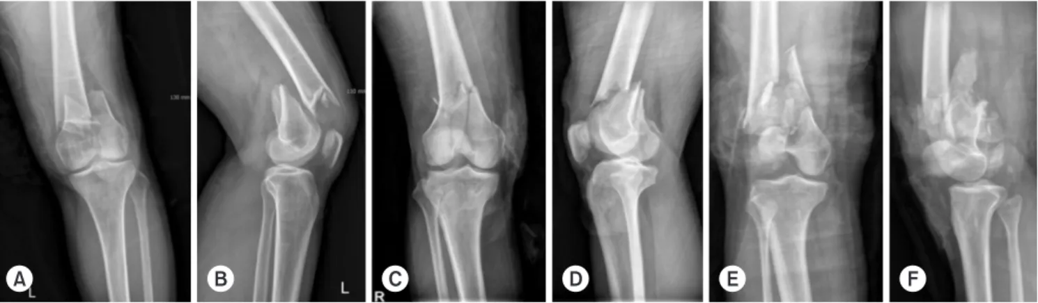
환자들은 AO/OTA 분류에 따라 나누었으며 C1, C2, 그리고 C3의 골절이 각각 2명, 4명, 13명이었다(Table 1, Fig. 1).
2. 수술 방법 및 수술 후 관리
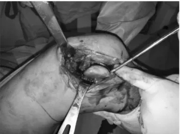
환자는 척추 마취하에 앙와위 자세로 취하고 근위 대퇴부 에 소독된 압박대를 적용하였다. 투시 촬영술 유도하에 원위 대퇴골의 골절 위치와 정렬축, 그리고 융기 간 절흔(intercon- dylar notch) 위치를 확인 후 표준형 외측 슬개 주위 도달법 (lateral parapatellar approach)으로 절개하여 원위 대퇴골의 관절 부위 및 골절 부위를 노출시켰다(Fig. 2). 관절 부위 골 절을 직접 시각화한 상태에서 관혈적으로 해부학적 정복을 시행하였고 골 재위치 겸자(bone reposition forcep)를 이용하 여 정복 유지하였다. 투시 촬영술 유도하에 골절의 정복 상태 및 대퇴골의 정렬축을 확인한 후 관절 부위 골절에 4.0 mm
Table 1. Demographic Data
Characteristic Value
Age (yr) 55.9 (32-79)
Sex (male:female) 11:8
Injury mechanism (low:high energy) 4:15 Fracture type (AO/OTA classification)
C1 2
C2 4
C3 13
Values are presented as median (range) or number only.
A B C D E F
Fig. 1. Simple radiographs show anterior-posterior and lateral view of distal femur fractures: AO/OTA classification. (A, B) type C1 fracture, (C, D) type C2 fracture, (E, F) type C3 fracture.
해면 골 나사(cancellous screw; Depuy Synthes, Warsaw, IN, USA) 또는 3.5 mm 피질 골 나사(cortical screw; Depuy Synthes)를 이용하여 내고정하거나 K-강선을 이용하여 금 속판을 고정할 위치를 피하여 임시적 고정을 했다. 이후 장 경 인대(iliotibial band)를 섬유의 방향으로 분할시키고 골막 분리기를 이용하여 원위 대퇴부에 근육 하부 공간을 형성했 다. 원위 대퇴골 윤곽 잠김 금속판(locking compression plate distal femur plate, LCP® system; Depuy Synthes)을 원위 대 퇴골 외측에서 골막 상부로 원위 부위에서 근위 부위 방향으 로 미끄러지게 삽입했다(Fig. 3). 투시 촬영술 유도하에 금속 판의 위치를 확인하였고 원위 부위부터 잠김 나사를 삽입하 여 고정하였다. 근위 나사 삽입 위치를 확인 후 외측으로 횡 절개(3-5 cm)를 가하였고 피질 및 잠김 나사를 이용하여 금 속판의 근위 부위를 고정하였다(Fig. 4, 5).
Fig. 2. Intraoperative photograph showing the lateral parapatella ap- proach for articular exposure and reconstruction.
Fig. 3. Following reduction, an appropriate-sized plate was slid in a distal-to-proximal direction over the periosteum at the lateral aspect of the distal femur.
Fig. 4. Intraoperative radiograph (using C-arm) for check the alignment of the distal femur and positioning of the plate.
A B C D
Fig. 5. Preoperative and immediate post- operative X-rays: anteroposterior (A, C), lateral view (B, D).
수술 후 환자는 2주간 장하지 부목 고정 및 비 체중부하를 시행하며 창상 및 부종 관리 후 2주째부터 대퇴사두근 강화 운동 및 슬관절 관절운동을 시작하였고 술 후 6주째까지는 비 체중부하를 교육하였다.
3. 평가
원위 대퇴골의 관절 내 골절에 대한 최소 침습적 잠김 금 속판 고정술에 대한 임상적 및 영상학적 결과 평가는 최종 추 시일에 이루어졌으며 설문조사 와 문진, 영상 촬영에 대해서 는 모두 환자들의 동의하에 진행하였다.
1) 임상적 결과 평가
최종 추시일에 수술부위의 visual analogue scale (VAS)과 슬관절의 운동 범위를 평가하고 Knee Society 점수를 측정하 였다.
2) 영상학적 결과 평가
최종 추시일에 시행한 단순 방사선 촬영를 이용하여 평 가하였으며 관상면 및 시상면의 정렬축 변형 각도를 측정하 고 역학적 외측 원위 대퇴골 각도(mechanical lateral distal femoral angle)도 측정하였다. 또한 연골하 경화증 또는 낭성 변화, 다발성 골극, 관절강 협착을 평가하여 외상 후 골 관절 염의 발생 여부에 대해서도 평가하였다.
4. 분석 방법
통계 분석은 IBM SPSS ver. 21.0 통계 소프트웨어(IBM Co., Armonk, NY, USA)를 이용하여 수행되었다. 기술 분석 은 인구 통계 및 결과 변수의 평균만 계산하기 위해 수행되었
다. 환자의 수가 제한되어 있기 때문에 결과 변수에 대한 하 위 집단 분석은 수행되지 않았다.
결 과
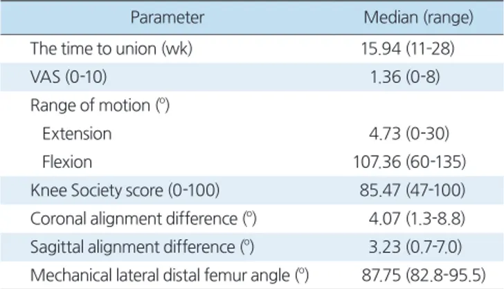
평균 추적 기간은 26.4개월(범위, 12-72개월)이었으며 골 유합까지 평균 기간은 15.94주(범위, 11-28주)였으며 추적 기간 동안 금속물 실패, 불유합, 재골절 등의 합병증 사례는 없었다. 임상적 결과는 최종 추시일에 VAS가 평균 1.36 (범 위, 0-8)이었으며 슬관절 관절 운동 범위 신전 4.73도(범위, 0-30도), 굴곡 107.36도(범위, 60-135도)이었다. Knee So- ciety score는 평균 85.47점(범위, 47-100)점이었다. 영상학적 결과는 최종 추시일에 촬영한 단순 방사선 촬영상 보이는 관 상면의 정렬축 변형 각도는 평균 4.07도(범위, 1.3-8.8도), 시 상면의 정렬축 변형 각도는 평균 3.23도(범위, 0.7-7.0도)였으 며 역학적 외측 원위 대퇴골 각도는 평균 87.75도(범위, 82.8- 95.5도)로 측정되었다(Table 2). 또한 6명의 환자에서 외상 후 골관절염이 관찰되었다(Fig. 6).
고 찰
원위 대퇴골의 완전 관절내 골절은 아직까지도 치료하기에 어렵고 복잡한 골절이다. 특히 이를 치료하기 위해서는 특히 복잡한 골절에서 관절의 골편들을 시각화하고 정복하기 위 해 광범위한 절개 및 접근이 필요하다. 관절 주위의 연조직의
Table 2. Clinical and Radiologic Outcomes at the Final Follow-Up
Parameter Median (range)
The time to union (wk) 15.94 (11–28)
VAS (0-10) 1.36 (0-8)
Range of motion (o)
Extension 4.73 (0-30)
Flexion 107.36 (60-135)
Knee Society score (0-100) 85.47 (47-100) Coronal alignment difference (o) 4.07 (1.3-8.8) Sagittal alignment difference (o) 3.23 (0.7-7.0) Mechanical lateral distal femur angle (o) 87.75 (82.8-95.5)
Fig. 6. Radiological changes in osteoarthritis at the final follow-up after the minimally invasive plate osteosynthesis technique for a distal femur complete intra-articular fracture.
손상은 초기 외상과 수술적 접근 모두에 의해서 발생하며 이 로 인해 골유합 및 재활에 어려움을 초래하여 슬관절 강직을 야기하게 된다. 따라서 원위 대퇴골의 관절 내 골절의 결과는 예측할 수 없으며 이는 불유합, 이상 회전, 강직 및 외상 후 골 관절염과 같은 합병증을 초래하게 된다. Ehlinger 등18)은 원위 대퇴골 골절에 대하여 금속판을 이용한 수술을 시행하고 6 년 추시 결과상 50%에서 외상 후 골관절염이 발생했다고 보 고한 바 있다. Rademakers 등10)은 원위 대퇴골 완전 관절내 골절에 대해 수술적 치료 이후 1년 이상 추시한 결과 36%의 환자에서 외상 후 골관절염으로 진행했고, 10%에서 창상 감 염의 합병증이 나타났다고 하였다. 또한, Hutson과 Zych1)는 원위 대퇴골 완전 관절내 분쇄 골절에서 제한된 내고정 및 긴 장대 강선 외고정을 시행한 결과 두 명의 환자가 지연유합으 로 재수술을 시행받았으며 수술 후 평균 슬관절 운동 범위는 0-92도로 측정되었고, 한 환자는 화농성 관절염과 골수염으 로 진행하였다고 하였다.
최소 침습적 금속판 고정술은 천공 동맥 및 영양 동맥을 보존하여 골막의 혈류 공급을 증가시키게 된다.19) 전위된 골 절의 경우, 천공 동맥의 공급을 받는 주변 근육으로부터 기 시하는 증가된 골막의 혈류 순환이 초기 혈액 공급원이 된
다.20,21) 최근 연구에 따르면 원위 대퇴골 주위 골절에서 잠김
금속판을 이용한 최소 침습적 금속판 골유합술의 임상적 유 용성이 검증되었다.22) 본 연구에서는 감염, 창상 문제, 불유 합 등과 같은 합병증이 없었다. 또한 생물학적 보존이 증가하 여 골절의 유합이 비교적 빠른 시기에 이루어졌다. 하지만 최 소 침습적 금속판 골유합술은 부정확한 정복이라는 단점이 있고 이로 인한 이상 회전이 발생할 가능성이 관혈적 정복술 에 비해 높다. 또한 골절부위를 단단히 압박하지 못하기 때문 에 지연유합과 불유합과 같은 문제가 발생한다. 본 연구에서 는 이상 회전에 대해서 추가적인 평가를 하지 못하였지만 통 증 또는 관절 운동 범위, 슬관절 점수, 일부 방사선적 계측의 측면에서 우수한 결과를 보였다. 비록 마지막 추시에서 19명 의 환자 중 6명(31.6%)의 환자가 방사선 촬영상 외상 후 골관 절염의 소견을 보였으나 이는 원위 대퇴골의 심한 완전 관절 내 골절의 치료에 관한 다른 연구들과 비슷한 수치이거나 더 낮은 수치에 해당한다.10,18) 또한 불유합, 부정유합, 지연유합, 감염 등의 다른 합병증을 보인 환자도 없었다.
본 연구의 주요 제한점은 적은 수의 표본이라는 것이다. 원 위 대퇴골의 관절내 골절만 포함시켰기 때문에 다른 연구들1-6) 과 마찬가지로 환자의 전체 수가 제한이 있었다. 또 다른 제한 점은 짧은 추적기간이다. 외상 후 골관절염의 발병률은 사실
상 오랜 추적 기간에 의해서 평가되어야 하지만 본 연구는 초 기 경향만 반영이 되었다. 마지막으로 수술 이후 컴퓨터 단층 촬영술을 시행하지 않았기 때문에 최소 침습적 골유합술의 가장 중요한 합병증인 이상 회전에 대해서는 정확한 평가가 없었다는 것이 본 연구의 제한점이라고 할 수 있다. 하지만 원 위 대퇴골 완전 관절내 골절에서 관절내 골절은 관혈적 정복 술을 통하여 해부학적 정복을 얻고 견고한 내고정을 통해 관 절 외 골절로 변형시킨 후 잠김 금속판을 이용하여 고정시킴 으로써 만족스러운 임상적 결과를 얻었다는 것에 본 연구의 의의가 있을 것으로 생각된다.
결 론
본 연구는 원위 대퇴골 완전 관절내 골절에서 관절내 골절 은 관혈적 정복술을 통하여 해부학적 정복을 얻고 견고한 내 고정을 통해 관절 외 골절로 전환시켜 잠김 금속판을 고정하 는 개념으로 술식을 진행하였고 수술 후 마지막 추시 기간에 서 임상적 평가 및 영상학적 평가는 비교적 만족스러운 결과 를 보였다. 또한 최소 침습적 술식으로 생물학적 손상을 많이 주지 않아 짧은 유합 기간을 보였으며 불유합, 부정유합, 감염 등의 합병증은 관찰되지 않았다.
요 약
목적:
본 연구는 원위 대퇴골의 완전 관절내 골절에서 최소 침습적 금속판 고정술의 임상적 결과에 대해 분석하였다.대상 및 방법:
2010년 6월부터 2016년 4월까지 원위 대퇴골 완전 관절내 골절 환자 중 최소 침습적 금속판 고정술을 시 행한 19명의 환자를 후향적으로 분석하였다. 19명의 환자 중 남자는 11명, 여자는 8명이었으며, 평균 나이는 55.9세였다.AO/OTA 분류상 C1이 2명, C2가 4명, C3가 13명이었다. 임 상적 평가는 visual analogue scale (VAS), 슬관절 운동 범위 (굴곡, 신전), Knee Society score를 통하여 시행하였고, 영상 학적 평가는 단순 방사선 촬영을 이용하여 관상면 및 시상면 의 정렬축 변형 각도, 역학적 외측 원위 대퇴골 각도를 측정 하였다.
결과:
평균 추적 기간은 26.4개월(범위, 12-72개월), 골유합 까지 평균 기간은 15.94주(범위, 11-28주)였다. VAS 평균 1.36 (범위, 0-8)이었으며 슬관절 관절 운동 범위 신전 4.73 도(범위,0-30), 굴곡 107.36도(범위, 60-135도)였다. Knee Society score는 평균 85.47점(범위, 47-100)점이었다. 영상학적 결과는 관상면의 정렬축 변형 각도가 평균 4.07도(범 위, 1.3-8.8도), 시상면 정렬축 변형 각도는 평균 3.23도(범위, 0.7-7.0도)였으며 역학적 외측 원위 대퇴골 각도는 평균 87.75 도(범위, 82.8-95.5도)로 측정되었다. 또한 6명의 환자에서 최종 추시상 외상성 골관절염이 관찰되었다.
결론:
본 연구는 원위 대퇴골 완전 관절내 골절에서 관절내 골절은 관혈적 정복술을 통하여 해부학적 정복을 얻고 견고 한 내고정을 통해 관절 외 골절로 전환시켜 잠김 금속판 고정 술을 시행하였고 수술 이후 마지막 추시에서 시행한 임상적 및 영상학적으로 비교적 만족스러운 결과를 나타내었다.색인 단어:
원위 대퇴골, 완전 관절내 골절, 최소 침습적 금속 판 골유합술ORCID
김성현, https://orcid.org/0000-0002-7514-3714 윤성현, https://orcid.org/0000-0003-4379-8174 박희곤, https://orcid.org/0000-0002-1028-2545 정재욱, https://orcid.org/0000-0002-6296-5129
References
1. Hutson JJ Jr, Zych GA: Treatment of comminuted intraarticular distal femur fractures with limited internal and external ten- sioned wire fixation. J Orthop Trauma, 14: 405-413, 2000.
2. Ramesh LJ, Rajkumar SA, Rajendra R, Rajagopal HP, Phanee- sha MS, Gaurav S: Ilizarov ring fixation and fibular strut graft- ing for C3 distal femoral fractures. J Orthop Surg (Hong Kong), 12: 91-95, 2004.
3. Lin D, Chen C, Lian K, Zhai W: Treatment of type C3.3 distal femoral fractures with double-plating fixation via U-shaped in- cision. Zhongguo Xiu Fu Chong Jian Wai Ke Za Zhi, 24: 683- 686, 2010.
4. Zhang ZM, Liu J, Huang CX, Zhao ZF, Wang G, Qin CC:
Treatment of type C3 distal femoral fractures with double-plat- ing fixation via anteriormiddle approach. Zhongguo Gu Shang, 25: 1049-1052, 2012.
5. Khalil AE, Ayoub MA: Highly unstable complex C3-type distal femur fracture: can double plating via a modified Olerud ex- tensile approach be a standby solution? J Orthop Traumatol, 13:
179-188, 2012.
6. Sîrbu PD, Asaftei R, Petreuş T, Lupaşcu C, Puha B, Luncă S:
Transarticular approach and retrograde plate osteosynthesis (TARPO) using implants with angular stability: a series of 17 cases of complex distal femoral fractures type C3/AO. Chirurgia
(Bucur), 109: 233-237, 2014.
7. Krettek C, Schandelmaier P, Miclau T, Bertram R, Holmes W, Tscherne H: Transarticular joint reconstruction and indirect plate osteosynthesis for complex distal supracondylar femoral fractures. Injury, 28 Suppl 1: A31-41, 1997.
8. Kregor PJ: Distal femur fractures with complex articular in- volvement: management by articular exposure and submuscular fixation. Orthop Clin North Am, 33: 153-175, ix, 2002.
9. Egund N, Kolmert L: Deformities, gonarthrosis and function after distal femoral fractures. Acta Orthop Scand, 53: 963-974, 1982.
10. Rademakers MV, Kerkhoffs GM, Sierevelt IN, Raaymakers EL, Marti RK: Intra-articular fractures of the distal femur: a long- term follow-up study of surgically treated patients. J Orthop Trauma, 18: 213-219, 2004.
11. Zehntner MK, Marchesi DG, Burch H, Ganz R: Alignment of supracondylar/intercondylar fractures of the femur after internal fixation by AO/ASIF technique. J Orthop Trauma, 6: 318-326, 1992.
12. Krettek C, Müller M, Miclau T: Evolution of minimally invasive plate osteosynthesis (MIPO) in the femur. Injury, 32 Suppl 3:
SC14-23, 2001.
13. Frigg R, Appenzeller A, Christensen R, Frenk A, Gilbert S, Schavan R: The development of the distal femur less invasive stabilization system (LISS). Injury, 32 Suppl 3: SC24-31, 2001.
14. Markmiller M, Konrad G, Südkamp N: Femur-LISS and distal femoral nail for fixation of distal femoral fractures: are there differences in outcome and complications? Clin Orthop Relat Res, (426): 252-257, 2004.
15. Schandelmaier P, Blauth M, Krettek C: Osteosynthese distaler femurfrakturen mit dem less invasive stabilizing system (LISS).
Oper Orthop Traumatol, 13: 178-197, 2001.
16. Schütz M, Schäfer M, Bail H, Wenda K, Haas N: Neue osteo- syntheseverfahren bei distalen femurfrakturen. Zentralbl Chir, 130: 307-313, 2005.
17. Goesling T, Frenk A, Appenzeller A, Garapati R, Marti A, Krettek C: LISS PLT: design, mechanical and biomechanical characteristics. Injury, 34 Suppl 1: A11-15, 2003.
18. Ehlinger M, Ducrot G, Adam P, Bonnomet F: Distal femur frac- tures. Surgical techniques and a review of the literature. Orthop Traumatol Surg Res, 99: 353-360, 2013.
19. Farouk O, Krettek C, Miclau T, Schandelmaier P, Guy P, Tscherne H: Minimally invasive plate osteosynthesis: does per- cutaneous plating disrupt femoral blood supply less than the traditional technique? J Orthop Trauma, 13: 401-406, 1999.
20. Colborn GL, Mattar SG, Taylor B, Skandalalkis JE, Lumsden AB: The surgical anatomy of the deep femoral artery. Am Surg, 61: 336-346, 1995.
21. Rhinelander FW: The normal microcirculation of diaphyseal cortex and its response to fracture. J Bone Joint Surg Am, 50:
784-800, 1968.
22. Ehlinger M, Adam P, Di Marco A, Arlettaz Y, Moor BK, Bon- nomet F: Periprosthetic femoral fractures treated by locked plat-
ing: feasibility assessment of the mini-invasive surgical option. A prospective series of 36 fractures. Orthop Traumatol Surg Res, 97: 622-628, 2011.