Introduction
Metabolic syndrome (MS) is becoming a major health con- cern particularly in developed countries, with a prevalence of over 20% of the general adult population in the United States.1) Several studies from South Korea have reported that, as in Western countries, the prevalence of MS in the general adult population approximates 20%.2)3) MS is a known risk factor for the development of various cardiovascular diseases, includ- ing heart failure.4-8) Recent studies have shown that MS is di- rectly related to relative left ventricular hypertrophy (LVH) and to systolic or diastolic dysfunction when assessed by two-dimen- sional or Doppler echocardiography.9-13) However, because in most of those studies, majority of patients had underlying dia- betes mellitus (DM) or hypertension (HT), the influence of early MS on myocardial function has not been thoroughly evaluated.
In addition, past studies have usually assessed diastolic function
in MS patients by conventional Doppler echocardiography.
Thus, we conducted this study to evaluate myocardial function in non-hypertensive MS patients by tissue Doppler Imaging (TDI) as well as conventional Doppler.
Methods
Study population
We screened subjects who visited the Healthcare Service Center of the Dong-A University Hospital. Complete cardio- vascular examinations, including history taking, physical ex- amination, and blood chemistry sampling, were performed in the outpatient clinic to screen for MS. Patients with known cardiovascular disease, HT, or stroke were excluded during the baseline examination. Weight, height, and waist circumfer- ence were obtained. Blood pressure (BP) was determined in ORIGINAL ARTICLE J Cardiovasc Ultrasound 2011;19(3):134-139
Subclinical Myocardial Dysfunction in Metabolic Syndrome Patients
without Hypertension
Jeong-Min Seo, MD1, Tae-Ho Park, MD1, Dong-Yeol Lee, MD1, Young-Rak Cho, MD1, Hee-Kyung Baek, MD1, Jong-Seong Park, MD1, Moo-Hyun Kim, MD1, Young-Dae Kim, MD1, Sun-Young Choi, MT1, Sun-Mi Lee, MD2 and Young-Seoub Hong, MD3
1Departments of Cardiology, 2Family Medicine, 3Preventive Medicine, Dong-A University College of Medicine, Busan, Korea
Background: The aim of this study was to evaluate myocardial function in patients with non-hypertensive metabolic syndrome.
Methods: We selected metabolic syndrome patients (n = 42) without evidence of hypertension and compared them to age- matched control individuals (n = 20). All patients were evaluated by two-dimensional and tissue Doppler echocardiography including tissue Doppler derived strain and strain rate measurements.
Results: There were no significant differences between the two groups in mitral E and A inflow velocities or the E/A ratio.
However, systolic and early diastolic myocardial velocities, and strain rate were significantly lower in patients with metabolic syndrome than in the control group (all p < 0.05). Multiple stepwise regression analyses revealed that age, waist circumference, and systolic blood pressure were independently associated with peak systolic myocardial velocity.
Conclusion: These results indicate that metabolic syndrome patients without hypertension may have decrease of myocardial systolic and early diastolic velocities on tissue Doppler imaging, even if they appear to have normal systolic and diastolic function on conventional echocardiography.
KEY WORDS: Metabolic syndrome · Doppler echocardiography.
• Received: March 28, 2011 • Revised: June 2, 2011 • Accepted: August 17, 2011
• Address for Correspondence: Tae-Ho Park, Department of Cardiology, Dong-A University College of Medicine, 1 Dongdaesin-dong 3-ga, Seo-gu, Busan 602-715, Korea Tel: +82-51-240-2964, Fax: +82-51-244-1713, E-mail: [email protected]
• This is an Open Access article distributed under the terms of the Creative Commons Attribution Non-Commercial License (http://creativecommons.org/licenses/by-nc/3.0) which permits unrestricted non-commercial use, distribution, and reproduction in any medium, provided the original work is properly cited.
the outpatient clinic using the conventional cuff method. Fast- ing serum glucose (FSG), total cholesterol, triglyceride (TG), high-density lipoprotein (HDL), and low-density lipoprotein levels were measured after fasting 12 hours overnight. For pa- tients who were diagnosed with MS and agreed to participate in this study, signed informed consent was obtained, and com- prehensive 2D and Doppler echocardiography were per- formed. We also selected control subjects who did not have MS. We applied the 2005 National Cholesterol Education Program (NCEP)/Adult Treatment Panel (ATP) III criteria to define MS. Subjects were diagnosed as having MS if they met three or more of the following criteria: waist circumference of
≥ 90 cm in men and ≥ 80 cm in women according to the NCEP/ATP III recommendations for the Asian population, TG level ≥ 150 mg/dL, serum HDL cholesterol level < 40 mg/
dL in men and < 50 mg/dL in women, systolic blood pressure (SBP) ≥ 130 mmHg or diastolic blood pressure (DBP) ≥ 85 mmHg, and FSG level ≥ 100 mg/dL.14) However, patients with HT (systolic BP ≥ 140 mmHg or diastolic BP ≥ 90 mmHg by the Joint National Committee 7th report crite- ria)15) or DM (FSG level ≥ 126 mg/dL by the American Dia- betes Association)16) were excluded for the purpose of this study. Patients who had systolic dysfunction, cardiomyopathy, or significant valvular heart disease on 2D echocardiography were also excluded. Being overweight or obese was defined as having a body mass index (BMI) between 25-29.9 kg/m2 or
≥ 30 kg/m2, respectively, according to the World Health Or- ganization criteria.17) For this study, impaired glucose metabo- lism was defined as a FSG level of 100-125.9 mg/dL. Finally, we selected 42 consecutive MS patients without HT and 20 age-matched control individuals without MS.
Echocardiography
Echocardiography was performed using the iE33 ultra- sound system and 2.5 MHz transducers (Philips Ultrasound Company, Cleveland, OH, USA). Standard parasternal and apical views were acquired. Complete 2D and M-mode echo- cardiogram, conventional Doppler, and TDI were obtained for all enrolled individuals. We measured the following left ven- tricle (LV) parameters by M-mode echocardiography: inter- ventricular septal wall thickness (IVS), posterior wall thick- ness (PW), and LV end diastolic dimension (EDD) at the chordae tendinae level. The LV mass was calculated according to the following equation: LV mass = 0.8 [1.04 × (PW + IVS + LVEDD)3 - (LVEDD)3] + 0.6.18)
LV mass index was calculated as the LV mass divided by body surface area. LVH was defined as LV mass index ≥ 116 g/m2 for men and ≥ 104 for women.19) LV ejection fraction was measured by the modified Simpson method.20) Left atrial (LA) volumes were calculated using biplane Simpson meth- od. LV diastolic function was evaluated by the measurements of early diastolic mitral inflow (E) velocity, late diastolic mi- tral inflow (A) velocity, E/A ratio, and mitral E wave deceler-
ation time (DT) using conventional pulsed wave Doppler echocardiography. LV diastolic function was also estimated by TDI. Early diastolic mitral annular (Ea) velocity was mea- sured by tissue Doppler placing sample volume at the lateral annulus.21)
To obtain longitudinal myocardial velocity, strain rate, and strain images with high quality, a narrow sector angle was used, and image depth was adjusted to allow for a high frame rate (> 120 frames/s) with care taken to avoid angulations.
The myocardial time-velocity and time-deformation curves were reconstructed off-line from color coded 2D tissue Dop- pler image loops. The peak systolic (Sm), early diastolic (Em), and late diastolic longitudinal myocardial velocities were mea- sured at basal and mid segments of the septal, lateral, inferior, and anterior walls from apical 2- and 4-chamber views. Like- wise, peak systolic (Ssr), early diastolic (Esr), late diastolic strain rate, and peak systolic strain (PSS) were measured at the same segments and expressed as absolute values. Their average values were used to compare regional and global LV functions of MS patients to those of control subjects. A single investiga- tor who was blinded to clinical data performed echocardio- graphic measurements.
Statistical analyses
Statistical analyses were performed using the Statistical Package for Social Sciences version 12 (SPSS Inc., Chicago, IL, USA). Continuous values were presented as mean ± standard deviations. Group means were compared using independent t- tests. Proportional values of the two groups were compared using Pearson’s Chi-Square test. Pearson’s correlation analyses were performed to assess the associations between clinical pa- rameters of MS (age, waist circumference, SBP, DBP, TG, HDL, and FSG) and echocardiographic parameters of myocardial function (tissue Doppler myocardial velocity, strain rate, and strain). Multiple stepwise regression analyses were performed to evaluate the influence of clinical factors on the differences in echocardiographic findings. A p-value of less than 0.05 was re- garded as having statistical significance.
Results
Study population
Demographic and clinical characteristics of the study pop- ulations (metabolic and non-metabolic control groups) are summarized in Table 1. Age and gender distributions were similar in both groups. Although there was no significant difference in BMI, waist circumference was significantly larg- er in the MS than in the control group. Another remarkable variation was demonstrated in terms of BP and TG levels be- tween the two groups. The FSG and HDL levels were not significantly different between the two groups. Thus, MS pa- tients included in the present study had minimal risk factors for MS.
M-mode, two-dimensional, and conventional doppler echocardiography
M-mode, 2D, and conventional Doppler echocardiographic measurements are shown in Table 2. LV size, LV mass, LA size, and LVEF were not significantly different between the two groups. LV diastolic function estimated by conventional Dop- pler criteria was similar in both groups. Thus, there were no significant differences in the prevalence of systolic and diastol- ic dysfunction when assessed by 2D and conventional Doppler echocardiography.
Tissue doppler imaging
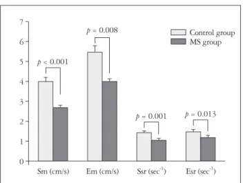
Echocardiographic measurements by TDI are summarized in Table 3 and Fig. 1. Tissue Doppler velocities of the lateral annulus were 8.8 ± 2.4 and 11.8 ± 1.9 cm/s (p < 0.001) in the MS and control groups, respectively. Average values of Sm and Em measured at 8 myocardial segments were significantly
Table 2. Two-dimensional and pulsed wave Doppler echocardiography
MS group (n = 42) Control group (n = 20) p-value
LVEDD (mm) 47.6 ± 3.8 45.8 ± 2.5 NS
VS (mm) 7.9 ± 1.2 8.1 ± 0.8 NS
PW (mm) 8.0 ± 0.9 8.2 ± 0.9 NS
LVM (g) 123.2 ± 25.4 116.2 ± 21.7 NS
LVMI (g/m2) 75.8 ± 12.7 70.6 ± 12.7 NS
LAV (mL) 49.0 ± 12.0 45.0 ± 11.4 NS
LAVI (mL/m2) 29.9 ± 6.7 27.6 ± 7.5 NS
LVEF (%) 63.8 ± 3.7 62.5 ± 2.6 NS
E velocity (cm/sec) 68.5 ± 13.8 69.3 ± 14.0 NS
A velocity (cm/sec) 72.1 ± 12.4 68.3 ± 18.2 NS
E/A 0.9 ± 0.3 1.1 ± 0.3 NS
DT (msec) 233.9 ± 33.0 217.9 ± 43.4 NS
LVEDD: left ventricular end-diastolic diameter, VS: left ventricular septal wall thickness, PW: left ventricular posterior wall thickness, LVM: left ventricular mass, LVMI: left ventricular mass index, LAV: left atrial volume, LAVI: left atrial volume index, LVEF: left ventricular ejection fraction, E: early diastolic transmitral inflow, A: late diastolic transmitral inflow, DT: mitral E wave deceleration time, NS: not significant
Table 1. Demographic and clinical characteristics
MS group (n = 42) Control group (n = 20) p-value
Number of women (%) 34 (81%) 12 (60%) NS
Age (years) 58.1 ± 7.0 57.9 ± 6.2 NS
Body mass index (kg/m2) 24.7 ± 1.9 23.7 ± 3.0 NS
Number of overweight 21 (50%) 5 (25.0%) NS
Waist circumference (cm) 86.8 ± 4.4 78.8 ± 4.8 < 0.001
Systolic blood pressure (mmHg) 124.1 ± 11.7 117.0 ± 10.3 0.024
Diastolic blood pressure (mmHg) 79.7 ± 7.1 75.0 ± 8.3 0.023
Fasting serum glucose level (mg/dL) 99.0 ± 11.7 97.6 ± 8.9 NS
Number of impaired glucose metabolism 19 (45.2%) 9 (45.0%) NS
Total cholesterol level (mg/dL) 202.0 ± 36.4 175.0 ± 37.9 0.009
Triglyceride level (mg/dL) 180.1 ± 74.7 98.8 ± 34.0 < 0.001
HDL cholesterol level (mg/dL) 45.6 ± 8.7 48.3 ± 7.0 NS
LDL cholesterol level (mg/dL) 120.4 ± 33.7 101.1 ± 32.5 0.038
HDL: high-density lipoprotein, LDL: low-density lipoprotein, NS: not significant
Fig. 1. Mean values of myocardial velocities and strain rate by tissue Doppler imaging in control and MS group. MS: metabolic syndrome, Sm:
peak systolic, Em: early diastolic, Ssr: peak systolic, Esr: early diastolic.
0 1 2 3 4 5 6 7
Sm (cm/s) Em (cm/s) Ssr (sec-1) Esr (sec-1) p < 0.001
p = 0.008
p = 0.001 p = 0.013 Control group MS group
lower in the MS group than in the control group (2.7 ± 0.4 vs.
4.0 ± 1.0 cm/s, p < 0.001; 4.0 ± 1.3 vs. 5.5 ± 1.4 cm/s, p = 0.008, respectively). In addition, average values of Ssr, Esr, and PSS were also significantly lower in the MS group than in the control group (1.1 ± 0.3 vs. 1.4 ± 0.3 s-1, p = 0.001; 1.2 ± 0.3 vs. 1.5 ± 0.3 s-1, p = 0.013; and 16.9 ± 3.7 vs. 20.5 ± 3.2%, p = 0.001, respectively).
Relationship of clinical and echocardiographic parameters
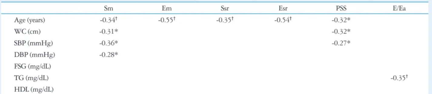
Linear regression analysis was performed to examine the re- lationship of echocardiographic measurements to clinical pa- rameters in patients with MS and non-MS (Table 4). Age sig- nificantly correlated with all echocardiographic parameters representing myocardial function. Waist circumference, SBP, and FSG had significant correlations with two of five echocar- diographic parameters. Among the echocardiographic param- eters, Sm is the most representative paramenter which is able to evaluate myocardial dysfunction in MS in present study. A correlation index of Sm greater than 0.3 was seen in waist cir- cumference and SBP.
TG and HDL levels did not have significant correlations with any of the echocardiographic parameters. Stepwise mul- tiple regression analysis was performed to examine the clinical
parameters that influence global Sm (Table 5). The results in- dicated that age (β coefficient = -0.313, p = 0.006), waist cir- cumference (β coefficient = -0.296, p = 0.012), and SBP (β co- efficient = -0.253, p = 0.031) were independently associated with Sm.
Discussion
The results of our study showed that non-hypertensive MS patients had subclinical myocardial dysfunction that was made apparent by TDI.
Previous studies have shown echocardiographic evidence of myocardial dysfunction in MS patients.11)12) However, most patients enrolled in those studies had overt DM or HT. Thus, diastolic dysfunction was evident by conventional parameters (E, A, and E/A) as well as TDI indices. In contrast, the present study shows that myocardial dysfunction in patients with an
Table 4. Correlation coefficients between clinical and echocardiographic parameters
Sm Em Ssr Esr PSS E/Ea
Age (years) -0.34† -0.55† -0.35† -0.54† -0.32*
WC (cm) -0.31* -0.32*
SBP (mmHg) -0.36* -0.27*
DBP (mmHg) -0.28*
FSG (mg/dL)
TG (mg/dL) -0.35†
HDL (mg/dL)
*< 0.05, †< 0.01. WC: waist circumference, SBP: systolic blood pressure, DBP: diastolic blood pressure, FSG: fasting serum glucose, TG: triglyceride, HDL:
high-density lipoprotein, Sm: peak systolic, Em: early diastolic, Ssr: peak systolic, Esr: early diastolic, PSS: peak systolic strain, E: early diastolic transmitral inflow, Ea:
peak mitral annular velocity during early diastole
Table 5. Multiple regression analyses to examine the clinical param- eters that influence global Sm
β coefficient p-value
Age -0.313 0.006
WC -0.296 0.012
SBP -0.253 0.031
Sm: peak systolic, WC: waist circumference, SBP: systolic blood pressure Table 3. Myocardial velocities, strain rates, and peak systolic strain
All segments MS group (n = 42) Control group (n = 20) p-value
Lateral Ea velocity (cm/sec) 8.8 ± 2.4 11.8 ± 1.9 < 0.001
Mitral E/Ea 8.2 ± 2.1 6.5 ± 1.5 0.003
Global Sm (cm/sec) 2.7 ± 0.4 4.0 ± 1.0 < 0.001
Global Em (cm/sec) 4.0 ± 1.3 5.5 ± 1.4 0.008
Global Am (cm/sec) 3.7 ± 0.9 4.1 ± 1.1 NS
Global Ssr (sec-1) 1.1 ± 0.3 1.4 ± 0.3 0.001
Global Esr (sec-1) 1.2 ± 0.3 1.5 ± 0.3 0.013
Global Asr (sec-1) 1.0 ± 0.3 1.1 ± 0.2 NS
Global PSS (%) 16.9 ± 3.7 20.5 ± 3.2 0.001
Ea: peak mitral annular velocity during early diastole, Global Sm: the average of peak left ventricular systolic velocities, Global Em: the average of peak left ventricular early diastolic velocities, Global Am: the average of peak left ventricular late diastolic velocities, Global Ssr: the average of peak left ventricular systolic strain rates, Global Esr: the average of peak left ventricular early diastolic strain rates, Global Asr: the average of peak left ventricular late diastolic strain rates, Global PSS: the average of peak left ventricular systolic strain, NS: not significant
early stage of MS was detected by TDI not conventional pa- rameters. We designed this study to test the hypothesis that early MS patients might have myocardial dysfunction. We ex- cluded HT patients to estimate the true impact of early stage MS on myocardial function, which was the most important aspect of the present study. In addition, a study of MS patients without DM or HT lends itself to a better examination of the relationship between each metabolic parameter to myocardial function, because HT by itself is strong enough to cause a de- cline in diastolic function.
Peak longitudinal myocardial velocities derived from pulsed wave TDI are useful indicators of global or regional LV dys- function. The early diastolic TDI velocity of the mitral annulus is generally thought to be a preload independent index for LV relaxation, and it is used to estimate LV filling pressures.22)23) Strain and strain rate are other tools that can reflect myocardi- al function.24)25) We applied tissue Doppler myocardial veloci- ty, strain, and strain rate together to detect subtle changes in global myocardial function.
Conventional Doppler echocardiography revealed a tenden- cy for MS patients to have more diastolic LV dysfunction.
However, that tendency did not achieve statistical signifi- cance. In contrast, TDI studies showed significant differences that were not revealed by conventional pulsed wave Doppler studies. Lateral Ea velocity, Sm, Em, Ssr, Esr, and PSS were more profoundly decreased in the MS group compared to con- trol, and the differences achieved statistical significance. The results of our study suggest that TDI is superior to a conven- tional pulsed wave Doppler study in detecting early myocar- dial dysfunction.
The 34 patients with MS enrolled in our study were diag- nosed by 3 criteria: waist circumference, low HDL levels, and high TG levels. Waist circumference reflects central obesity and risk of cardiovascular disease. Furthermore, it is consid- ered to be the best predictor of MS among other diagnostic criteria.26) On the other hand, the direct influence of dyslipid- emia on myocardial function is not well known. A recent study showed that short-term control of dyslipidemia using statin improved myocardial dysfunction assessed by the Tei index and tissue Doppler myocardial velocities. In that study, as in our current one, overt DM and HT patients were excluded;
however, waist circumference was not measured. The results suggested that dyslipidemia itself may be a risk factor for myocardial dysfunction.27)
In this study, we expected that waist circumference and lip- id levels would exhibit good correlations with echocardio- graphic parameters because most MS patients were diagnosed by waist circumference and dyslipidemia. However, all meta- bolic parameters had a weak correlation with echocardio- graphic indices. Especially, dyslipidemia itself was not related to echocardiographic parameters, because the patients enrolled in this study had mild abnormalities of lipid profile. These data are different from previous study.27) Based on our results,
age was the best parameter to predict myocardial dysfunction.
Of the metabolic parameters, waist circumference and SBP were more important than FSG, TG and HDL in influencing myocardial dysfunction in patients with early MS. From mul- tiple comparisons of echocardiographic and metabolic param- eters, the highest correlation was observed between age and Em (r = -0.551, p < 0.001); however, metabolic parameters were more closely related to Sm than Em. The data in the present study indicate that waist circumference and SBP are independently associated with myocardial dysfunction (Sm).
It is common knowledge that even a mild degree of diastol- ic dysfunction may be associated with poor prognosis on long- term follow-up;29) therefore, early detection of myocardial dys- function may provide MS patients with a chance to modify their lifestyles, thereby preventing future heart disease. The use of TDI might detect early systolic and diastolic myocardi- al dysfunction in MS patients, even if they do not have overt DM, HT, or any structural abnormalities.
There are a few limitations in present study. Firstly, the size of this study to assess the relationship of each MS factor with myocardial dysfunction was relatively small. Secondly, it is dif- ficult to explain the exact pathophysiologic mechanisms of how early MS without overt HT influences myocardial func- tion, although we postulate that insulin resistance that was not measured in this study might underlie decreased myocardial function. Thirdly, although all subjects in our study did not have any signs or symptoms of angina, and had normal find- ings on electrocardiogram and on 2-D echocardiography, we could not completely exclude asymptomatic coronary artery disease.
In conclusion, these results indicate that patients with MS without HT may have myocardial dysfunction apparent by tis- sue Doppler imaging, even if they appear to have normal find- ings on two-dimensional and conventional Doppler studies.
• Acknowledgements
This study was supported by research funds from Dong-A University.
References
1. Ford ES, Giles WH, Dietz WH. Prevalence of the metabolic syndrome among US adults: findings from the third National Health and Nutrition Examination Survey. JAMA 2002;287:356-9.
2. Choi KM, Kim SM, Kim YE, Choi DS, Baik SH, Lee J; Interna- tional Diabetes Federation. Prevalence and cardiovascular disease risk of the metabolic syndrome using National Cholesterol Education Program and International Diabetes Federation definitions in the Korean population. Me- tabolism 2007;56:552-8.
3. Choi SH, Ahn CW, Cha BS, Chung YS, Lee KW, Lee HC, Huh KB, Kim DJ. The prevalence of the metabolic syndrome in Korean adults:
comparison of WHO and NCEP criteria. Yonsei Med J 2005;46:198- 205.
4. Isomaa B, Almgren P, Tuomi T, Forsén B, Lahti K, Nissén M, Taskinen MR, Groop L. Cardiovascular morbidity and mortality associ- ated with the metabolic syndrome. Diabetes Care 2001;24:683-9.
5. Lakka HM, Laaksonen DE, Lakka TA, Niskanen LK, Kumpusalo E,
Tuomilehto J, Salonen JT. The metabolic syndrome and total and cardio- vascular disease mortality in middle-aged men. JAMA 2002;288:2709- 16.
6. Malik S, Wong ND, Franklin SS, Kamath TV, L’Italien GJ, Pio JR, Williams GR. Impact of the metabolic syndrome on mortality from coro- nary heart disease, cardiovascular disease, and all causes in United States adults. Circulation 2004;110:1245-50.
7. McNeill AM, Rosamond WD, Girman CJ, Golden SH, Schmidt MI, East HE, Ballantyne CM, Heiss G. The metabolic syndrome and 11-year risk of incident cardiovascular disease in the atherosclerosis risk in communities study. Diabetes Care 2005;28:385-90.
8. Ingelsson E, Arnlöv J, Lind L, Sundström J. Metabolic syndrome and risk for heart failure in middle-aged men. Heart 2006;92:1409-13.
9. de Simone G, Devereux RB, Chinali M, Roman MJ, Lee ET, Resn- ick HE, Howard BV. Metabolic syndrome and left ventricular hypertrophy in the prediction of cardiovascular events: the Strong Heart Study. Nutr Metab Cardiovasc Dis 2009;19:98-104.
10. Chinali M, Devereux RB, Howard BV, Roman MJ, Bella JN, Liu JE, Resnick HE, Lee ET, Best LG, de Simone G. Comparison of cardi- ac structure and function in American Indians with and without the meta- bolic syndrome (the Strong Heart Study). Am J Cardiol 2004;93:40-4.
11. Masugata H, Senda S, Goda F, Yoshihara Y, Yoshikawa K, Fujita N, Daikuhara H, Nakamura H, Taoka T, Kohno M. Left ventricular dia- stolic dysfunction as assessed by echocardiography in metabolic syndrome.
Hypertens Res 2006;29:897-903.
12. de las Fuentes L, Brown AL, Mathews SJ, Waggoner AD, Soto PF, Gropler RJ, Dávila-Román VG. Metabolic syndrome is associated with abnormal left ventricular diastolic function independent of left ventricular mass. Eur Heart J 2007;28:553-9.
13. Wong CY, O’Moore-Sullivan T, Fang ZY, Haluska B, Leano R, Marwick TH. Myocardial and vascular dysfunction and exercise capacity in the metabolic syndrome. Am J Cardiol 2005;96:1686-91.
14. Grundy SM, Cleeman JI, Daniels SR, Donato KA, Eckel RH, Franklin BA, Gordon DJ, Krauss RM, Savage PJ, Smith SC Jr, Spertus JA, Costa F; American Heart Association; National Heart, Lung, and Blood Institute. Diagnosis and management of the metabolic syndrome: an American Heart Association/National Heart, Lung, and Blood Institute Scientific Statement. Circulation 2005;112:2735-52.
15. Chobanian AV, Bakris GL, Black HR, Cushman WC, Green LA, Izzo JL Jr, Jones DW, Materson BJ, Oparil S, Wright JT Jr, Roccella EJ;
National Heart, Lung, and Blood Institute Joint National Commit- tee on Prevention, Detection, Evaluation, and Treatment of High Blood Pressure; National High Blood Pressure Education Program Coordinating Committee. The Seventh Report of the Joint National Committee on Prevention, Detection, Evaluation, and Treatment of High Blood Pressure: the JNC 7 report. JAMA 2003;289:2560-72.
16. Expert Committee on the Diagnosis and Classification of Diabetes Mellitus. Report of the expert committee on the diagnosis and classification of diabetes mellitus. Diabetes Care 2003;26 Suppl 1:S5-20.
17. Obesity: preventing and managing the global epidemic. Report of a WHO consultation. World Health Organ Tech Rep Ser 2000;894:i-xii, 1-253.
18. Lang RM, Bierig M, Devereux RB, Flachskampf FA, Foster E, Pel-
likka PA, Picard MH, Roman MJ, Seward J, Shanewise J, Solomon S, Spencer KT, St John Sutton M, Stewart W; American Society of Echocardiography’s Nomenclature and Standards Committee; Task Force on Chamber Quantification; American College of Cardiology Echocardiography Committee; American Heart Association; Euro- pean Association of Echocardiography, European Society of Cardiol- ogy. Recommendations for chamber quantification. Eur J Echocardiogr 2006;7:79-108.
19. Lester SJ, Tajik AJ, Nishimura RA, Oh JK, Khandheria BK, Seward JB. Unlocking the mysteries of diastolic function: deciphering the Rosetta Stone 10 years later. J Am Coll Cardiol 2008;51:679-89.
20. Okin PM, Devereux RB, Nieminen MS, Jern S, Oikarinen L, Vii- tasalo M, Toivonen L, Kjeldsen SE, Julius S, Dahlöf B. Relationship of the electrocardiographic strain pattern to left ventricular structure and function in hypertensive patients: the LIFE study. Losartan Intervention For End point. J Am Coll Cardiol 2001;38:514-20.
21. Von Bibra H, Thrainsdottir IS, Hansen A, Dounis V, Malmberg K, Rydén L. Tissue Doppler imaging for the detection and quantitation of myocardial dysfunction in patients with type 2 diabetes mellitus. Diab Vasc Dis Res 2005;2:24-30.
22. Nagueh SF, Middleton KJ, Kopelen HA, Zoghbi WA, Quiñones MA. Doppler tissue imaging: a noninvasive technique for evaluation of left ventricular relaxation and estimation of filling pressures. J Am Coll Cardiol 1997;30:1527-33.
23. Nagueh SF. Tissue Doppler imaging for the assessment of left ventricular diastolic function. J Cardiovasc Ultrasound 2008;16:76-9.
24. Park TH, Lakkis NM, Middleton KJ, Franklin J, Zoghbi WA, Qui- ñones MA, Spencer WH 3rd, Nagueh SF. Acute effect of nonsurgical sep- tal reduction therapy on regional left ventricular asynchrony in patients with hypertrophic obstructive cardiomyopathy. Circulation 2002; 106:412-5.
25. Takemoto Y, Pellikka PA, Wang J, Modesto KM, Cauduro S, Be- lohlavek M, Seward JB, Thomson HL, Khandheria B, Abraham TP.
Analysis of the interaction between segmental relaxation patterns and global diastolic function by strain echocardiography. J Am Soc Echocardiogr 2005;
18:901-6.
26. Palaniappan L, Carnethon MR, Wang Y, Hanley AJ, Fortmann SP, Haffner SM, Wagenknecht L; Insulin Resistance Atherosclerosis Study. Predictors of the incident metabolic syndrome in adults: the Insulin Resistance Atherosclerosis Study. Diabetes Care 2004;27:788-93.
27. Talini E, Di Bello V, Bianchi C, Palagi C, Delle Donne MG, Penno G, Nardi C, Canale ML, Del Prato S, Mariani M, Miccoli R. Early impairment of left ventricular function in hypercholesterolemia and its revers- ibility after short term treatment with rosuvastatin A preliminary echocar- diographic study. Atherosclerosis 2008;197:346-54.
28. Schillaci G, Pasqualini L, Verdecchia P, Vaudo G, Marchesi S, Por- cellati C, de Simone G, Mannarino E. Prognostic significance of left ven- tricular diastolic dysfunction in essential hypertension. J Am Coll Cardiol 2002;39:2005-11.
29. Bella JN, Palmieri V, Roman MJ, Liu JE, Welty TK, Lee ET, Fab- sitz RR, Howard BV, Devereux RB. Mitral ratio of peak early to late diastolic filling velocity as a predictor of mortality in middle-aged and el- derly adults: the Strong Heart Study. Circulation 2002;105:1928-33.