대사증후군의 진단에 있어 생체 전기저항 분석법에 의해 추정된 비만지표들의 유용성
황인철
1, 김경곤
1, 이경식
1, 김승수
21가천의과학대학교 길병원 가정의학과, 2GSK 코리아
The Usefulness of Indices for Central Obesity Estimated by Bioelectrical Impedance Analysis in the Diagnosis of Metabolic Syndrome
In Cheol Hwang
1, Kyoung Kon Kim
1, Kyoung Sik Lee
1, Seung Soo Kim
21
Department of Family Medicine, Gachon University Gil Hospital, Incheon,
2GSK Korea, Seoul, Korea
Background: Recently-developed equipment based on bioelectrical impedance analysis (BIA) not only meas- ures total body fat but also displays several estimated indicators that reflect intra-abdominal fat, such as waist circumference (WC) and waist-to-hip ratio (WHR). This study examined the usefulness of these indicators in the diagnosis of metabolic syndrome (MS).
Methods: A total of 632 people over 20 years of age (355 men and 277 women, mean age 48.61±11.08 years, mean BMI 23.62±3.00 kg/m
2, 117 MS patients) were enrolled in the study. Measurements of WC and hip cir- cumference were measured by one individual, and WHR was calculated. BIA was performed to estimate waist circumference (BIAWC) and waist-to-hip ratio (BIAWHR). Receiver operating characteristic (ROC) curve analy- sis was used to examine the usefulness of BIAWC and BIAWHR in diagnosing MS.
Results: The areas under the curve (AUCs) were 0.836 (95% CI 0.805-0.864) for WC, 0.814 (95% CI 0.782-0.844) for BIAWC, 0.815 (95% CI 0.782-0.844) for WHR, and 0.805 (95% CI 0.772-0.835) for BIAWHR.
The difference between the AUCs of WC and BIAWC (0.022, 95% CI -0.004 to 0.048) and the difference be- tween the AUCs of WHR and BIAWHR (0.010, 95% CI -0.015 to 0.034) were not significant.
Conclusions: The indices for central obesity estimated by BIA had high agreement with the direct method, and they were not inferior to direct measured indices for predicting metabolic syndrome.
Korean J Health Promot 2011;11(2):64-71
Keywords: Bioelectrical impedance, Abdominal obesity, Metabolic syndrome, Waist circumference, Waist-to-hip ratio
■ Received:October 27, 2010 ■ Accepted:June 9, 2011
■ Corresponding author:Kyoung Kon Kim, MD, PhD
Department of Family Medicine, Gachon University Gil Hospital, 1198 Guwol-dong, Namdong-gu, Incheon 405-760, Korea
Tel: +82-32-460-3354, Fax: +82-32-460-3354 E-mail: [email protected]
INTRODUCTION
Because obesity is not defined as simple weight gain but rather as over-accumulation of body fat, body composi- tion assessment is a crucial step in weight management intervention. Knowing the body fat distribution, as well as
total amount of body fat, is important,
1)and visceral fat is a
stronger predictor of health-related risks than subcuta-
neous fat.
2-4)However, the ‘gold standard’ measures of vis-
ceral fat area, such as computed tomography (CT) or mag-
netic resonance imaging (MRI), are not practical for
large-scale research studies or as clinical tools, due to limi-
tations associated with cost, availability, and/or radiation
exposure. In contrast, anthropometric measurements,
such as waist circumference (WC) or waist-to-hip ratio
(WHR), for assessment of visceral fat accumulation are
simple and noninvasive. WC, in particular, holds a pivotal
position in current definitions of metabolic syndrome
(MS),
5)and is widely used in clinical practice as a surrogate for central obesity in assessing health status.
3)Bioelectrical impedance analysis (BIA), dual energy x-ray absorptiometry and underwater weighing are the useful methods for assessing human body composition.
Because BIA offers the advantages of being inexpensive, noninvasive, and operationally simple, it has been used in large-scale epidemiologic studies.
6)Segmental BIA has been introduced in recent decades, and indices (e.g., BIAWC, waist circumference calculated by BIA; BIAWHR, waist-to-hip ratio calculated by BIA) for regional fat dis- tribution, as well as total body fat measures have been presented.
7,8)However, to the best of our knowledge, very few stud- ies have investigated the clinical implications of the indices of BIA. We performed a preliminary study for gauging the clinical significance of BIAWHR in 132 persons, and ob- tained results that demonstrated that BIAWHR is not in- ferior to WC in diagnosing MS.
9)According to this pre- liminary result, we attempted to compare the usefulness of BIAWC and BIAWHR with that of actual measurements in diagnosing MS using a larger group.
METHODS
1. Subjects
Participants in the current study included 632 persons (355 men, 277 women) 20 years of age and older who vis- ited the health promotion centre at Gachon University Gil Hospital in March and April of 2009. All participants pro- vided written informed consent, and all aspects of this study were in accordance with the Declaration of Helsinki of the World Medical Association.
2. Estimation of WC and WHR by BIA
Subjects wore light indoor clothing, including shorts/
trousers to ensure that the legs were not in contact with skin at any point. Prior to measuring, subjects were asked to void in order to minimize measurement errors. They stood barefoot on the four foot electrodes on the platform of the analyzer, gripping the two palm and thumb electrodes. Foot electrodes were placed between the me- dial and lateral malleoli of both ankles of the subjects.
Estimates of body components
10)were derived from calcu- lations using the manufacturer’s software. A detailed de- scription of the sequence of measurements has been described.
11)WC and WHR estimated by InBody720 (Biospace Co., Seoul, Korea) were displayed by the ma- chine and printed out.
3. Laboratory and anthropometric measurements
All blood samples were obtained from antecubital veins on the morning after a 12-h overnight fast. Height and weight were measured using an automatic digital stadi- ometer (InBody BSM330, Biospace, Co., Seoul, Korea) while subjects wore a lightweight gown and stood bare foot. Based on the WHO (World Health Organization) protocol, WC was measured at the midpoint between the inferior costal margin and the superior border of the iliac crest along the mid-axillary line, and hip circumference was measured at the widest circumference of the hip.
12)WHR was then calculated. One individual performed all measurements of central obesity indices using a non- stretchable standard tape. Measurements were recorded to the nearest decimal place. To assess intra-observer varia- bility in measurement of waist and hip circumference, a pi- lot test was carried out in a small group (n=13), where measurements were performed by both the measurer and by the BIA equipment on separate occasions within 30 mi- nutes of the initial measurement.
4. Definition of MS
We used the revised NCEP (National Cholesterol Education Program) criteria proposed by the AHA/NHLBI (American Heart Association/National Heart, Lung, and Blood Institute),
13)which require at least three of five met- abolic components. The cutoff point for waist circum- ference was defined according to central obesity for Koreans, men ≥90 cm, women ≥85 cm.
14)5. Sample size estimation and statistical analyses
The sample size estimate of 114 for the MS group was
based on our preliminary results, which would lead to an
alpha level of 0.01 and a beta level of 0.10. In our prelimi-
nary study, the areas under the curves (AUCs) were 0.764
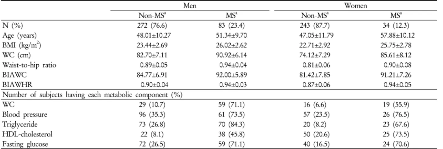
Table 1. Basic characteristics of study subjects
Men Women
Non-MSa MSa Non-MSa MSa
N (%) 272 (76.6) 83 (23.4) 243 (87.7) 34 (12.3)
Age (years) 48.01±10.27 51.34±9.70 47.05±11.79 57.88±10.12
BMI (kg/m2) 23.44±2.69 26.02±2.62 22.71±2.92 25.75±2.78
WC (cm) 82.70±7.11 90.92±6.14 74.12±7.29 85.61±8.12
Waist-to-hip ratio 0.89±0.05 0.94±0.04 0.81±0.06 0.90±0.08
BIAWC 84.77±6.91 92.00±5.89 81.42±7.85 91.21±7.26
BIAWHR 0.90±0.04 0.94±0.03 0.87±0.06 0.94±0.05
Number of subjects having each metabolic component (%)
WC 29 (10.7) 59 (71.1) 16 (6.6) 19 (55.9)
Blood pressure 96 (35.3) 61 (73.5) 57 (23.5) 26 (76.5)
Triglyceride 73 (26.8) 70 (84.3) 20 (8.2) 23 (67.6)
HDL-cholesterol 22 (8.1) 38 (45.8) 50 (20.6) 25 (73.5)
Fasting glucose 72 (26.5) 59 (71.1) 40 (16.5) 24 (70.6)
Abbreviations: MS, metabolic syndrome; BMI, body mass index; WC, waist circumference; BIAWC and BIAWHR, waist circumference and waist-to-hip ratio estimated by bioelectrical impedance analysis; HDL, high density lipoprotein. Significance differences were noted in whole variables by independent two sample t-test or χ2 test (P<0.01).
aDiagnosis was made using the 2005 AHA/NHLBI modified ATP III definition, and cutoff values of waist circumference were 90 cm for men and 85 cm for women based on the Korean society for the study of obesity.
for WC and 0.837 for BIAWHR. Spearman’s rank correla- tion coefficient of WC and BIAWHR was 0.616 in the MS group and 0.726 in the non-MS group.
Data were expressed as mean±S.D. or number (%).
Independent two sample t-test and χ
2test were used to compare factors associated with MS in patients with MS and those without.
Received operating characteristics ( ROC ) curve analy- sis was used to evaluate and compare four indices of cen- tral obesity, i.e. WC, WHR, BIAWC, and BIAWHR in diagnosing metabolic syndrome. AUC of each index was compared. Non-inferiority would be claimed if the upper two-sided 95% confidence intervals (CIs) of AUC of BIAWC and BIAWHR were more than the lower two-sided 95% CI of AUC of WC, and the difference of AUCs between BIAWC and WC was less than 0.05.
Statistical analyses were performed using R: A language and environment for statistical computing (version 2.9.2, R Development Core Team (2009). R Foundation for Statistical Computing, Vienna, Austria. ISBN 3-900051-07-0, URL http://www.R-project.org) and MedCalc for Windows, version 11.0.0 (MedCalc Software, Mariakerke, Belgium).
RESULTS
1. Reproducibility of measurements
The reproducibility of WC, WHR, BIAWC, and BIAWHR
was very high. Intra-class correlation coefficients (ICCs) for intra-observer reliability of WC, WHR, BIAWC, and BIAWHR were 0.959, 0.940, 1.000, and 0.998, respectively (Figure 1). Data on BIAWC and BIAWHR, in particular, show accurate reproducibility of WC and WHR estima- tion using BIA equipment.
2. General characteristics of subjects
Table 1 provides the general characteristics of the 632 subjects. Within the study sample, 117 (18.5%) were diag- nosed as MS. The prevalence rate of MS in our study was 23.4% for men and 12.3% for women, which is lower than the most recent report of the Korean population (men 30.7%, women 31.6%).
15)The mean age of participants was 48.61±11.08 years (48.79±10.22 years for men and 48.38±12.12 years for women). Age in the MS group was higher than in the non-MS group for both men and women. The age of women in the MS group (57.88±10.12 years) was higher than that of men (51.34±9.70 years, P=0.001). Mean body mass index of participants was 23.62±3.00 kg/m
2.
3. WC, WHR, BIAWC, and BIAWHR
The four indices of central obesity, WC and WHR, and
WC and WHR estimated by BIA, were higher in the MS
group than in the non-MS group for both men and women
Figure 1. Scatter plots of waist circumferences and waist-to-hip ratios for separate measurements taken by direct measurement and bioelectrical impedance analysis (A and B: measured waist circumference and waist-to-hip ratio, C and D: estimated waist circumference and waist-to-hip ratio by bioelectrical impedance analysis).
Abbreviations: ICC, intra-class correlation coefficient; CI, confidential interval; WC, waist circumference; WHR, waist-to-hip ratio; BIAWC and BIAWHR, waist circumference and waist-to-hip ratio estimated by bioelectrical impedance analysis.
(Table 1, P<0.001).
Agreement between WCs by direct measurement and BIA equipment was good. However, agreement between WHRs by direct measurement and BIA equipment was not as good. ICC between WC and BIAWC was 0.766 (95% CI 0.731-0.796) and ICC between WHR and BIAWHR was 0.561 (95% CI 0.505-0.612) (Figure 2).
The estimated value of WC by BIA was significantly higher than that of directly measured WC in all subjects (difference 4.14 (95% CI 3.79-4.49), P<0.001), the male group [difference 1.84 (95% CI 1.50-2.17), P<0.001], and
the female group [difference 7.09 (95% CI 6.61-7.57), P<0.001]. The estimated value of WHR by BIA was also significantly higher than that of directly measured WHR in all subjects [difference 0.032 (95% CI 0.028-0.037), P<0.001], the male group [difference 0.009 (95% CI 0.005-0.014), P=0.004], and the female group [difference 0.062 (95% CI 0.055-0.068), P<0.001].
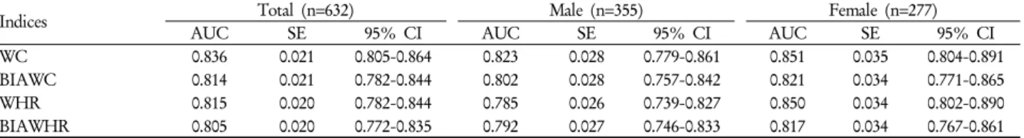
4. ROC analysis for diagnostic prediction of MS
AUCs of ROC curves were 0.836 for WC, 0.814 for
Figure 2. Scatter plots of waist circumference and waist hip ratio (A: measured waist circumference vs. estimated waist circumference by bioelectrical impedance analysis, B: waist-to-hip ratio obtained through direct measurement vs. estimated waist-to-hip ratio by bioelectrical impedance analysis).
Abbreviations: ICC, intra-class correlation coefficient; CI, confidential interval; WC, waist circumference; WHR, waist-to-hip ratio; BIA, bioelectrical impedance analysis.
Table 2. Comparison of 4 indices for central obesity in diagnosing metabolic syndrome
aIndices Total (n=632) Male (n=355) Female (n=277)
AUC SE 95% CI AUC SE 95% CI AUC SE 95% CI
WC 0.836 0.021 0.805-0.864 0.823 0.028 0.779-0.861 0.851 0.035 0.804-0.891
BIAWC 0.814 0.021 0.782-0.844 0.802 0.028 0.757-0.842 0.821 0.034 0.771-0.865
WHR 0.815 0.020 0.782-0.844 0.785 0.026 0.739-0.827 0.850 0.034 0.802-0.890
BIAWHR 0.805 0.020 0.772-0.835 0.792 0.027 0.746-0.833 0.817 0.034 0.767-0.861
Abbreviations: AUC, area under the curve; SE, standard error; BIAWC and BIAWHR, waist circumference and waist-to-hip ratio estimated by bioelectrical impedance analysis; WHR, waist-to-hip ratio; WC, waist circumference.
aMetabolic syndrome was diagnosed according to the 2005 AHA/NHLBI modified ATP III definition; cutoff values of waist circumference were 90 cm for men and 85 cm for women based on the Korean society for the study of obesity.
BIAWC, 0.815 for WHR, and 0.805 for BIAWHR in all subjects. In all subjects, the male group and the female
group, all AUCs of ROC curves for BIAWC and BIAWHR were above 0.805, which was the lowest value of the lower 95% CI of AUCs of WC (Table 2).
The difference between the AUCs of WC and BIAWC was not significant (0.022, 95% CI -0.004 to 0.049), and the difference between the AUCs of WHR and BIAWHR was also not significant (0.010, 95% CI -0.034 to 0.054) (Figure 3A). The difference between the AUCs of WC and BIAWC (0.021, 95% CI -0.008 to 0.050 for male; 0.030 95% CI -0.018 to 0.077 for female) and between the AUCs of WHR and BIAWHR (0.007, 95% CI -0.046 to 0.059 for male; 0.033, 95% CI -0.055 to 0.120 for female) were also not significant in the male and female groups (Figure 3B and C).
5. Comparison of ROC curves in each index for central obesity
The upper two-sided 95% CI of the AUC of BIAWC and BIAWHR were more than the lower two-sided 95%
CI of the AUC of WC. The difference between the AUCs of WC and BIAWC was less than 0.05. The difference of the AUCs between WHR and BIAWHR was also less than 0.05. This is not only true for all subjects, but also for the male and female subgroups (Table 2, Figure 3).
DISCUSSION
In this study, the indices for central obesity estimated by
BIA were not inferior to those of direct measurement in
predicting MS. BIA devices, including the InBody720
used in this study, have been used to estimate body com-
position in adults.
16-18)Scharfetter et al.
19)initially in-
troduced local bioimpedance analysis for assessing ab-
dominal fat in 2001. Ryo et al.
20)then reported excellent
Figure 3. Received operating characteristics (ROC) curve and area under the curve (AUC) of 4 indices of metabolic syndrome (A: total, B: male, C: female). Metabolic syndrome was diagnosed according to the 2005 AHA/NHLBI modified ATP III definition; cutoff value for waist circumference is 90 cm for men and 85 cm for women based on the Korean society for the study of obesity.
Abbreviations: BIAWC, waist circumference estimated by bioelectrical impedance analysis; BIAWHR, waist-to-hip ratio estimated by bioelectrical impedance analysis; WC, waist circumference; WHR, waist-to-hip ratio.
correlation between BIA and CT scan in estimating viscer- al fat accumulation. However, to the best of our knowl- edge, few studies have reported on matters of clinical im- plication for central obesity indices estimated by devices based on BIA methods.
WC measurement has limitations. WC is known as a strong predictor for obesity-related morbidity and mor- tality, and routine measurement of WC is strongly recom- mended in clinical practice.
21-23)Measurement protocols have been based principally on anatomical landmarks.
Commonly used landmarks include the midpoint between the lower border of the rib cage and the iliac crest (WHO guidelines), the superior border of the iliac crest (National Institutes of Health guidelines), and the umbilicus.
12,24,25)In some studies, researchers regarded the smallest circum- ference of the trunk as the WC.
26-28)No consensus, as of yet, exists on the optimal protocol for this measurement, and no scientific rationale has been provided for any of the measurement protocols recommended by leading health authorities.
29)On the other hand, the BIA method for estimating WC can be consistent. In contrast to the WC measurement for analysis of body composition, the BIA method is a highly reliable method with a very small inter- or intra-observer error.
30,31)Estimation of WC or WHR by BIA is some- what different from body fat composition analysis by BIA. However, data from this study show that intra-ob- server errors in estimating WC and WHR by BIA are very small.
The errors of direct measurement were small in this study. The aim of this study was to examine the usefulness of BIAWC and BIAWHR in diagnosing MS. Waste cir- cumference is a key element of MS diagnosis; therefore, accurate measurement is essential. According to some study results on the three methods of measuring WC, we consider the WHO protocol to be an appropriate method.
32,33)
Moreover, considering that inter-observer errors of waist and hip circumference are significant, we trained on- ly one observer to measure all waist and hip circum- ferences in this study.
34)ICCs of WC and WHR measure- ment were satisfactory.
Although agreement between direct measurement and
BIA assumption for WC and WHR was not good, the re-
producibility of BIA was excellent. Figure 2 A shows the
relationship between WC and BIAWC. In this study,
BIAWC was larger than WC. The difference between BIAWC and WC was 4.14±4.44 cm for all subjects, 1.84±3.19 cm for the male group, and 7.09±4.01 cm for the female group. CT and MRI are the ‘gold standard’ meth- ods for assessing visceral fat. The WC and WHR are used only as indices of central obesity, not as gold standards. In a recent study, Nagai et al. reported no difference between the mean visceral fat area (VFA) seen on CT and that esti- mated by multi-frequency BIA.
35)Moreover, regardless of high accuracy, test results that cannot be reproduced lose their value and usefulness.
36)The aims of this study were not to exhibit agreement between WC and BIAWC, but rather to compare their predictabilities for MS. Accurate estimation of VFA by the BIA method and its high re- producibility may make it more likely to predict MS. The results from this study show that we can use the BIA method to measure WC or WHR for diagnosing MS.
However, we do not insist that the BIA method is superior to direct measurement. Although BIA equipment is not expensive, the BIA method consumes more cost and time than direct WC measurement, particularly for large populations. Moreover, a large number of studies on MS and its cardio-vascular disease (CVD) risk assessment are based on direct WC measurement. However, fellow workers who use BIA equipment in obesity clinics or in research may use their central obesity indices data from BIA equipment in their work on CVD risk assessment.
There are two limitations to our study. First, most par- ticipants were middle-aged, and few were morbidly obese.
Second, although the BIA method itself has high reprodu- cibility, there are no data, as of yet, on the cross-reliability and cross-validity among the several types of BIA equip- ment in current use. Therefore, we are not certain whether our results are applicable to both younger and older peo- ple, or to morbidly obese people, or to other BIA equipment.
However, this is an uncommon study that investigates the clinical significance of assumed WC and WHR using the BIA method. In conclusion, central obesity indices es- timated by BIA were highly correlated with these of direct measuring, and they were not inferior to direct measured WC or WHR in predicting metabolic syndrome.
Acknowledgements
We would like to thank Eun-Jung Shin of the Department
of Family Medicine, Gil Hospital, for her help with meas- uring and collecting data.
요 약
연구배경: 최근에 개발된 생체전기저항 분석기기(Bioelectric impedance analysis, BIA)는 총지방량뿐 아니라, 허리둘레 나 허리-엉덩이 둘레비와 같이 내장지방을 반영하는 지표를 제시한다. 본 연구는 대사증후군을 진단하는 데 있어 그러한 지표들의 유용성에 대해 살펴보았다.
방법: 20세 이상의 남녀 632명을 대상으로 하였다. 허리둘 레(waist circumference, WC)와 엉덩이 둘레는 한 명의 연 구자에 의해 측정되었고, 허리-엉덩이 둘레비(waist to hip ratio, WHR)를 계산하였다. 대사증후군을 진단하는 데 있 어, InBody 720을 통해 유추된 허리둘레(BIAWC) 및 허리- 엉덩이 둘레비(BIAWHR)를 실제 측정한 비만지표들과 비 교하기 위해 received operating characteristics (ROC) 분석 법을 사용하였다.
결과: ROC분석에서 area under the curve (AUC)는 WC 0.836, BIAWC 0.814, WHR 0.815, 그리고 BIAWHR 0.805였다. 동일지표 간 AUC의 차이는 허리둘레의 경우 0.022 (95% CI -0.004-0.048)이었고, 허리-엉덩이 둘레비의 경우 0.010 (95% CI -0.015-0.034)이었다.
결론: BIA에 의해 유추된 복부비만 지표들은 직접 측정된
지표와 상관성이 높았고 , 대사증후군을 진단하는 데 있어 직
접 측정한 값에 비해 열등하지 않았다 .
중심단어: 생체전기저항 분석, 복부비만, 대사증후군, 허
리둘레 , 허리-엉덩이 둘레비
REFERENCES
1. Larsson B, Svärdsudd K, Welin L, Wilhelmsen L, Björntorp P, Tibblin G. Abdominal adipose tissue distribution, obesity, and risk of cardiovascular disease and death: 13 year follow up of participants in the study of men born in 1913. Br Med J (Clin Res Ed) 1984;288(6428):1401-4.
2. Janssen I, Katzmarzyk PT, Ross R. Body mass index, waist cir- cumference, and health risk: evidence in support of current National Institutes of Health guidelines. Arch Intern Med 2002;162(18):2074-9.
3. Janssen I, Katzmarzyk PT, Ross R. Waist circumference and not body mass index explains obesity-related health risk. Am J Clin Nutr 2004;79(3):379-84.
4. Simpson JA, MacInnis RJ, Peeters A, Hopper JL, Giles GG, English DR. A comparison of adiposity measures as predictors of all-cause mortality: the Melbourne Collaborative Cohort Study. Obesity (Silver Spring) 2007;15(4):994-1003.
5. Liberopoulos EN, Mikhailidis DP, Elisaf MS. Diagnosis and
management of the metabolic syndrome in obesity. Obes Rev 2005;6(4):283-96.
6. Sun SS, Chumlea WC, Heymsfield SB, Lukaski HC, Schoeller D, Friedl K, et al. Development of bioelectrical impedance anal- ysis prediction equations for body composition with the use of a multicomponent model for use in epidemiologic surveys. Am J Clin Nutr 2003;77(2):331-40.
7. Jackson AS, Pollock ML, Graves JE, Mahar MT. Reliability and validity of bioelectrical impedance in determining body composition. J Appl Physiol 1988;64(2):529-34.
8. Ogawa H, Fujitani K, Tsujinaka T, Imanishi K, Shirakata H, Kantani A, et al. InBody 720 as a new method of evaluating vis- ceral obesity. Hepatogastroenterology 2011;58(105):42-4.
9. Hwang IC, Jo YM, Kim KK. The usefulness of waist to hip ratio estimated by bioelectric impedance analysis in diagnosing meta- bolic syndrome based on NCEP-ATP III guideline. Korean J Obes 2009;18(3):79-86.
10. Cha K, Shin S, Shon C, Choi S, Wilmore DW. Evaluation of seg- mental bioelectrical impedance analysis (SBIA) for measuring muscle distribution. J ICHPER SD-ASIA 1997;1:11-4.
11. Biospace Co., Ltd. InBody720 for reasearch grade analysis [Internet]. Seoul: Biospace Co., Ltd; 2009. Available from:
http://www.e-inbody.com/Product/ib720.html.
12. Physical status: the use and interpretation of anthropometry.
Report of a WHO Expert Committee. World Health Organ Tech Rep Ser 1995;854:1-452.
13. American Heart Association; National Heart, Lung, and Blood Institue, Grundy SM, Cleeman JI, Daniels SR, Donato KA, Eckel RH, Franklin BA, et al. Diagnosis and management of the metabolic syndrome. An American Heart Association/National Heart, Lung, and Blood Institute Scientific Statement.
Executive summary. Cardiol Rev 2005;13(6):322-7.
14. Lee SY, Park HS, Kim DJ, Han JH, Kim SM, Cho GJ, et al.
Appropriate waist circumference cutoff points for central obe- sity in Korean adults. Diabetes Res Clin Pract 2007;75(1):72-80.
15. Park HS, Park CY, Oh SW, Yoo HJ. Prevalence of obesity and metabolic syndrome in Korean adults. Obes Rev 2008;9(2):
104-7.
16. Demura S, Sato S, Kitabayashi T. Percentage of total body fat as estimated by three automatic bioelectrical impedance analyzers.
J Physiol Anthropol Appl Human Sci 2004;23(3):93-9.
17. Gibson AL, Holmes JC, Desautels RL, Edmonds LB, Nuudi L.
Ability of new octapolar bioimpedance spectroscopy analyzers to predict 4-component-model percentage body fat in Hispanic, black, and white adults. Am J Clin Nutr 2008;87(2):332-8.
18. Malavolti M, Mussi C, Poli M, Fantuzzi AL, Salvioli G, Battistini N, et al. Cross-calibration of eight-polar bioelectrical impedance analysis versus dual-energy X-ray absorptiometry for the assessment of total and appendicular body composition in healthy subjects aged 21-82 years. Ann Hum Biol 2003;30(4):
380-91.
19. Scharfetter H, Schlager T, Stollberger R, Felsberger R, Hutten H, Hinghofer-Szalkay H. Assessing abdominal fatness with lo- cal bioimpedance analysis: basics and experimental findings. Int J Obes Relat Metab Disord 2001;25(4):502-11.
20. Ryo M, Maeda K, Onda T, Katashima M, Okumiya A, Nishida M, et al. A new simple method for the measurement of visceral
fat accumulation by bioelectrical impedance. Diabetes Care 2005;28(2):451-3.
21. Hu FB. Obesity and mortality: watch your waist, not just your weight. Arch Intern Med 2007;167(9):875-6.
22. Lau DC; Obesity Canada Clinical Practice Guidelines Steering Committee and Expert Panel. Synopsis of the 2006 Canadian clinical practice guidelines on the management and prevention of obesity in adults and children. CMAJ 2007;176(8):1103-6.
23. Smith SC Jr, Haslam D. Abdominal obesity, waist circum- ference and cardio-metabolic risk: awareness among primary care physicians, the general population and patients at risk--the Shape of the Nations survey. Curr Med Res Opin 2007;23(1):
29-47.
24. Clinical Guidelines on the Identification, Evaluation, and Treatment of Overweight and Obesity in Adults--The Evidence Report. National Institutes of Health. Obes Res 1998;6 Suppl 2:51S-209S.
25. Krishnan S, Rosenberg L, Djoussé L, Cupples LA, Palmer JR.
Overall and central obesity and risk of type 2 diabetes in U.S.
black women. Obesity (Silver Spring) 2007;15(7):1860-6.
26. Woo J, Ho SC, Yu AL, Sham A. Is waist circumference a useful measure in predicting health outcomes in the elderly? Int J Obes Relat Metab Disord 2002;26(10):1349-55.
27. Welborn TA, Dhaliwal SS. Preferred clinical measures of central obesity for predicting mortality. Eur J Clin Nutr 2007;61(12):
1373-9.
28. Bermudez OI, Tucker KL. Total and central obesity among eld- erly Hispanics and the association with Type 2 diabetes. Obes Res 2001;9(8):443-51.
29. Ross R, Berentzen T, Bradshaw AJ, Janssen I, Kahn HS, Katzmarzyk PT, et al. Does the relationship between waist cir- cumference, morbidity and mortality depend on measurement protocol for waist circumference? Obes Rev 2008;9(4):312-25.
30. Diaz EO, Villar J, Immink M, Gonzales T. Bioimpedance or an- thropometry? Eur J Clin Nutr 1989;43(2):129-37.
31. Segal KR, Burastero S, Chun A, Coronel P, Pierson RN Jr, Wang J. Estimation of extracellular and total body water by multiple-frequency bioelectrical-impedance measurement. Am J Clin Nutr 1991;54(1):26-9.
32. Lee YM, Park HS, Chun BC, Kim HS. Reliability of Measurements of Waist Circumference at 3 Different Site Korean J Obes 2002;11(2):123-30.
33. Sant'Anna Mde S, Tinoco AL, Rosado LE, Sant'Ana LF, Mello Ade C, Brito IS, et al. Body fat assessment by bioelectrical im- pedance and its correlation with different anatomical sites used in the measurement of waist circumference in children. J Pediatr (Rio J) 2009;85(1):61-6.
34. Ulijaszek SJ, Kerr DA. Anthropometric measurement error and the assessment of nutritional status. Br J Nutr 1999;82(3):165- 77.
35. Nagai M, Komiya H, Mori Y, Ohta T, Kasahara Y, Ikeda Y.
Development of a new method for estimating visceral fat area with multi-frequency bioelectrical impedance. Tohoku J Exp Med 2008;214(2):105-12.
36. Gordis L. Epidemiology. 3rd ed. Philadelphia (Pa): Elsevier Saunders; 2004.