J Korean Surg Soc 2012;83:298-306 http://dx.doi.org/10.4174/jkss.2012.83.5.298
ORIGINAL ARTICLE
Journal of the Korean Surgical Society
JKSS
pISSN 2233-7903ㆍeISSN 2093-0488
Received April 26, 2012, Revised September 15, 2012, Accepted October 4, 2012 Correspondence to: Kyung Sik Kim
Department of Surgery, Yonsei University College of Medicine, 50 Yonsei-ro, Seodaemun-gu, Seoul 120-749, Korea Tel: +82-2-2228-2125, Fax: +82-2-313-8289, E-mail: [email protected]
cc Journal of the Korean Surgical Society is an Open Access Journal. All articles are distributed under the terms of the Creative Commons Attribution Non-Commercial License (http://creativecommons.org/licenses/by-nc/3.0/) which permits unrestricted non-commercial use, distribution, and reproduction in any medium, provided the original work is properly cited.
Immediately transcripted genes in various hepatic ischemia models
Kang Kook Choi, Jin A Cho
1, Se Hoon Kim
2, Sang Woo Lee
3,4, Seon Ok Min
3,4, Kyung Sik Kim
4Department of Surgery, Samsung Medical Center, Sungkyunkwan University School of Medicine, Seoul, 1Gachon University of Medicine and Science, Incheon, 2Department of Pathology, Yonsei University College of Medicine, Seoul, 3Graduate Program of Nano Science and Technology, Graduate School of Yonsei University, Seoul, 4Department of Surgery, Yonsei University College of Medicine, Seoul, Korea
Purpose: To elucidate the characteristic gene transcription profiles among various hepatic ischemia conditions, immediately transcribed genes and the degree of ischemic injury were compared among total ischemia (TI), intermittent clamping (IC), and ischemic preconditioning (IPC). Methods: Sprague-Dawley rats were equally divided into control (C, sham-operated), TI (ischemia for 90 minutes), IC (ischemia for 15 minutes and reperfusion for 5 minutes, repeated six times), and IPC (ischemia for 15 minutes, reperfusion for 5 minutes, and ischemia again for 90 minutes) groups. A cDNA microarray analysis was performed using hepatic tissues obtained by partial hepatectomy after occluding hepatic inflow. Results: The cDNA mi- croarray revealed the following: interleukin (IL)-1β expression was 2-fold greater in the TI group than in the C group. In the IC group, IL-1α/β expression increased by 2.5-fold, and Na+/K+ ATPase β1 expression decreased by 2.4-fold. In the IPC group, interferon regulatory factor-1, osteoprotegerin, and retinoblastoma-1 expression increased by approximately 2-fold compared to that in the C group, but the expression of Na+/K+ ATPase β1 decreased 3-fold. Conclusion: The current find- ings revealed characteristic gene expression profiles under various ischemic conditions. However, additional studies are needed to clarify the mechanism of protection against IPC.
Key Words: Reperfusion injury, Ischemic preconditioning, Necrosis, Apoptosis, Microarray analysis
INTRODUCTION
Major complications of hepatic resection are closely re- lated to intraoperative hemorrhaging. Therefore, re- ducing intraoperative hemorrhaging is essential for pro- moting a favorable postoperative prognosis. Intermittent clamping (IC, Pringle maneuver) and ischemic pre- conditioning (IPC) have generally been used for this purpose. IC involves the repetition of long ischemic peri-
ods combined with short reperfusion periods. IPC consists of an ischemic period followed by a short reperfusion be- fore a second, prolonged ischemic period.
Hepatic ischemia-reperfusion injury (IRI), which refers to damage occurring when the blood supply returns to the liver after a period of ischemia, is an inevitable problem af- ter IC and IPC. Hepatic IRI consists of a series of complex pathophysiological processes [1,2]. However, the precise mechanism of hepatic IRI remains unclear. Temporal is-
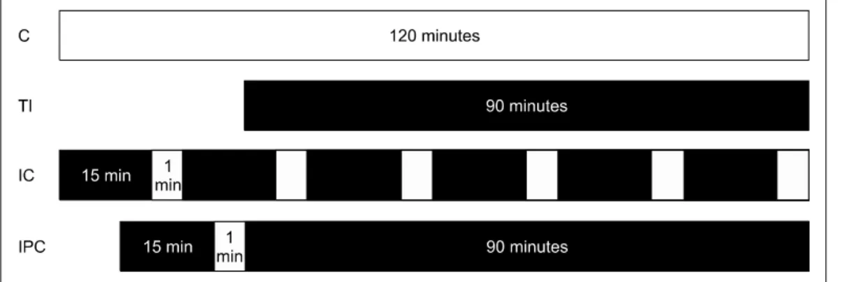
Fig. 1. Experimental design. The white area represents reperfusion time, and the black area represents ischemia time. C, control; TI, total ischemia; IC, intermittent clamping; IPC, ischemic preconditioning. The C group underwent sham operations (laparotomy with exposure of the liver but without vascular occlusion), the TI group underwent continuous clamping with 90 minutes of ischemia, the IC group underwent six cycles of intermittent clamping with 15 minutes of ischemia and 5 minutes of reperfusion, and the IPC group underwent clamping with 90 minutes of ischemia after IPC (15 minutes of clamping followed by 5 minutes reperfusion).
chemia is not necessarily a cause of IRI, but it can protect against hepatic IRI [3]. For example, an endogenous pro- tective effect of IPC against IRI after sublethal ischemia and reperfusion was reported for myocardial tissue [1].
Extensive studies have been performed to reveal the mechanism of IRI and the protective effect of temporal is- chemic conditions [2,4-9]. Unfortunately, traditional bio- chemical approaches have been unsuccessful.
cDNA microarray is a novel genomic research method that permits simultaneous parallel expression analysis of multiple genes. In addition, it provides useful genetic in- formation for several physiologic conditions [10,11].
Several reports have examined the association of genomic information with IRI and the protective effects of IC and IPC [10-12].
The focus of the current study was to compare 1) the ex- pression of immediately transcribed genes using cDNA microarray and 2) the degree of ischemic injury by histo- pathologic examination among various rat hepatic ische- mia models that mimic the conditions of clinical partial hepatectomy in humans.
METHODS
Rats
Experimental animals were maintained in a pyogen- free (SPF) animal facility at the Clinical Research Center,
Yonsei University College of Medicine, under constant room temperature (22oC) and humidity (55%), with a sup- ply of sterile drinking water and sterile animal feed ac- cording to the standards of the Association of Assessment and Accreditation of Laboratory Animal Care Interna- tional. In addition, animal experiments were conducted after approval (approval number; 04-086) of the experi- ment procedures by the ethical animal experiment guide- line established by the Department of Experimental Animals, Clinical Medical Research Center, Yonsei University College of Medicine.
All experiments were performed using 5-week-old male Sprague-Dawley rats weighing 150 to 200 g. The rats were housed under constant environmental conditions with a 12-hour light-dark cycle and fed a laboratory diet of water and rat chow. Twelve rats were randomly and even- ly divided into 4 groups as follows: control (C), total ische- mia (TI), IC, and IPC.
Protocols of hepatic ischemic conditions
The C group underwent a sham operation without vas- cular clamping. The TI group underwent continuous vas- cular clamping for 90 minutes. The IC group underwent six cycles of intermittent vascular clamping with 15 mi- nutes of ischemia and 5 minutes of reperfusion. The IPC group underwent vascular clamping for 90 minutes after 15 minutes of clamping followed by 5 minutes of re- perfusion (Fig. 1).
Vascular clamping operation
All surgical procedures were performed under general anesthesia using 30 mg/kg Zoletil (Virbac, Carros, France) via intraperitoneal injections. During anesthesia, body temperature was maintained between 36.5oC and 37.5oC using a heating pad. After a midline laparotomy, the falci- form ligament was sectioned. For visualization of the hep- atic hilum, traction of the duodenum and the middle lobe (ML)/left lateral lobe (LLL) was performed towards the left and cranial direction in that order. After dissection of the hepatic hilum, branches of the portal triad to the ML/LLL were identified and prepared for clamping. All structures in the portal triad to the ML/LLL were occluded by a microvascular clamp (Aesculap, San Francisco, CA, USA) according to specified protocols. Color changes of the ML/LLL were confirmed after clamping the triad to the ML/LLL. During the ischemic period, the abdomen was temporarily closed, and portal blood flow to the right and caudate lobes was maintained. Immediately after each round of ischemia, the ML/LLL were resected in all rats and prepared for histopathologic examination and micro- array.
Liver function test
Blood samples were taken from caudal vessels for liver function tests after hepatic resections. Supernatant was sampled after an immediate centrifugation of blood sam- ples at 4,500 rpm for 10 minutes. Using an automated bio- chemical analyzer (30FR, Toshiba, Tokyo, Japan), total se- rum protein and albumin levels were measured using biuret and bromocresol green methods, respectively.
Bilirubin levels were evaluated using the Jendrassik-Grof method at 600 nm. Alkaline phosphatase levels were esti- mated using the p-nitrophenyl phosphate substrate meth- od at 405 nm. Aspartate transaminase and alanine trans- aminase levels were measured using an ultraviolet-rate method at 340 nm.
Histopathologic examination
The excised ischemic lobes were fixed using 10% for- malin and embedded in paraffin. Tissues were cut into 5-μm-thick sections and stained using either hematox- ylin-eosin (H-E) or terminal deoxyuridine triphosphate
nick end labeling (TUNEL). All histopathologic assess- ments were performed by a pathologist who was blinded to the treatment protocols.
Assessment of H-E staining
The degree of liver damage was analyzed using light microscopy under ×400 magnification by a point-counting method as follows: grade 0, minimal or no evidence of in- jury; grade 1, mild injury consisting of cytoplasmic vacuo- lation and focal nuclear pyknosis; grade 2, moderate to se- vere injury with extensive nuclear pyknosis, cytoplasmic hypereosinophilia, and loss of intercellular borders; grade 3, severe necrosis with the disintegration of hepatic cords, hemorrhage, and neutrophil infiltration [13].
TUNEL staining
Cleavage of genomic DNA during apoptosis results in DNA strand breaks that can be identified by labeling free 38-hydroxyl ends with modified nucleotides in an enzy- matic reaction involving terminal deoxynucleotidyl trans- ferase [14-16]. The prepared paraffin-embedded sections were stained using a commercially available kit (Apop Tag Peroxidase In Situ Apoptosis Detection Kit S7100;
Chemicon International Inc., Billerica, MA, USA) accord- ing to the manufacturer’s protocols. Hepatocytes were ex- amined using light microscopy under ×400 magnification.
Morphometric analysis of the fluorescent cells was per- formed to determine the number of TUNEL-positive (apoptotic) cells in 25 random fields.
Microarray
RNA isolation
Each fragment of rat liver was lysed and homogenized.
RNA was extracted from each lysate by using RNeasy kits (Qiagen, Basel, Switzerland) according to the manu- facturer’s protocols. RNA quality was assessed using ul- traviolet absorption at 260/280 nm by spectrophotometry and gel electrophoresis. After confirming that all samples had 18S and 28S rRNA peaks without degradation, pre- pared RNA from each rat was pooled [17].
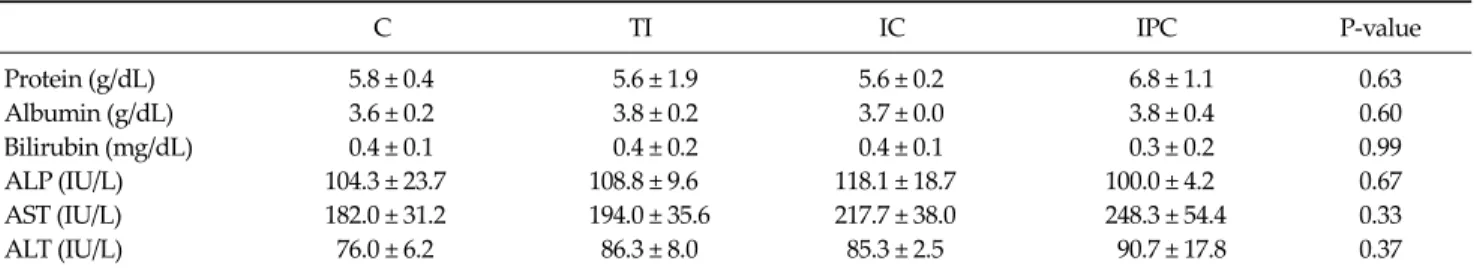
C TI IC IPC P-value
Protein (g/dL) 5.8 ± 0.4 5.6 ± 1.9 5.6 ± 0.2 6.8 ± 1.1 0.63
Albumin (g/dL) 3.6 ± 0.2 3.8 ± 0.2 3.7 ± 0.0 3.8 ± 0.4 0.60
Bilirubin (mg/dL) 0.4 ± 0.1 0.4 ± 0.2 0.4 ± 0.1 0.3 ± 0.2 0.99
ALP (IU/L) 104.3 ± 23.7 108.8 ± 9.6 118.1 ± 18.7 100.0 ± 4.2 0.67
AST (IU/L) 182.0 ± 31.2 194.0 ± 35.6 217.7 ± 38.0 248.3 ± 54.4 0.33
ALT (IU/L) 76.0 ± 6.2 86.3 ± 8.0 85.3 ± 2.5 90.7 ± 17.8 0.37
Values are presented as mean ± SD.
C, control; TI, total ischemia; IC, intermittent clamping; IPC, ischemic preconditioning; ALP, alkaline phosphatase; AST, aspartate transaminase; ALT, alanine transaminase.
Table 1. Liver function test cDNA microarray
cDNA microarray was performed using four Code- LinkTM Bioarray UniSet Rat I Chips (Amersham Bioscien- ces, Piscataway, NJ, USA) that were designed to report on 10,000 genes in the rat genome. Every procedure was per- formed according to the manufacturer's protocols as follows. Total RNA was reverse-transcribed into sin- gle-strand cDNA. Afterward, the second strand was used to synthesize double-stranded cDNA. Priming with anch- ored oligo(dT) directed the start of cDNA synthesis from the 5' end of the poly(A) tail. During reverse transcription, the cDNA strands of the C group and experimental groups were labeled by cyanine (Cy)-3 and Cy-5 fluorescent dyes, respectively. The labeled cDNA was hybridized to the pre- pared cDNA chip (CodeLink Bioarray UniSet Rat I) at 58oC in water for 16 hours and then washed with hybrid- ization wash buffer, followed by a subsequent wash with chemiluminescence rinse buffer.
Data analysis for the cDNA microarray
Fluorescence intensities were captured and converted to numerical values using a ScanArray Lite Microarray Analysis System (Packard Biochip Technologies, Billerica, MA, USA). Signal intensities were imported into GenPlex ver. 3.0 (Istech, Goyang, Korea) and normalized to reduce interarray variation in hybridization intensity after filter- ing out probe sets with unreliable intensity values.
Expression changes were presented as fold changes in comparison with the control group intensities. Then, the expression intensities were transformed into log2 values.
Finally, the genes with greater than 2-fold changes in tran- scription in the cDNA microarray were identified.
Statistical analysis for assays other than micro- array
All data were expressed as means ± standard deviation and analyzed using SPSS ver. 17.0 (SPSS Inc., Chicago, IL, USA). The Kruskal-Wallis test was used for comparing liv- er function tests and histopathologic findings. Tukey’s post hoc test was used for analysis between groups. A val- ue of P < 0.05 (two-sided tests) was considered statisti- cally significant.
RESULTS
Liver function test
There were no significant differences in liver function among the groups for all parameters tested (P > 0.05) (Table 1).
Histopathology examinations
H-E staining
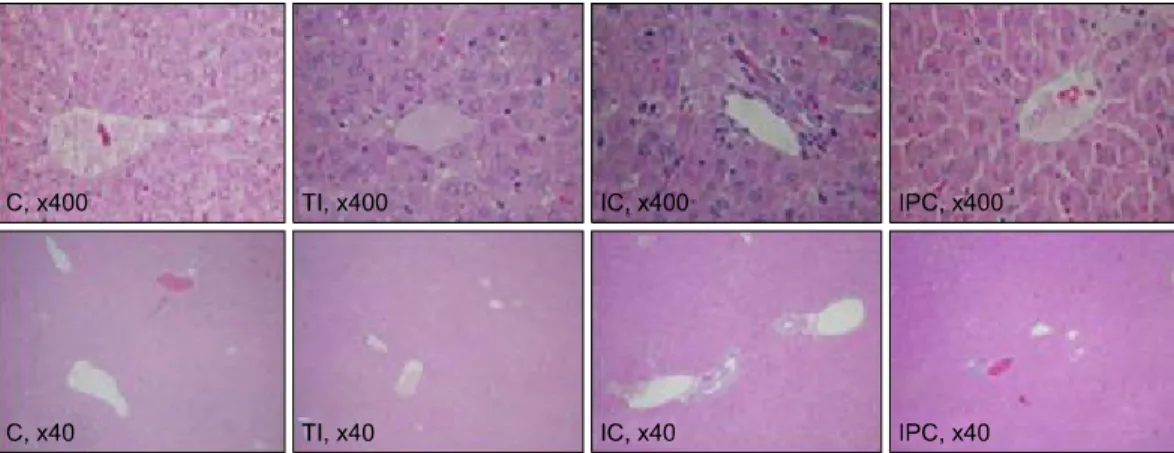
The C group had minimal or no evidence of injury (grade 0), whereas the experimental groups exhibited grade 1 injury (Fig. 2).
TUNEL staining
The number of TUNEL-positive cells was significantly higher in the experimental groups than in the C group (2.0
± 1.4, 57.0 ± 23.9, 7.7 ± 9.4, and 21.0 ± 22.3 in C, TI, IC, and IPC groups, respectively; P < 0.05 for all three groups [Figs. 3, 4]). Among the experimental groups, the TI group had significantly more positive hepatocytes than the other
Fig. 3. Terminal deoxyuridine triphosphate nick end labeling (TUNEL) staining (×40 or ×400 magnification). The TI group had significantly more TUNEL-positive hepatocytes than the C and IC groups. There were no significant differences in TUNEL-positive hepatocytes among the C, IC, and IPC groups. C, control; TI, total ischemia; IC, intermittent clamping; IPC, ischemic preconditioning.
Fig. 2. Hematoxylin-eosin staining of resected livers (×40 or ×400 magnification). The degree of liver damage was determined at ×400 magnification by using a point-counting method as follows: grade 0, minimal or no evidence of injury; grade 1, mild injury consisting of cytoplasm vacuolation and focal nuclear pyknosis; grade 2, moderate to severe injury with extensive nuclear pyknosis, cytoplasmichypereosinophilia, and loss of intercellular borders; grade 3, severe necrosis with disintegration of hepatic cords, hemorrhage, and neutrophil infiltration. The C group had minimal or no evidence of injury (grade 0). All three treatment groups exhibited grade 1 liver damage. C, control; TI, total ischemia; IC, intermittent clamping; IPC, ischemic preconditioning.
two groups.
cDNA microarray
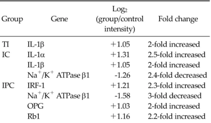
Interleukin (IL)-1β expression was 2-fold higher in the TI groups than in the C group (log2 TI/C = 1.05). Compared to the expression in the C group, IL-1α/IL-1β expression was 2-fold higher in the IC group (log2 IC/C = 1.31 and 1.05), but Na+/K+ ATPase β1 expression was 2-fold lower (log2 IC/C = -1.26).
In the IPC group, the expression of interferon regu- latory factor 1 (IRF-1) was 2-fold higher than that in the C group (log2 IPC/C = 1.21; Table 2, Fig. 5); conversely, Na+/K+ ATPase β1 expression was decreased by approximately
50% (log2 IPC/C = -1.58). The expression of both tumor ne- crosis factor receptor superfamily (member 11b; osteopro- tegerin (OPG); log2 IPC/C = 1.03) and retinoblastoma (Rb)1 expression (log2 IPC/C = 1.16) was approximately 2-fold higher in the IPC group than in the C group.
DISCUSSION
This study was performed to identify the degree of is- chemic injury and changes in gene transcription in various rat hepatic ischemia models. These models were chosen because they reflect the ischemic conditions of human
Fig. 4. Number of terminal deoxyuridine triphosphate nick end labeling (TUNEL)-positive cells. The number of TUNEL- positive cells was significantly higher in the experimental groups than in the C group. Among the experimental groups, the total ischemia (TI) group had significantly more positive hepatocytes than the other two groups. IC, intermittent clamping; IPC, ischemic precon- ditioning. *Statistical significance was tested using Kruskal-Wallis test of variance among groups. The comparison between the groups was performed using Tukey's post hoc test.
Fig. 5. Hierarchical clustering of the intermittent clamping (IC), ischemic preconditioning (IPC), and total ischemia (TI) groups. The hierarchical clustering of the IC, IPC, and TI group reveals different genetic expression patterns among the groups regarding imme- diate gene transcription in the livers of rats.
Group Gene
Log2
(group/control intensity)
Fold change
TI IL-1β +1.05 2-fold increased
IC IL-1α +1.31 2.5-fold increased
IL-1β +1.05 2-fold increased
Na+/K+ ATPase β1 -1.26 2.4-fold decreased
IPC IRF-1 +1.21 2.3-fold increased
Na+/K+ ATPase β1 -1.58 3-fold decreased
OPG +1.03 2-fold increased
Rb1 +1.16 2.2-fold increased
TI, total ischemia; IL, interleukin; IC, intermittent clamping; IPC, ischemic preconditioning; IRF, interferon regulatory factor; OPG, osteoprotegerin; Rb, retinoblastoma.
Table 2. Genes with greater than 2-fold changes in transcription in the cDNA microarray
hepatectomy including IC and IPC. In liver function tests and histopathologic examinations in the current study, there were no significant differences among the different ischemic conditions. We believe it is reasonable outcome because every sample was obtained just after the end of stimuli. However, even though there was no significant difference between groups, higher liver enzyme level was expected in TI group. Moreover the liver enzyme level of control C group was slightly higher than reference value of Sprague-Dawley Rats. Even though it is not considerable
difference, we cannot completely rule out the bias related with hepatotoxicity caused by hypoxemia or anesthetics.
However, TUNEL staining may distinguish differences in the degree of ischemia. It implies IC and IPC model may militate in favor of hepatocyte survival in comparison to TI model.
In previous studies, liver function tests and histopatho- logic examinations were useful for comparing the effects of IPC and TI [18]. Likewise, TUNEL staining has been considered a feasible method to assess the degree of apop- tosis in hepatic ischemia [19]. However, those studies had reperfusion periods of 1 to 6 hours before sampling speci- mens after the ischemic periods, and that period may al- low for more cellular changes than observed in the current study. This type of study design uses another reperfusion period before sampling specimens, which is different from the procedures used clinically in humans. Therefore, the appropriateness of biochemical and histopathologic ex- aminations and TUNEL staining to assess immediate dif- ferences between IC and IPC through 90 minutes of hep- atic ischemia remains to be evaluated in future studies.
The cDNA microarray used in the current study is a powerful method for revealing altered gene transcription
in early and delayed IPC [20]. Early IPC protects the tissue within minutes of reperfusion, but the protection is only maintained for 1 to 2 hours. It is considered a process in- dependent of protein synthesis and relies on preexisting substances as effectors [20]. The delayed preconditioning exerts its effect 24 hours after reperfusion and this effect may last for several days. It is thought that the delayed IPC relies on altered gene expression, resulting in the pro- duction of new proteins. A few reports have investigated gene transcription in IPC using cDNA microarrays [10,11,21,22]. Chen et al. [21] compared gene transcription between C and IPC groups using rats. Similarly as the cur- rent study, they only analyzed differences of 2-fold or greater in gene expression. Forty-three genes with sig- nificantly altered expression patterns were discovered in their report. The current study revealed the following al- terations in gene expression: IL-1β in the TI group, IL-1α/β and Na+/K+ ATPase β1 in the IC group, and IRF-1, OPG, Rb1, and Na+/K+ ATPase β1 in the IPC group. There were no common alterations of gene transcription between our study and Chen’s study [21]. Chen et al. [21] used 150 mi- nutes of reoxygenation time following IPC. Conversely, in the current study, the liver lobes were resected immedi- ately after the stimulus was discontinued. The time differ- ence between the two studies should be considered when comparing the results. The resulting changes in gene ex- pression in both studies may contribute to delayed IPC, but the genes with altered expression in the current study are more likely to act as triggers rather than mediators of IPC. IRI is believed to result from the activation of the proinflammatory cascade. Although no common alter- ation in gene expression was observed in the two studies, IL-10, a well-known anti-inflammatory cytokine, was sig- nificantly overexpressed in Chen's report [21]. In the cur- rent study, IL-1α and IL-1β, two proinflammatory cyto- kines, were overexpressed in the TI and IC groups, which is consistent with Chen’s results. The transcription of IL-1α in the TI group was 1.7-fold greater than that in the C group in the current study.
In the current study, the β subunit of Na+/K+ ATPase was underexpressed. Na+/K+ ATPase was also underex- pressed in a previous study with a design similar to that of the current study. Navarro-Sabate et al. [11] profiled the
gene expression patterns associated with long, cold ische- mia and IPC. In that study, a number of genes were in- volved in cellular physiologic processes that were downregulated. Decreased Na+/K+ ATPase activity may contribute to apoptosis in ischemic hepatocytes, preserv- ing ATP [23]. ATP preservation prevents cell necrosis but appears to provide the necessary substrate for cells to un- dergo apoptosis instead [24]. This switch from necrosis to apoptosis may be beneficial for cell survival. However, this hypotheses requires further investigation.
Originally, apoptosis was believed to be completely dif- ferent from necrosis. However, they were recently identi- fied to have a shared intracellular pathway called necroa- poptosis [15,25]. Therefore, proapoptotic gene tran- scription is not necessarily protective against IRI and is now thought to be a potential risk. In fact, increased ex- pression of B cell lymphoma (Bcl)-2, a well-known an- ti-apoptotic gene, was observed after IPC in the previous study [10]. Conversely, the expression of Rb1 was prom- inent in the current study. Rb1 was known as a proa- poptotic, tumor suppressor gene [26]. However, the recent evidences have demonstrated the existence of additional, cell type specific Rb1-encoded protein functions in cellular differentiation and survival [26]. Therefore the effect of over-expression of Rb1 can vary depending on the cellular circumstances. Especially, under stress such as hypoxia, apoptosis decreased caused by Rb1 loss [26].
The results of the current study, including over- expression of IL-1α and IL-1β in the TI and IC groups, ap- pear consistent with those of Barrier’s study [10]. Barrier et al. [10] used cDNA microarrays to compare global gene expression in liver biopsies from living human liver do- nors who did or did not undergo IPC just before liver devascularization. The IPC group exhibited significant overexpression of an IL-1 receptor antagonist (IL-1Ra) and Rb1. IL-1R is known to inhibit the effects of IL-1α and IL-1β by competing for type I and type II IL-1 receptors, result- ing in reduced inflammation [27].
OPG is a member of the tumor necrosis factor receptor superfamily [28]. High OPG level is believed to be an im- portant factor of atherosclerosis associated with vascular calcification [29]. However, OPG could promote cell sur- vival by inhibiting tumor necrosis factor-related apoptosis
[30]. The over-expression of OPG in current study is con- sistent with the previous studies.
The results of the current study were different from pre- vious studies with regard to methodology. However, the characteristics of altered gene expression were consistent with those of other studies. In both the current and pre- vious studies, genes that were overexpressed in the IPC group commonly exerted antiapoptotic and antiinflam- matory effects.
It is not simple to determine whether IPC or IC is the more beneficial procedure for liver surgery. Each method has benefits and drawbacks. IPC has advantages in terms of less intraoperative blood loss and a shorter operation time than IC. However, for longer periods of operation, IC was superior in several studies [3,19]. In addition, the pro- tective effect of IC was superior to that of IPC in steatotic patients [2]. One cannot determine the superiority of these procedures based on the current study because it was not designed for that purpose. Nevertheless, the overexpres- sion of IL-1α and IL-1β in the IC group is considered a re- sult of IRI. Conversely, the OPG, Rb1, and Na+/K+ ATPase β1 genes, which had altered expression in the IPC group, were related to the mechanism protecting against IRI. It can be postulated that the IC group underwent IRI due to repeated reperfusion, but this requires further evaluation.
The current study has several limitations. The cDNA microarray analysis was performed only once because of the relatively small number of rats. Repeated microarrays are needed for reliability. Moreover, clustering analysis was not performed due to lack of repetitive microarrays.
However, we consider this study as a pilot study, and we expect better outcomes in additional study based on this study. In addition, we used 2-fold gene expression change as a cutoff. We think outcomes come from the higher cutoff value are potentially more important genes. Moreover we used pooled method [17]. We believe that may be a factor to improve the limitation from the small number of speci- mens.
In conclusion, the current study provided valuable in- formation about the clinical temporal hepatic occlusion method for studying immediate hepatic changes in vari- ous rat ischemia models, including 90 minutes of ische- mia, because of the similarity of the protocols.
CONFLICTS OF INTEREST
No potential conflict of interest relevant to this article was reported.
REFERENCES
1. Murry CE, Jennings RB, Reimer KA. Preconditioning with ischemia: a delay of lethal cell injury in ischemic myocar- dium. Circulation 1986;74:1124-36.
2. Petrowsky H, McCormack L, Trujillo M, Selzner M, Jochum W, Clavien PA. A prospective, randomized, con- trolled trial comparing intermittent portal triad clamping versus ischemic preconditioning with continuous clamp- ing for major liver resection. Ann Surg 2006;244:921-8.
3. Clavien PA, Selzner M, Rudiger HA, Graf R, Kadry Z, Rousson V, et al. A prospective randomized study in 100 consecutive patients undergoing major liver resection with versus without ischemic preconditioning. Ann Surg 2003;
238:843-50.
4. Fernandez L, Carrasco-Chaumel E, Serafín A, Xaus C, Grande L, Rimola A, et al. Is ischemic preconditioning a useful strategy in steatotic liver transplantation? Am J Transplant 2004;4:888-99.
5. Hong F, Radaeva S, Pan HN, Tian Z, Veech R, Gao B.
Interleukin 6 alleviates hepatic steatosis and ischemia/re- perfusion injury in mice with fatty liver disease. Hepatol- ogy 2004;40:933-41.
6. Koti RS, Yang W, Glantzounis G, Quaglia A, Davidson BR, Seifalian AM. Effect of ischaemic preconditioning on hep- atic oxygenation, microcirculation and function in a rat model of moderate hepatic steatosis. Clin Sci (Lond) 2005;108:55-63.
7. Selzner N, Selzner M, Jochum W, Clavien PA. Ischemic preconditioning protects the steatotic mouse liver against reperfusion injury: an ATP dependent mechanism. J Hepatol 2003;39:55-61.
8. Serafín A, Rosello-Catafau J, Prats N, Xaus C, Gelpi E, Peralta C. Ischemic preconditioning increases the tolerance of Fatty liver to hepatic ischemia-reperfusion injury in the rat. Am J Pathol 2002;161:587-601.
9. Vendemiale G, Grattagliano I, Caraceni P, Caraccio G, Domenicali M, Dall'Agata M, et al. Mitochondrial oxida- tive injury and energy metabolism alteration in rat fatty liver: effect of the nutritional status. Hepatology 2001;33:
808-15.
10. Barrier A, Olaya N, Chiappini F, Roser F, Scatton O, Artus C, et al. Ischemic preconditioning modulates the ex- pression of several genes, leading to the overproduction of IL-1Ra, iNOS, and Bcl-2 in a human model of liver ische- mia-reperfusion. FASEB J 2005;19:1617-26.
11. Navarro-Sabate A, Peralta C, Calvo MN, Manzano A, Massip-Salcedo M, Rosello-Catafau J, et al. Mediators of rat ischemic hepatic preconditioning after cold preserva-
tion identified by microarray analysis. Liver Transpl 2006;12:1615-25.
12. Butte A. The use and analysis of microarray data. Nat Rev Drug Discov 2002;1:951-60.
13. Bilbao G, Contreras JL, Eckhoff DE, Mikheeva G, Krasnykh V, Douglas JT, et al. Reduction of ischemia-reperfusion in- jury of the liver by in vivo adenovirus-mediated gene transfer of the antiapoptotic Bcl-2 gene. Ann Surg 1999;230:
185-93.
14. Gao W, Bentley RC, Madden JF, Clavien PA. Apoptosis of sinusoidal endothelial cells is a critical mechanism of pres- ervation injury in rat liver transplantation. Hepatology 1998;27:1652-60.
15. Gujral JS, Bucci TJ, Farhood A, Jaeschke H. Mechanism of cell death during warm hepatic ischemia-reperfusion in rats: apoptosis or necrosis? Hepatology 2001;33:397-405.
16. Jin LM, Liu YX, Zhou L, Xie HY, Feng XW, Li H, et al.
Ischemic preconditioning attenuates morphological and biochemical changes in hepatic ischemia/reperfusion in rats. Pathobiology 2010;77:136-46.
17. Pan W, Lin J, Le CT. How many replicates of arrays are re- quired to detect gene expression changes in microarray ex- periments? A mixture model approach. Genome Biol 2002;3:research0022.
18. Ishii S, Abe T, Saito T, Tsuchiya T, Kanno H, Miyazawa M, et al. Effects of preconditioning on ischemia/reperfusion injury of hepatocytes determined by immediate early gene transcription. J Hepatobiliary Pancreat Surg 2001;8:461-8.
19. Jang JH, Kang KJ, Kang Y, Lee IS, Graf R, Clavien PA.
Ischemic preconditioning and intermittent clamping con- fer protection against ischemic injury in the cirrhotic mouse liver. Liver Transpl 2008;14:980-8.
20. Banga NR, Homer-Vanniasinkam S, Graham A, Al-Mukh- tar A, White SA, Prasad KR. Ischaemic preconditioning in transplantation and major resection of the liver. Br J Surg 2005;92:528-38.
21. Chen W, Qiu JF, Zhang ZQ, Luo HF, Rosello-Catafau J, Wu ZY. Gene expression changes after hypoxic precondition- ing in rat hepatocytes. Hepatobiliary Pancreat Dis Int 2006;5:416-21.
22. Raza A, Dikdan G, Desai KK, Shareef A, Fernandes H, Aris V, et al. Global gene expression profiles of ischemic pre- conditioning in deceased donor liver transplantation. Liver Transpl 2010;16:588-99.
23. Peralta C, Bartrons R, Riera L, Manzano A, Xaus C, Gelpí E, et al. Hepatic preconditioning preserves energy metabo- lism during sustained ischemia. Am J Physiol Gastrointest Liver Physiol 2000;279:G163-71.
24. Kim JS, Ohshima S, Pediaditakis P, Lemasters JJ. Nitric ox- ide protects rat hepatocytes against reperfusion injury mediated by the mitochondrial permeability transition.
Hepatology 2004;39:1533-43.
25. Jaeschke H. Reperfusion injury after warm ischemia or cold storage of the liver: role of apoptotic cell death.
Transplant Proc 2002;34:2656-8.
26. Goodrich DW. The retinoblastoma tumor-suppressor gene, the exception that proves the rule. Oncogene 2006;25:
5233-43.
27. Gabay C, Smith MF, Eidlen D, Arend WP. Interleukin 1 re- ceptor antagonist (IL-1Ra) is an acute-phase protein. J Clin Invest 1997;99:2930-40.
28. Fili S, Karalaki M, Schaller B. Therapeutic implications of osteoprotegerin. Cancer Cell Int 2009;9:26.
29. Venuraju SM, Yerramasu A, Corder R, Lahiri A. Osteopro- tegerin as a predictor of coronary artery disease and car- diovascular mortality and morbidity. J Am Coll Cardiol 2010;55:2049-61.
30. Jensen JK, Ueland T, Atar D, Gullestad L, Mickley H, Aukrust P, et al. Osteoprotegerin concentrations and prog- nosis in acute ischaemic stroke. J Intern Med 2010;267:
410-7.