Incidence of underlying biliary neoplasm in patients after major hepatectomy for preoperative benign hepatolithiasis
Hyeong Min Park, Young Hoe Hur, Chol Kyoon Cho, Yang Seok Koh, Hee Joon Kim, and Eun Kyu Park
Department of Surgery, Chonnam National University College of Medicine, Hwasun, Korea
Backgrounds/Aims: Despite hepatolithiasis being a risk factor for biliary neoplasm including cholangiocarcinoma, the incidence of underlying biliary neoplasm is unknown in patients with preoperative benign hepatolithiasis. The aim of this study was to evaluate the incidence of underlying biliary neoplasm in patients who underwent major hepatectomy for preoperative benign hepatolithiasis. Methods: Between March 2005 and December 2015, 73 patients who under- went major hepatectomy for preoperative benign hepatolithiasis were enrolled in this study. The incidence and patho- logical differentiation of concomitant biliary neoplasm were retrospectively determined by review of medical records.
Postoperative complications after major hepatectomy were evaluated. Results: Concomitant biliary neoplasm was pathologically confirmed in 20 patients (27.4%). Biliary intraepithelial neoplasia (BIN) was detected in 12 patients (16.4%), and 1 patient (1.4%) had intraductal papillary mucinous neoplasm (IPMN), as the premalignant lesion.
Cholangiocarcinoma was pathologically confirmed in 7 patients (9.6%). Preoperative imaging of the 73 patients re- vealed biliary stricture at the first branch of bile duct in 31 patients (42.5%), and at the second branch of bile duct in 39 patients (53.4%). Postoperative complications developed in 14 patients (19.1%). Almost all patients recovered from complications, including intra-abdominal abscess (9.6%), bile leakage (4.1%), pleural effusion (2.7%), and wound infection (1.4%). Only 1 patient (1.4%) died from aspiration pneumonia. Conclusions: The incidence of underlying biliary neoplasm was not negligible in the patients with hepatolithiasis, despite meticulous preoperative evaluations. (Ann Hepatobiliary Pancreat Surg 2016;20:173-179)
Key Words: Hepatolithiasis; Biliary neoplasm; Cholangiocarcinoma; Major hepatectomy
Received: July 6, 2016; Revised: September 22, 2016; Accepted: September 26, 2016 Corresponding author: Young Hoe Hur
Department of Surgery, Chonnam National University Hwasun Hospital, 322 Seoyang-ro, Hwasun 58128, Korea Tel: +82-61-379-7646, Fax: +82-61-379-7661, E-mail: [email protected]
Copyright Ⓒ 2016 by The Korean Association of Hepato-Biliary-Pancreatic Surgery
This is an Open Access article distributed under the terms of the Creative Commons Attribution Non-Commercial License (http://creativecommons.org/
licenses/by-nc/4.0) which permits unrestricted non-commercial use, distribution, and reproduction in any medium, provided the original work is properly cited.
Annals of Hepato-Biliary-Pancreatic Surgery ∙ pISSN: 2508-5778ㆍeISSN: 2508-5859
INTRODUCTION
Hepatolithiasis is defined as the presence of stones within the intrahepatic bile ducts, proximal to the right and left hepatic ducts. Hepatolithiasis has a poor prog- nosis due to other associated complications such as re- current cholangitis, biliary strictures, liver abscess, liver atrophy, or cirrhosis.1,2 Additionally, hepatolithiasis is an important leading cause of intrahepatic cholangio- carcinoma.3,4
Hepatolithiasis treatment includes both non-surgical and surgical approaches. Non-surgical procedures, such as per- cutaneous transhepatic cholangioscopic lithotripsy, show a high clearance rate of intrahepatic stones; however, these treatments are not effective for preventing recurrence of
hepatolithiasis and removal of benign biliary strictures.5-7 Surgical treatment can be divided into minor and major hepatectomies, depending on the resection range. The range of minor hepatectomy is generally limited to atro- phic segments and intrahepatic stones. In this context, a residual intrahepatic biliary stricture may remain, which is a risk factor for hepatolithiasis. Unlike minor hep- atectomy, the range of major hepatectomy includes all in- trahepatic duct stones and a wider segment, including the intrahepatic biliary strictures. Thus, major hepatectomy can be the fundamental treatment for hepatolithiasis by eliminating not only the stones but also the benign biliary stricture, which is a crucial cause of the stones.
Previous studies have reported the rate of unidentified underlying cholangiocarcinoma before hepatectomy in pa-
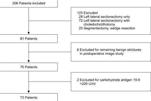
Fig. 1. Flow chart of patient se- lection for hepatic resection.
tients with hepatolithiasis.2,8 In these reports, it is un- certain whether the resection range includes all intra- hepatic biliary strictures and their segments. To confirm the incidence of underlying biliary neoplasm in patients with hepatolithiasis, we need to investigate patients who underwent major hepatectomy for both hepatolithiasis and intrahepatic biliary strictures.
Here, we evaluated the incidence of underlying biliary neoplasm in patients who underwent major hepatectomy for benign hepatolithiasis, based on preoperative diagnosis.
MATERIALS AND METHODS
Patients and outcome evaluation
The medical records of Chonnam National University Hospital were examined between March 2005 and December 2015. We identified 206 patients with possible benign hepatolithiasis detected on preoperative computed tomography (CT) or magnetic resonance (MR) imaging, who subsequently underwent hepatic resection. Of the 206 patients, 125 patients underwent minor hepatectomy, in- cluding left lateral sectionectomy for hepatolithiasis.
These were excluded because of possible residual stricture of the bile ducts related to hepatolithiasis after resection.
A total of 81 patients underwent major hepatectomy for hepatolithiasis. Of the 81 patients, 6 patients were ex-
cluded because of residual intrahepatic biliary stricture identified on the postoperative CT imaging after the procedure.
For the diagnosis of cholangiocarcinoma, the value of tumor markers remains controversial. Various cut-off val- ues of carbohydrate antigen 19-9 (CA 19-9) have been proposed.9 One study proposed 253 U/ml for CA 19-9 as the mean serum level for hilar cholangiocarcinoma in cas- es of International Union against Cancer tumor stage I.10 It is generally considered that CA 19-9 levels over 200 U/ml indicate a possibility of cholangiocarcinoma. In our study, 2 patients with serum CA 19-9 values above 200 U/ml on preoperative laboratory tests were excluded due to suspected underlying biliary malignancy. Finally, 73 patients who underwent major hepatectomy for benign hepatolithiasis as diagnosed on preoperative imaging and laboratory tests, were enrolled in this study (Fig. 1).
At our hospital, endoscopic retrograde chol- angiopancreatography (ERCP) is performed in patients having stones or strictures localized in the first branch of bile ducts. Patients with definite stones or strictures in other peripheral branches of intrahepatic bile ducts under- go major hepatectomy, regardless of the symptoms and ir- respective of the presence of atrophy.
The locations of hepatolithiasis and intrahepatic biliary strictures were evaluated on preoperative CT or MR chol- angiography in all patients. Since multiple strictures can
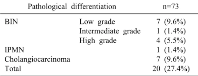
Table 1. The incidence of underlying biliary neoplasm Pathological differentiation n=73 BIN
IPMN
Cholangiocarcinoma Total
Low grade Intermediate grade High grade
7 (9.6%) 1 (1.4%) 4 (5.5%) 1 (1.4%) 7 (9.6%) 20 (27.4%) BIN, biliary intraepithelial neoplasia; IPMN, intraductal pap- illary mucinous neoplasm
be present in the intrahepatic ducts, we designated the biliary stricture located farthest distal in the direction of the bile flow as the main stricture site.
The incidence and the pathological differentiation of a concomitant biliary neoplasm were retrospectively eval- uated through review of the medical records for all patients. The postoperative complications after major hep- atectomy were also evaluated.
Definitions
Benign hepatolithiasis was defined as hepatolithiasis with a concomitant stricture of the intrahepatic bile duct, with no clinical evidence of malignancy on CT or MRI, or on preoperative laboratory tests.
Major hepatectomy was defined as the resection of 3 or more Couinaud’s liver segments, while segmentectomy of 1 or 2 segments and non-anatomical wedge resection were classified as minor hepatectomy.11
The first branch of the intrahepatic bile duct was de- fined from the confluence of the right and left hepatic ducts to the bifurcation of the right anterior sectoral duct and right posterior sectoral duct, or to the bifurcation of the segment IV duct and the left lateral sectoral duct. The second branch of the intrahepatic bile duct was defined from the proximal end of the first branch with bile flow to the confluence of the segment V duct and VIII duct, or to the confluence of the segment VI duct and VII duct, or to the confluence of the segment II duct and III duct.
The third branch was defined from the proximal end of the second branch to the proximal end of each segmental duct, according to the direction of bile flow.
Comparison of the benign stricture group and the biliary neoplasm group
Patient demographics including sex, age, preoperative symptoms (including abdominal pain, fever, jaundice, or weight loss), preoperative serum CA 19-9, presence of atrophy in preoperative image, and serum total bilirubin before the operation, were compared between the benign stricture group and pathologically proven biliary neoplasm group. The locations of the main intrahepatic biliary stric- tures and the type of surgical procedure applied were fur- ther evaluated between the two groups.
Statistics
Continuous variables were analyzed using the Mann-Whitney analysis. Categorical variables were com- pared using the Chi-squared test or Fisher’s exact test.
Age, preoperative serum CA 19-9, and preoperative total bilirubin were included as continuous variables, while sex, preoperative symptoms, locations of the main biliary stric- ture, type of operative procedure, presence of atrophy in preoperative image were considered as categorical variables. A p-value of <0.05 was considered statistically significant. The Statistical Package for Social Science ver- sion 21.0 was used for the analysis.
RESULTS
Of the 73 patients with preoperative benign hep- atolithiasis, 20 patients (27.4%) were confirmed as patho- logical biliary neoplasm, and 53 patients as chronic in- flammation with bile duct stones. Of the 20 patients, 12 (16.4%) presented with biliary intraepithelial neoplasia (BIN) and 1 (1.4%) with intraductal papillary mucinous neoplasm (IPMN) as the premalignant lesions. Cholangio- carcinoma was pathologically confirmed in 7 patients (9.6%) (Table 1).
The main intrahepatic biliary strictures were catego- rized according to their location at the confluence of the right hepatic duct and the left hepatic duct, at the first branch of the intrahepatic bile duct, at the second branch of the bile duct, or at the third branch of bile duct. Biliary stricture at the first branch of bile duct was observed in 23 patients (43.4%) of the benign stricture group and 8 patients (40.0%) of the biliary neoplasm group. A biliary stricture was identified at the second branch of the bile duct in 28 patients (52.8%) in the benign stricture group and in 11 patients (55%) in the biliary neoplasm group.
Table 3. Comparison of clinical characteristics between the benign stricture group and the biliary neoplasm group
Variables
Benign stricture n=53
Biliary neoplasm
n=20
p-value
Age [range] (years) Sex
Male Female
Operative procedure Left hepatectomy Right hepatectomy Left hepatectomy+S1 Right hepatectomy+S1 Symptom
No symptom Abdominal pain Fever
Jaundice Weight loss
Total bilirubin [range]
(mg/dl)
CA 19-9 [range] (U/ml) Atrophy
Absence of atrophy Presence of atrophy
61.13 [46-78]
16 (30.2%) 37 (69.8%) 35 (66.0%) 9 (17.0%) 8 (15.1%) 1 (1.9%) 15 (28.3%) 32 (60.4%) 5 (9.4%) 1 (1.9%) 0 (0%)
0.82 [0.2-7.9]
35.9 [0.5-157.2]
34 (64.2%) 19 (35.8%)
62.15 [25-75]
11 (55.0%) 9 (45.0%) 12 (60.0%) 3 (15.0%) 5 (25.0%)
0 (0%) 7 (35.0%) 10 (50.0%) 2 (10.0%)
0 (0%) 1 (5.0%)
0.65 [0.4-1.3]
21.545 [4.3-114.3]
8 (40.0%) 12 (60.0%)
0.36
0.05
0.772
0.571
0.482 0.85
0.063
S1, Liver segment I segmentectomy; CA 19-9, carbohydrate antigen 19-9
Table 2. Locations of the main biliary strictures in the benign stricture group and the biliary neoplasm group
Location of stricture
Benign stricture n=53
Biliary neoplasm
n=20
p-value Confluence of right and
left ducts
First-order branch of bile duct
Second-order branch of bile duct
Third-order branch of bile duct
1 (1.9%) 23 (43.4%) 28 (52.8%) 1 (1.9%)
1 (5.0%) 8 (40.0%) 11 (55.0%) 0 (0%)
0.784
Table 4. Complications after major hepatectomy according to Clavien-Dindo classification
Grade Complication n=73
IIIa
IIIb
V Total
Intra-abdominal abscess Bile leakage
Pleural effusion Wound infection Bile duct injury
Common bile duct primary repair site dehiscence
Aspiration pneumonia
7 (9.6%) 1 (1.4%) 2 (2.7%) 1 (1.4%) 1 (1.4%) 1 (1.4%) 1 (1.4%) 14 (19.1%) One patient each in the benign stricture group and in the
biliary neoplasm group had a biliary stricture at the con- fluence of the right and left hepatic ducts. A stricture at the third branch of bile duct was seen in 1 patient in the biliary stricture group. Postoperative pathological exami- nation confirmed that the location of the intrahepatic stric- tures was consistent with that of concomitant biliary neo- plasms (Table 2).
The variables considered were sex, age, the type of sur- gical procedure, location of the biliary stricture, pre- operative symptoms, preoperative serum CA 19-9, pres- ence of atrophy in preoperative image, and preoperative serum total bilirubin level. The main symptoms of patients with hepatolithiasis were abdominal pain, fever, jaundice, and weight loss. Results from the statistical analysis showed no significant difference when comparing the be- nign stricture group and the pathologically proven biliary neoplasm group. Although sex appeared to be significant, it was not a definitive factor (Table 3).
According to the Clavien-Dindo classification, post- operative complications above grade IIIa developed in 14 patients (19.1%). Of these, 10 patients (13.7%) were treat- ed by procedures such as pig-tail catheter insertion, for intra-abdominal abscess (9.6%), bile leakage (1.4%), or pleural effusion (2.7%). Three patients (4.1%) were re-op- erated for wound infection, bile duct injury, and common bile duct primary repair site dehiscence. Only 1 patient (1.4%) expired during the hospital stay as a result of aspi- ration pneumonia, and not by hepatic failure or surgical complications (Table 4).
DISCUSSION
Epidemiological, pathological, and genetic studies dem- onstrate a relationship between hepatolithiasis and cholan- giocarcinoma.12-14 The overall incidence of hepatoli- thiasis-related cholangiocarcinoma has been reported as 5% to 13%.15-17 In a cohort study, 65 years or older (hazard ratio, 3.029; p-value, 0.017) and having stone re- moval only as the initial treatment (hazard ratio, 2.873;
p-value, 0.012) were found to be significant risk factors
for the development of cholangiocarcinoma.18 The authors reported that hepatectomy could significantly reduce the risk of developing cholangiocarcinoma (hazard ratio, 0.066; p-value, 0.010). In another cohort study, it was re- ported that hepatectomy significantly reduced the risk of developing cholangiocarcinoma.19 In general, hepatic re- section might offer an advantage in eliminating the risk of developing cholangiocarcinoma because of complete removal of both the intrahepatic stones and the bile ducts involved, which are likely to have a hidden malignancy.
In this regard, hepatic resection could be considered as a primary treatment in hepatolithiasis.
Despite preoperative evaluations, it is still difficult to detect underlying biliary neoplasms or chlangiocarcinoma in hepatolithiasis. Catena et al.20 reported that the rate of unrecognized cholangiocarcinoma was quite high at 11.7%, and that it might be underestimated. Although se- rum CA 19-9 is known as a tumor biomarker for chol- angiocarcinoma, its levels could be normal or increased in benign diseases, such as in bacterial cholangitis or choledocholithiasis.21,22 In preoperative images or even during the surgery, a diagnosis of cholangiocarcinoma as- sociated with hepatolithiasis is difficult, because the af- fected liver segment is often fibrotic and scarred.2 Chen et al.2 reported 2 patients (2.0%) having the tumor diag- nosed during the operation, and 4 patients (3.9%) having cholangiocarcinoma diagnosed only after pathological examination. They also reported that in 1 patient (1.0%), a cholangiocarcinoma was diagnosed 7 months after the hepatic resection for hepatolithiasis, and the tumor was likely present during the surgery, but was unidentified, and the undetected cholangiocarcinoma was present in the remnant liver even after hepatectomy.
Hepatic resection is regarded as an established treat- ment, recommended for its ability to resolve not only the stones but also the strictures.23,24 Biliary strictures, found in 42% to 96% of patients with hepatolithiasis,25-27 may be related to biliary carcinogenesis as an aspect of re- current cholangitis, bile stasis, and bacterial infections.
Biliary stricture is associated with recurrent and chronic inflammations causing prolonged inflammation of the bile duct epithelium, leading to the subsequent development of cholangiocarcinoma.28,29 Up to 50% of patients with ma- lignant biliary strictures may have coexistent hepatolithia- sis.30 To prevent the development of cholangiocarcino-
genesis, hepatic resection, including the biliary stricture, would be considered in cases of hepatolithiasis.
Minor hepatectomy, which involves segmentectomy of 1 or 2 segments and non-anatomical wedge resection, does not include either the first branch or the second branch of the bile duct in the range of resection. In left lateral sectionectomy, where hepatic resection including segments II and III, the resection range includes only the third branch of the bile duct in segments II and III, and does not include the confluence of the segments II duct and III duct, or the second branch of the bile duct. In our study, biliary strictures in hepatolithiasis were mostly lo- cated in the first and second branches of the intrahepatic bile duct, and the location of the concomitant biliary neo- plasms was consistent with the location of the biliary strictures. Among all the patients who underwent major hepatectomy in benign hepatolithiasis, 31 patients (42.5%) had a biliary stricture at the first branch of bile duct, and 39 patients (53.4%) had a biliary stricture at the second branch of bile duct. Only 1 patient (1.4%) had a biliary stricture at the third branch of the bile duct. In order to completely remove biliary strictures and thus eliminate the hidden biliary neoplasms, major hepatectomy, including the first and second branches of bile duct, would be an appropriate approach in the treatment of hepatolithiasis.
Immunohistochemical studies have identified 2 types of neoplastic lesions preceding invasive intrahepatic chol- angiocarcinoma in hepatolithiasis: BIN and IPNM.31,32 Ohta et al.33 observed hyperplasia in the epithelium of the bile ducts associated with chronic cholangitis related to hepatolithiasis, and suggested that mucosal dysplasia may be a precursor to the cholangiocarcinoma. It seems that elimination of BIN and IPNM, potential precursors of cholangiocarcinoma, may be required to prevent the de- velopment of cholangiocarcinoma in hepatolithiasis.
There also exist concerns regarding surgical complica- tions after major hepatectomy. The surgical techniques of hepatectomy and the perioperative management of pa- tients have sufficiently developed in the past decade, re- sulting in a marked decrease in the morbidity and mortal- ity following liver surgery.34-36 In one study, the cumu- lative survival rates, excluding unrelated deaths, did not differ significantly between the hepatic resection group and the cholangioscopic lithotomy group.25 In another study evaluating the outcome of hepatectomy for hep-
atolithiasis, 7 patients (6.8%) underwent re-operation and 2 patients (1.9%) died during hospital stay.2 In our study, 3 patients (4.1%) underwent re-operation and only 1 pa- tient (1.4%) died during hospital stay. The rate of post- operative complication in our study was not significantly different compared to that of other studies.
Our study presents some limitations, including its retro- spective nature and the relatively small number of patients involved. Also, there was no comparison of the post- operative complications in major hepatectomy and minor hepatectomy in patients with hepatolithiasis. Finally, no long-term follow-up was performed to confirm that there would be a significant difference in the development of cholangiocarcinoma between major hepatectomy and mi- nor hepatectomy in benign hepatolithiasis.
In our study, the incidence of biliary neoplasm (including cholangiocarcinoma) not detected before hep- atectomy was 27.4% in benign hepatolithiasis. Although preoperative evaluations were performed in patients with hepatolithiasis, in order to exclude underlying biliary neo- plasm, the incidence of biliary neoplasm was not negligible. Major hepatectomy as the definite treatment was safely performed for these patients. The presence of cholangiocarcinoma was a significant factor influencing the long-term survival in hepatolithiasis patients, as dem- onstrated by multivariate analysis.2,14 Major hepatectomy, including not only hepatolithiasis but also concomitant strictures of intrahepatic bile duct, would be one of the appropriate treatment options for preoperative benign hepatolithiasis. However, further research is required to compare major hepatectomy with other treatments, for a prolonged period.
REFERENCES
1. Lee TY, Chen YL, Chang HC, Chan CP, Kuo SJ. Outcomes of hepatectomy for hepatolithiasis. World J Surg 2007;31:479-482.
2. Chen DW, Tung-Ping Poon R, Liu CL, Fan ST, Wong J.
Immediate and long-term outcomes of hepatectomy for hepatolithiasis. Surgery 2004;135:386-393.
3. Blechacz B, Komuta M, Roskams T, Gores GJ. Clinical diag- nosis and staging of cholangiocarcinoma. Nat Rev Gastroenterol Hepatol 2011;8:512-522.
4. Tyson GL, El-Serag HB. Risk factors for cholangiocarcinoma.
Hepatology 2011;54:173-184.
5. Lee SK, Seo DW, Myung SJ, Park ET, Lim BC, Kim HJ, et al. Percutaneous transhepatic cholangioscopic treatment for hep- atolithiasis: an evaluation of long-term results and risk factors for recurrence. Gastrointest Endosc 2001;53:318-323.
6. Jeng KS, Sheen IS, Yang FS. Percutaneous transhepatic chol- angioscopy in the treatment of complicated intrahepatic biliary strictures and hepatolithiasis with internal metallic stent. Surg Laparosc Endosc Percutan Tech 2000;10:278-283.
7. Yeh YH, Huang MH, Yang JC, Mo LR, Lin J, Yueh SK.
Percutaneous trans-hepatic cholangioscopy and lithotripsy in the treatment of intrahepatic stones: a study with 5 year follow-up.
Gastrointest Endosc 1995;42:13-18.
8. Kim HJ, Kim JS, Suh SJ, Lee BJ, Park JJ, Lee HS, et al.
Cholangiocarcinoma risk as long-term outcome after hepatic re- section in the hepatolithiasis patients. World J Surg 2015;39:
1537-1542.
9. Van Beers BE. Diagnosis of cholangiocarcinoma. HPB (Oxford) 2008;10:87-93.
10. Juntermanns B, Radunz S, Heuer M, Hertel S, Reis H, Neuhaus JP, et al. Tumor markers as a diagnostic key for hilar cholangiocarcinoma. Eur J Med Res 2010;15:357-361.
11. Fan ST. Hepatocellular carcinoma--resection or transplant? Nat Rev Gastroenterol Hepatol 2012;9:732-737.
12. Zhou Q, Gong Y, Huang F, Lin Q, Zeng B, Li Z, et al.
Expression levels and significance of nuclear factor-κB and epi- dermal growth factor receptor in hepatolithiasis associated with intrahepatic cholangiocarcinoma. Dig Surg 2013;30:309-316.
13. Lee KT, Chang WT, Wang SN, Chuang SC, Chai CY, Hu SW.
Expression of DPC4/Smad4 gene in stone-containing intrahepatic bile duct. J Surg Oncol 2006;94:338-343.
14. Kuroki T, Tajima Y, Kanematsu T. Hepatolithiasis and intra- hepatic cholangiocarcinoma: carcinogenesis based on molecular mechanisms. J Hepatobiliary Pancreat Surg 2005;12:463-466.
15. Uenishi T, Hamba H, Takemura S, Oba K, Ogawa M, Yamamoto T, et al. Outcomes of hepatic resection for hepatolithiasis. Am J Surg 2009;198:199-202.
16. Cheon YK, Cho YD, Moon JH, Lee JS, Shim CS. Evaluation of long-term results and recurrent factors after operative and nonoperative treatment for hepatolithiasis. Surgery 2009;146:
843-853.
17. Lin CC, Lin PY, Chen YL. Comparison of concomitant and sub- sequent cholangiocarcinomas associated with hepatolithiasis:
clinical implications. World J Gastroenterol 2013;19:375-380.
18. Suzuki Y, Mori T, Abe N, Sugiyama M, Atomi Y. Predictive factors for cholangiocarcinoma associated with hepatolithiasis determined on the basis of Japanese Multicenter study. Hepatol Res 2012;42:166-170.
19. Suzuki Y, Mori T, Yokoyama M, Nakazato T, Abe N, Nakanuma Y, et al. Hepatolithiasis: analysis of Japanese nation- wide surveys over a period of 40 years. J Hepatobiliary Pancreat Sci 2014;21:617-622.
20. Catena M, Aldrighetti L, Finazzi R, Arzu G, Arru M, Pulitanò C, et al. Treatment of non-endemic hepatolithiasis in a Western country. The role of hepatic resection. Ann R Coll Surg Engl 2006;88:383-389.
21. Khan SA, Davidson BR, Goldin R, Pereira SP, Rosenberg WM, Taylor-Robinson SD, et al. Guidelines for the diagnosis and treatment of cholangiocarcinoma: consensus document. Gut 2002;51 Suppl 6:VI1-VI9.
22. Dodson RM, Weiss MJ, Cosgrove D, Herman JM, Kamel I, Anders R, et al. Intrahepatic cholangiocarcinoma: management options and emerging therapies. J Am Coll Surg 2013;217:
736-750.e4.
23. Jan YY, Chen MF, Wang CS, Jeng LB, Hwang TL, Chen SC.
Surgical treatment of hepatolithiasis: long-term results. Surgery 1996;120:509-514.
24. Sato M, Watanabe Y, Horiuchi S, Nakata Y, Sato N, Kashu Y, et al. Long-term results of hepatic resection for hepatolithiasis.
HPB Surg 1995;9:37-41.
25. Otani K, Shimizu S, Chijiiwa K, Ogawa T, Morisaki T, Sugitani A, et al. Comparison of treatments for hepatolithiasis: hepatic resection versus cholangioscopic lithotomy. J Am Coll Surg 1999;189:177-182.
26. Jeng KS, Yang FS, Ohta I, Chiang HJ. Dilatation of intrahepatic biliary strictures in patients with hepatolithiasis. World J Surg 1990;14:587-592.
27. Jeng KS, Yang FS, Chiang HJ, Ohta I. Bile duct stents in the management of hepatolithiasis with long-segment intrahepatic biliary strictures. Br J Surg 1992;79:663-666.
28. Nakanuma Y, Terada T, Tanaka Y, Ohta G. Are hepatolithiasis and cholangiocarcinoma aetiologically related? A morphological study of 12 cases of hepatolithiasis associated with cholangio- carcinoma. Virchows Arch A Pathol Anat Histopathol 1985;406:
45-58.
29. Rizvi S, Gores GJ. Pathogenesis, diagnosis, and management of cholangiocarcinoma. Gastroenterology 2013;145:1215-1229.
30. Wakabayashi H, Akamoto S, Yachida S, Okano K, Izuishi K, Nishiyama Y, et al. Significance of fluorodeoxyglucose PET imaging in the diagnosis of malignancies in patients with biliary stricture. Eur J Surg Oncol 2005;31:1175-1179.
31. Itatsu K, Zen Y, Ohira S, Ishikawa A, Sato Y, Harada K, et
al. Immunohistochemical analysis of the progression of flat and papillary preneoplastic lesions in intrahepatic cholangiocarcino- genesis in hepatolithiasis. Liver Int 2007;27:1174-1184.
32. Zen Y, Sasaki M, Fujii T, Chen TC, Chen MF, Yeh TS, et al.
Different expression patterns of mucin core proteins and cytoker- atins during intrahepatic cholangiocarcinogenesis from biliary in- traepithelial neoplasia and intraductal papillary neoplasm of the bile duct--an immunohistochemical study of 110 cases of hepatolithiasis. J Hepatol 2006;44:350-358.
33. Ohta T, Nagakawa T, Ueda N, Nakamura T, Akiyama T, Ueno K, et al. Mucosal dysplasia of the liver and the intraductal var- iant of peripheral cholangiocarcinoma in hepatolithiasis. Cancer 1991;68:2217-2223.
34. Sun WB, Han BL, Cai JX. The surgical treatment of isolated left-sided hepatolithiasis: a 22-year experience. Surgery 2000;127:493-497.
35. Midorikawa Y, Kubota K, Takayama T, Toyoda H, Ijichi M, Torzilli G, et al. A comparative study of postoperative complica- tions after hepatectomy in patients with and without chronic liver disease. Surgery 1999;126:484-491.
36. Fan ST, Lo CM, Liu CL, Lam CM, Yuen WK, Yeung C, et al. Hepatectomy for hepatocellular carcinoma: toward zero hos- pital deaths. Ann Surg 1999;229:322-330.