274
Copyrights © 2021 The Korean Society of RadiologyCase Report
J Korean Soc Radiol 2021;82(1):274-277 https://doi.org/10.3348/jksr.2020.0010 eISSN 2288-2928Perineal Metastatic Clear Cell Renal Cell Carcinoma:
A Case Report
회음부 전이성 신장 투명세포암: 증례 보고
Seung Wook Lee, MD , Boem Ha Yi, MD* , Min Hee Lee, MD , Seo-Youn Choi, MD , Ji Eun Lee, MD
Department of Radiology, Soonchunhyang University Bucheon Hospital, Bucheon, Korea
Perineal involvement by metastatic renal cell carcinoma (RCC) is very rare, and there are only few reports on its radiological findings in the literature. Here, we present a case of a 76-year old female who presented with perineal pain caused by metastatic clear cell RCC. We discuss the radiological changes of the tumor before and after targeted therapy.
Index terms Perineum; Metastasis; Renal Cell Carcinoma
서론
투명세포 신세포암은 가장 흔한 신장 악성 종양으로 진단 당시 전이가 동반되는 경우가 드물지 않으며 주로 전이되는 장기는 폐(45.2%), 뼈(29.5%), 림프절(21.8%), 간(20.3%), 부 신(8.9%) 그리고 뇌(8.1%)로 알려져 있다(1). 또한 진단 당시 전이 장기가 2곳 이상인 경우도 39%로 다발성 전이가 드물지 않다(2).
증례 보고
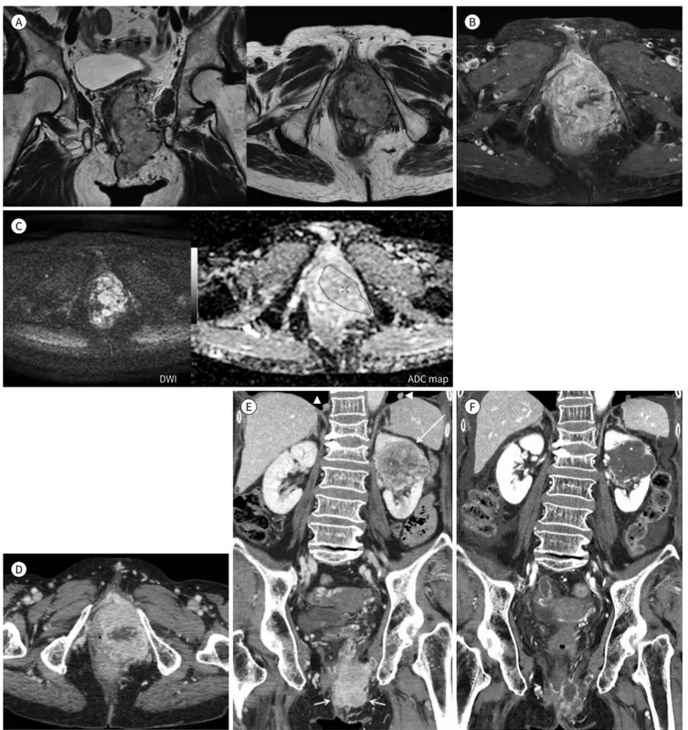
76세 여자 환자가 좌측 회음부의 통증성 종괴를 주소로 내원하였다. 환자는 회음부 통증 과 함께 배뇨통을 호소하였으나 질 출혈은 없었다. 자기공명영상에서 좌측 회음부에 경계가 분명한 7 cm × 8 cm × 4.6 cm 크기의 엽상 경계를 보이는 종괴가 관찰되었고, T2 강조영 상과 T1 강조영상에서 모두 인접한 근육보다 높은 신호강도를 보였다(Fig. 1A). 조영증강 후 영상에서 강한 가돌리늄 조영증강을 보였고, 인접한 요도, 방광과 질을 침범하였으며(Fig.
1B), 확산계수영상에서 확산 제한이 동반되어 있었다(Fig. 1C). 골반강 내부와 대혈관 주위 에 유의하게 커진 림프절은 보이지 않았고, 회음부 조직 검사에서 투명세포암(clear cell
Received January 20, 2020 Revised April 7, 2020 Accepted May 2, 2020
*Corresponding author Boem Ha Yi, MD
Department of Radiology, Soonchunhyang University Bucheon Hospital, 170 Jomaru-ro, Bucheon 14584, Korea.
Tel 82-32-621-5851 Fax 82-32-621-5874 E-mail [email protected] This is an Open Access article distributed under the terms of the Creative Commons Attribu- tion Non-Commercial License (https://creativecommons.org/
licenses/by-nc/4.0) which permits unrestricted non-commercial use, distribution, and reproduc- tion in any medium, provided the original work is properly cited.
ORCID iDs Seung Wook Lee https://
orcid.org/0000-0001-8532-9777 Boem Ha Yi
https://
orcid.org/0000-0003-2917-260X Min Hee Lee
https://
orcid.org/0000-0001-9198-0814 Seo-Youn Choi
https://
orcid.org/0000-0002-2434-8779 Ji Eun Lee
https://
orcid.org/0000-0002-4442-4441
https://doi.org/10.3348/jksr.2020.0010
275
대한영상의학회지 2021;82(1):274-277
carcinoma)으로 진단되었다. 복부 전산화단층촬영(이하 CT)에서 좌측 회음부의 종양 외에 좌측 신장에 6 cm × 6.8 cm × 7.2 cm 크기의 종양이 발견되었다. 이 신장의 종양은 동맥기에 불균질 한 조영증강을 보이며 지연 영상에서 감약계수의 감소를 보이고 내부에 조영증강이 잘되지 않는 낭성 변성이 동반되어 있어 신장의 투명세포암으로 생각하였으며, 신장의 종양이 회음부로 전이 한 것으로 진단하였다. 또한 양측 폐의 혈행성 전이도 동반되어 있었으며 그 외 다른 전이는 발견 되지 않았다(Fig. 1D, E).
환자는 전이성 신세포암의 표적 치료인 경구 항암제(Pazopanib 800 mg) 복용을 시작하였으며 3개월 후에 시행한 복부 CT에서 왼쪽 신장의 종양과 왼쪽 회음부의 전이성 종양 모두 이전보다 크 기가 감소하였고, 종양 대부분이 조영증강이 되지 않는 괴사가 진행되었다(Fig. 1F). 환자의 폐 전 이 병변도 크기가 감소하였으며 일부는 괴사가 동반되었다. 이후 6개월 추적검사에서도 좌측 신 장 및 좌측 회음부의 종양은 조영증강이 거의 되지 않는 상태로 남아 있었으며 폐병변도 3개월 추 적검사와 비슷한 양상을 보였다.
고찰
신세포암의 아형에 따라 원격 전이의 호발 부위가 다르다. 투명세포 신세포암은 폐전이가 흔하 고, 유두상 신세포암에서는 림프절 전이가 흔하다(3, 4). 그 외 드문 전이 장소로는 두경부, 침샘, 피부 등이 있고, 생식기 및 회음부 전이도 매우 드물게 보고되었다. 현재까지 보고된 생식기 전이 성 신세포암의 2/3은 좌측 생식기에서 발견되어 신정맥에서 생식선 정맥(gonadal vein)을 통한 역행성 전이에 의한 종양파급으로 생각된다(5, 6). 또한 신세포암의 전이는 원발 종양과 같이 발견 되기도 하나 대부분 수술 후 1~3년 후에 발견되는 경우도 많고, 드물게 신세포암 절제술 이후 10년 후에 발견되는 경우도 있다. 투명세포암은 신세포암의 아형 중 수술 이후에 발견되는 전이암의 비 율이 다른 아형에 비하여 높다는 보고가 있다(3).
신세포암의 전이 병변은 대개 원발 신세포암과 비슷한 영상 소견을 보이는데, 투명세포암은 동 맥기 또는 피질-수질기(corticomedullary phase)에서 신장의 피질과 비슷한 정도의 조영증강을 보인다(7). 본 증례에서 회음부 종양은 자기공명영상에서 전체적으로 조영증강이 잘 되고 확산 제 한이 되었으며, CT에서 피질-수질기에 불균질한 조영증강이 되고 분비기(excretory phase)에서 는 주변 조직에 비하여 감약계수가 감소하는 양상으로, 신장의 원발성 투명세포암에서 보이는 소 견과 일치하였다.
전이성 신세포암 치료는 다양한 표적 치료제가 사용된다(8). Vascular endothelial growth fac-
tor (이하 VEGF) 억제제 및 mammalian target of rapamycin (mTOR) 억제제들이 있으며, suni-
tinib, sorafenib, pazopanib 등은 VEGF pathway에 작용하는 표적 치료제들이다. Pazopanib은
종양의 혈관형성억제 치료제(antiangiogenic drug)로 세포독성(cytotoxic)이 아닌 세포증식 억제
제(cytostatic)의 성격을 갖고 있어 종양의 혈액 관류(perfusion)를 감소시킨다. 이로 인해 치료 후
영상검사에서 크기 감소보다는 CT 계수의 감소가 주로 보인다. 조영증강이 잘 되는 종괴는 혈관생
성억제 약물 치료 이후에는 균질하고 낮은 CT 계수 병변으로 바뀌는데 이는 점액성 퇴행(myxoid
jksronline.org
276
회음부 전이성 신장세포암
Fig. 1. 76-year-old female with a palpable left perineal metastatic renal cell carcinoma.
A. T2 weighted coronal and axial scans show lobulating high signal intensity mass abutting urethra in left perineum.
B. Gd-enhanced fat-saturated T1 weighted axial image shows heterogeneous, strong enhancement with intratumoral non-enhancing area.
C. DWI at a b value of 1000 s/mm2 shows a hyperintense mass in left perineum. ADC map shows a low ADC value (mean, 1.63 × 10⁻³ mm2/sec).
D. Contrast enhanced axial CT image of perineum shows a heterogeneously enhancing perineal mass.
E. Coronal reformatted CT image shows large heterogeneously enhancing left renal mass (long arrow) as well as left perineal mass (short ar- rows), indicating renal cell carcinoma of left kidney with perineal metastasis. Lung metastases are seen on both basal lungs (arrowheads).
F. CT scan performed 3 months after targeted therapy shows decrease in tumor size in left kidney and left perineum. Almost of the tumors has cystic appearance with necrotic change of primary and metastatic clear cell carcinomas.
ADC = apparent diffusion coefficient, DWI = diffusion-weighted image A
C
E F
B
D
DWI ADC map
https://doi.org/10.3348/jksr.2020.0010
277
대한영상의학회지 2021;82(1):274-277
degeneration), 출혈, 괴사를 반영하는 것으로 알려져 있다(9). 혈관형성 억제제의 치료에 동반될 수 있는 합병증으로는 심장 독성, 고혈압, 장관 기종(pneumatosis intestinalis), 장천공 등이 있다.
본 증례에서도 항암제 복용 3개월 후에 시행한 복부 CT에서 종양의 크기가 감소하였을 뿐 아니라 종양의 대부분에서 조영증강이 되지 않는 소견을 보여 치료 효과가 있음을 알 수 있었다. 6개월 후 추적검사에서 큰 변화를 보이지 않았으며 치료 기간 중 알려진 합병증들은 나타나지 않았다.
Author Contributions
Conceptualization, Y.B.H., L.S.W.; resources, L.M.H., C.S., L.J.E.; supervision, Y.B.H.; wiritng—origi- nal draft, L.S.W., Y.B.H.; and writing—review & editing, L.M.H., C.S., L.J.E.
Conflicts of Interest
The authors have no potential conflicts of interest to disclose.
REFERENCES
1. Siegel R, Naishadham D, Jemal A. Cancer statistics, 2012. CA Cancer J Clin 2012;62:10-29
2. Bianchi M, Sun M, Jeldres C, Shariat SF, Trinh QD, Briganti A, et al. Distribution of metastatic sites in renal cell carcinoma: a population-based analysis. Ann Oncol 2012;23:973-980
3. Renshaw AA, Richie JP. Subtypes of renal cell carcinoma. Different onset and sites of metastatic disease. Am J Clin Pathol 1999;111:539-543
4. Sivaramakrishna B, Gupta NP, Wadhwa P, Hemal AK, Dogra PN, Seth A, et al. Pattern of metastases in renal cell carcinoma: a single institution study. Indian J Cancer 2005;42:173-177
5. Sountoulides P, Metaxa L, Cindolo L. Atypical presentations and rare metastatic sites of renal cell carcinoma:
a review of case reports. J Med Case Rep 2011;5:429
6. Mendese GW, Ayvazian PJ, Li C. Renal cell carcinoma presenting as a perineal mass: case report and review of the literature. Urology 2006;67:847.e1-2
7. Kim JK, Kim TK, Ahn HJ, Kim CS, Kim KR, Cho KS. Differentiation of subtypes of renal cell carcinoma on helical CT scans. AJR Am J Roentgenol 2002;178:1499-1506
8. Motzer RJ, Hutson TE, Cella D, Reeves J, Hawkins R, Guo J, et al. Pazopanib versus sunitinib in metastatic renal- cell carcinoma. N Engl J Med 2013;369:722-731
9. Griffin N, Gore ME, Sohaib SA. Imaging in metastatic renal cell carcinoma. AJR Am J Roentgenol 2007;189:360- 370
회음부 전이성 신장 투명세포암: 증례 보고
이승욱 · 이범하* · 이민희 · 최서연 · 이지은
신세포암의 회음부 전이는 매우 드물고 영상 소견은 거의 보고되지 않았다. 저자들은 투명세 포 신세포암의 회음부 전이에 의한 회음부 통증을 주소로 내원한 76세 여자 환자의 표적 치 료 전후의 영상 소견을 보고하고자 한다.
순천향대학교 부천병원 영상의학과