The Effect of Admission at Weekends on Clinical Outcomes in Patients with Non-ST-segment Elevation Acute Coronary
Syndrome and Its Contributing Factors
We investigated the effects of weekend admission on adverse cardiac events in patients with non-ST-segment elevation acute coronary syndrome (NSTE-ACS). Patients with NSTE- ACS treated with percutaneous coronary intervention (PCI) were divided into a “weekend group” and a “weekday group” according to the emergency room arrival time. The primary outcome was 30-day major adverse cardiac events (MACE) including cardiac death, recurrent myocardial infarction, repeat revascularization, and urgent PCI. Of 577 patients, 168 patients were allocated to the weekend and 409 patients to the weekday group. The incidence of 30-day MACE was significantly higher in the weekend group (Crude: 15.5%
vs. 7.3%, P = 0.005; propensity score matched: 12.8% vs. 4.8%, P = 0.041). After adjustment for all the possible confounding factors, in Cox proportional hazard regression analysis, weekend admission was associated with a 2.1-fold increased hazard for MACE (HR, 2.13; 95% CI, 1.26-3.60, P = 0.005). These findings indicate that weekend admission of patients with NSTE-ACS is associated with an increase in 30-day adverse cardiac event.
Keywords: Coronary Artery Disease; Acute Coronary Syndrome; Percutaneous Coronary Intervention
Hyun-Jin Kim,1,2 Kwang-Il Kim,1 Young-Seok Cho,1 Jeehoon Kang,1,2 Jin Joo Park,1 Il-Young Oh,1 Chang-Hwan Yoon,1 Jung-Won Suh,1 Tae-Jin Youn,1 In-Ho Chae,1 and Dong-Ju Choi1
1Department of Internal Medicine, Seoul National University Bundang Hospital, Seongnam;
2Department of Internal Medicine, Seoul National University Hospital, Seoul, Korea
Received: 6 July 2014 Accepted: 5 December 2014 Address for Correspondence:
Young-Seok Cho, MD
Department of Internal Medicine, Seoul National University Bundang Hospital, and Department of Internal Medicine, Seoul National University College of Medicine, 82 Gumi-ro 173-gil, Bundang-gu, Seongnam 463-707, Korea
Tel: +82.31-787-7018, Fax: +82.31-787-4290 E-mail: [email protected]
http://dx.doi.org/10.3346/jkms.2015.30.4.414 • J Korean Med Sci 2015; 30: 414-425
INTRODUCTION
It has been well established that the immediate reperfusion ther- apy lowers mortality in patients with ST-segment elevation myo- cardial infarction (STEMI) (1). In contrast, for patients present- ing with non-ST-segment elevation acute coronary syndrome (NSTE-ACS), guidelines do not recommend an immediate re- vascularization (2, 3). Instead, in patients with NSTE-ACS, re- vascularization is typically performed 12-72 hr after medical stabilization, except in those with hemodynamic instability, elec- trical instability, or refractory ischemia who require urgent re- vascularization (4).
With regard to the hospital arrival time, during the weekend, invasive revascularization procedures can be delayed inappro- priately (5-7). Hospitals have reduced staffing levels on the week- end and an interventional cardiologist may not be immediately available, possibly leading to a delay in the revascularization procedure for patients with NSTE-ACS (8-11).
Therefore, we sought to examine the effects of weekend ad- mission on clinical outcomes in patients with NSTE-ACS under- going percutaneous coronary intervention (PCI). The hypothe- sis was that the incidence of major adverse cardiac events (MA - CE) is increased in patients who are admitted to the hospital
during the weekend than those admitted during the weekday.
MATERIALS AND METHODS Study design and study population
Patients who were admitted to the coronary care unit (CCU) via the emergency room (ER) from January 2007 to December 2010 in Seoul National University Bundang Hospital were retrospec- tively identified for the inclusion in this study. The inclusion criteria included the diagnosis of unstable angina pectoris (UA) or non-ST-segment elevation myocardial infarction (NSTEMI).
The exclusion criteria were patients diagnosed with STEMI or stable angina, patients admitted to the general ward or via an outpatient clinic, and patients experiencing UA or NSTEMI dur- ing hospitalization for other medical conditions. Patients who had undergone coronary artery bypass graft surgery (CABG) after diagnostic coronary angiography (CAG) were also exclud- ed. Since medically-managed ACS patients comprised a het- erogeneous population including patients without severe coro- nary stenosis, those with coronary artery spasm, and patients who refused PCI or CABG due to economic reason, those pa- tients were also excluded from the study.
The patient’s demographic and clinical characteristics, labo- Cardiovascular Disorders
ratory tests and angiographic characteristics were reviewed. Car- diac biomarkers including creatine kinase –myocardial band (CK-MB) and Troponin-I were checked every 8 hr from the ad- mission until PCI, and twice a day after PCI for one day, and daily until their normalization. In addition, we collected the ex- act time, day, and date of symptom onset, admission to ER and then CCU, and the first balloon inflation time during PCI. In our hospital, NSTE-ACS patients visiting ER from Sunday to Thursday after 6 p.m. undergo PCI in the next-day morning and those who visit ER between Friday 6 p.m. to Sunday 5:59 p.m.
undergo PCI in coming Monday morning. The enrolled patients were divided into two groups according to ER admission time.
Patients who were admitted to the ER from Friday 6 p.m. to Sun- day 5:59 p.m. or from the day before holiday 6 p.m. to holiday 5:59 p.m. were allocated to the ‘weekend group’. Patients who were admitted to the ER during all other time periods were clas- sified as the ‘weekday group’. Event adjudication was done by two experienced reviewers. If the two reviewers’ opinions dis- agree, then a third reviewer evaluated the clinical events and established the adjudication.
Definition
Patients were diagnosed as NSTE-ACS if there were appropriate clinical manifestations including chest discomfort or angina- equivalent suggesting UA with or without positive biomarkers of necrosis in the absence of electrocardiographic ST-segment elevation (12). UA was defined as angina pectoris with at least one of three features: 1) chest pain occurring at rest and usually lasting > 20 min; 2) chest pain being severe and usually described as frank pain; or 3) chest pain occurring with a crescendo pat- tern. We differentiated between NSTEMI and UA based on car- diac troponin I with at least one value above the 99th percentile of the upper reference level (13, 14). The risk stratification was done using thrombolysis in myocardial infarction (TIMI) risk scores and the scores classified as low (TIMI risk score 0-2), in- termediate (TIMI risk score 3-4), and high risk (TIMI risk score 5-7) (15, 16). We defined symptom-to-admission time as the time from current angina symptom onset to admission to the ER. Also, admission-to-PCI time referred to the time interval between admission to the ER and time of the first balloon infla- tion to culprit lesion.
Study outcomes
The primary outcome was the incidence of MACE during the 30-day follow-up period after admission to the ER, including cardiac death, recurrent myocardial infarction (MI), repeat re- vascularization and urgent PCI. MI was defined according to the universal definition of myocardial infarction, and recurrent MI was defined to MI which occurred after an incident event with 20% or greater increase of the cardiac troponin I in the sec- ond sample compared to the sample at the time of suspected
recurrent MI (13). Repeat revascularization included target or non-target vessel revascularization including CABG. We also examined the incidence of urgent PCI which was performed in cases of refractory angina, hemodynamic instability, or electri- cal instability, during the period of medical stabilization.
To determine whether the PCI was urgent one or not, for pa- tients who experienced unstable clinical condition during the period of medical stabilization, we reviewed the vital sign chart, nursing records, doctors’ records including physical examina- tion, and the test results including electrocardiogram for ST-seg- ment change or ventricular tachyarrhythmia and cardiac en- zymes. The secondary outcomes included the incidence of car- diac death, recurrent MI, repeat revascularization, urgent PCI, peak CK-MB level during hospital stay, and peak troponin-I level during hospital stay. Also, we investigated the daily distri- bution of MACE according to ER admission day. Moreover, we evaluated the clinical outcomes including cardiac death, myo- cardial infarction, and repeat revascularization at 18 months.
Statistical analysis
All categorical data were summarized as frequencies and per- centages, whereas statistics for continuous variables are present- ed as means and standard deviations. The Pearson’s chi-square test was used for comparison of categorical variables and the Fisher’s exact test was used for comparison of categorical vari- ables with 20% or more of the expected cell frequencies below 5. The Student’s t-test was used for comparison of continuous variables and the Mann-Whitney U-test was used for sample sizes below 30 in at least one group. Linear-by-linear association was used to extract trend of clinical characteristics and MACE according to ER admission day. The time interval trend accord- ing to ER admission day was evaluated using ANOVA. In addi- tion, we selected a propensity score matched population to ad- just for uneven distribution of baseline characteristics; a 1:1 mat- ched analysis without replacement was performed using pro- pensity score. Logistic regression model was conducted to gen- erate propensity score which was probability that a patient ad-
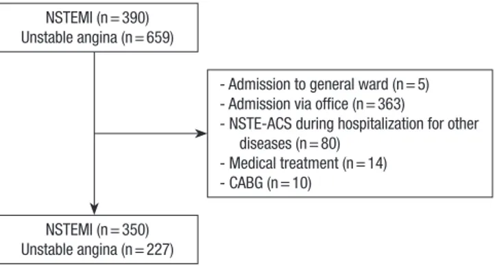
NSTEMI (n = 390) Unstable angina (n = 659)
NSTEMI (n = 350) Unstable angina (n = 227)
- Admission to general ward (n = 5) - Admission via office (n = 363)
- NSTE-ACS during hospitalization for other diseases (n = 80)
- Medical treatment (n = 14) - CABG (n = 10)
Fig. 1. Selection of patients. NSTEMI, non-ST-segment elevation myocardial infarc- tion; NSTE-ACS, non-ST-segment elevation acute coronary syndrome; CABG, coro- nary artery bypass grafting.
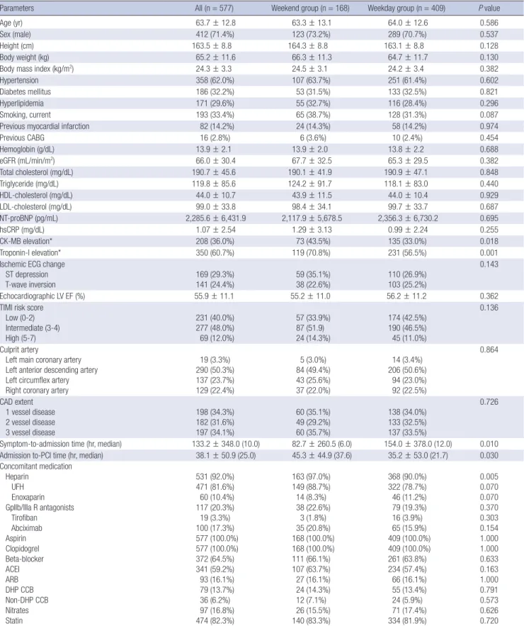
Table 1. Baseline patient characteristics
Parameters All (n = 577) Weekend group (n = 168) Weekday group (n = 409) P value
Age (yr) 63.7 ± 12.8 63.3 ± 13.1 64.0 ± 12.6 0.586
Sex (male) 412 (71.4%) 123 (73.2%) 289 (70.7%) 0.537
Height (cm) 163.5 ± 8.8 164.3 ± 8.8 163.1 ± 8.8 0.128
Body weight (kg) 65.2 ± 11.6 66.3 ± 11.3 64.7 ± 11.7 0.130
Body mass index (kg/m2) 24.3 ± 3.3 24.5 ± 3.1 24.2 ± 3.4 0.382
Hypertension 358 (62.0%) 107 (63.7%) 251 (61.4%) 0.602
Diabetes mellitus 186 (32.2%) 53 (31.5%) 133 (32.5%) 0.821
Hyperlipidemia 171 (29.6%) 55 (32.7%) 116 (28.4%) 0.296
Smoking, current 193 (33.4%) 65 (38.7%) 128 (31.3%) 0.087
Previous myocardial infarction 82 (14.2%) 24 (14.3%) 58 (14.2%) 0.974
Previous CABG 16 (2.8%) 6 (3.6%) 10 (2.4%) 0.454
Hemoglobin (g/dL) 13.9 ± 2.1 13.9 ± 2.0 13.8 ± 2.2 0.688
eGFR (mL/min/m2) 66.0 ± 30.4 67.7 ± 32.5 65.3 ± 29.5 0.382
Total cholesterol (mg/dL) 190.7 ± 45.6 190.1 ± 41.9 190.9 ± 47.1 0.848
Triglyceride (mg/dL) 119.8 ± 85.6 124.2 ± 91.7 118.1 ± 83.0 0.440
HDL-cholesterol (mg/dL) 44.0 ± 10.7 43.9 ± 11.5 44.0 ± 10.4 0.929
LDL-cholesterol (mg/dL) 99.0 ± 33.8 98.4 ± 34.1 99.7 ± 33.7 0.687
NT-proBNP (pg/mL) 2,285.6 ± 6,431.9 2,117.9 ± 5,678.5 2,356.3 ± 6,730.2 0.695
hsCRP (mg/dL) 1.07 ± 2.54 1.29 ± 3.13 0.99 ± 2.24 0.255
CK-MB elevation* 208 (36.0%) 73 (43.5%) 135 (33.0%) 0.018
Troponin-I elevation* 350 (60.7%) 119 (70.8%) 231 (56.5%) 0.001
Ischemic ECG change ST depression T-wave inversion
169 (29.3%) 141 (24.4%)
59 (35.1%) 38 (22.6%)
110 (26.9%) 103 (25.2%)
0.143
Echocardiographic LV EF (%) 55.9 ± 11.1 55.2 ± 11.0 56.2 ± 11.2 0.362
TIMI risk score Low (0-2) Intermediate (3-4) High (5-7)
231 (40.0%) 277 (48.0%) 69 (12.0%)
57 (33.9%) 87 (51.9) 24 (14.3%)
174 (42.5%) 190 (46.5%) 45 (11.0%)
0.136
Culprit artery
Left main coronary artery Left anterior descending artery Left circumflex artery Right coronary artery
19 (3.3%) 290 (50.3%) 137 (23.7%) 129 (22.4%)
5 (3.0%) 84 (49.4%) 43 (25.6%) 37 (22.0%)
14 (3.4%) 206 (50.6%)
94 (23.0%) 92 (22.5%)
0.864
CAD extent 1 vessel disease 2 vessel disease 3 vessel disease
198 (34.3%) 182 (31.6%) 197 (34.1%)
60 (35.1%) 49 (29.2%) 60 (35.7%)
138 (34.0%) 133 (32.5%) 137 (33.5%)
0.726
Symptom-to-admission time (hr, median) 133.2 ± 348.0 (10.0) 82.7 ± 260.5 (6.0) 154.0 ± 378.0 (12.0) 0.010 Admission to-PCI time (hr, median) 38.1 ± 50.9 (25.0) 45.3 ± 44.9 (37.6) 35.2 ± 53.0 (21.7) 0.030 Concomitant medication
Heparin 531 (92.0%) 163 (97.0%) 368 (90.0%) 0.005
UFH 471 (81.6%) 149 (88.7%) 322 (78.7%) 0.070
Enoxaparin 60 (10.4%) 14 (8.3%) 46 (11.2%) 0.070
GpIIb/IIIa R antagonists 117 (20.3%) 38 (22.6%) 79 (19.3%) 0.370
Tirofiban 19 (3.3%) 3 (1.8%) 16 (3.9%) 0.303
Abciximab 100 (17.3%) 35 (20.8%) 65 (15.9%) 0.154
Aspirin 577 (100.0%) 168 (100.0%) 409 (100.0%) 1.000
Clopidogrel 577 (100.0%) 168 (100.0%) 409 (100.0%) 1.000
Beta-blocker 372 (64.5%) 111 (66.1%) 261 (63.8%) 0.633
ACEI 341 (59.2%) 107 (63.7%) 234 (57.4%) 0.163
ARB 93 (16.1%) 27 (16.1%) 66 (16.1%) 1.000
DHP CCB 79 (13.7%) 24 (14.3%) 55 (13.4%) 0.791
Non-DHP CCB 36 (6.2%) 12 (7.1%) 24 (5.9%) 0.573
Nitrates 97 (16.8%) 26 (15.5%) 71 (17.4%) 0.626
Statin 474 (82.3%) 140 (83.3%) 334 (81.9%) 0.720
*> 1 × upper reference limit; CABG, coronary artery bypass grafting; eGFR, estimated glomerular filtration rate; HDL, high-density lipoprotein; LDL, low-density lipoprotein; NT- proBNP, N-terminal-proB-type natriuretic peptide; hsCRP, high-sensitivity C-reactive protein; CK-MB, creatine kinase-myocardial band; ECG, electrocardiography; LV EF, left ven- tricular ejection fraction; TIMI, thrombolysis in myocardial infarction; CAD, coronary artery disease; PCI, percutaneous coronary intervention; UFH, unfractionated heparin; GpIIb/
IIIa R, glycoprotein IIb/IIIa receptor; ACEI, angiotensin-converting-enzyme inhibitor; ARB, angiotensin receptor blocker; DHP CCB, dihydropyridine-calcium channel blocker.
Fig. 2. Thirty-day major adverse cardiac events (MACE) for patients with NSTE-ACS who were admitted on weekday or weekend. Of 577 patients, 26 patients in the week- end group (15.5%) and 30 patients in the weekday group (7.3%) had MACE within 30 days. The difference between the two groups was statistically significant (P = 0.005).
30-day MACE (%)
Weekend Weekday
20
15
10
5
0
P = 0.005
15.5%
7.3%
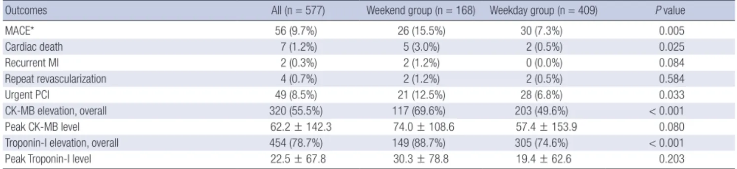
Table 2. Thirty-day clinical outcomes
Outcomes All (n = 577) Weekend group (n = 168) Weekday group (n = 409) P value
MACE* 56 (9.7%) 26 (15.5%) 30 (7.3%) 0.005
Cardiac death 7 (1.2%) 5 (3.0%) 2 (0.5%) 0.025
Recurrent MI 2 (0.3%) 2 (1.2%) 0 (0.0%) 0.084
Repeat revascularization 4 (0.7%) 2 (1.2%) 2 (0.5%) 0.584
Urgent PCI 49 (8.5%) 21 (12.5%) 28 (6.8%) 0.033
CK-MB elevation, overall 320 (55.5%) 117 (69.6%) 203 (49.6%) < 0.001
Peak CK-MB level 62.2 ± 142.3 74.0 ± 108.6 57.4 ± 153.9 0.080
Troponin-I elevation, overall 454 (78.7%) 149 (88.7%) 305 (74.6%) < 0.001
Peak Troponin-I level 22.5 ± 67.8 30.3 ± 78.8 19.4 ± 62.6 0.203
*MACE (major adverse cardiac event) including cardiac death, recurrent MI, repeat revascularization, and unplanned urgent PCI. MI, myocardial infarction; PCI, percutaneous coronary intervention; CK-MB, creatine kinase-myocardial band.
Table 3. Distribution of the occurrence of MACE MACE duration All Weekend group
(n = 26) Weekday group
(n = 30) P value
Day 1-5 52 (92.9%) 23 (88.4%) 29 (96.7%) 0.490
Day 6-10 2 (3.6%) 1 (3.8%) 1 (3.3%) -
Day 11-20 1 (1.8%) 1 (3.8%) 0 (0%) -
Day 21-30 1 (1.8%) 1 (3.8%) 0 (0%) -
mitted in weekend. The adjusted variables were as follows: age, sex, height, body weight, body mass index, hypertension, dia- betes mellitus, hyperlipidemia, smoking, previous MI history, previous CABG history, symptom-to-admission time, admis- sion-to-PCI time, hemoglobin, estimated glomerular filtration rate, total cholesterol, triglyceride, HDL-cholesterol, LDL-cho- lesterol, NT-proBNP, hsCRP, elevation of CK-MB, elevation of troponin-I, ischemic change of ECG, left ventricular ejection fraction, TIMI risk scores, culprit artery, and extent of coronary artery disease. The Greedy 5→1 digit match algorithm was used for matching. We were able to match 125 patients in weekend group to 125 patients in weekday group. McNemar’s test and marginal homogeneity test were used for comparison of cate- gorical variables between the matched patient groups, paired t- test for continuous variables. Multivariate Cox proportional haz- ards regression analyses were performed to evaluate the risk of 30-day MACE with adjustment for individual risk factors. We obtained hazard ratio of weekend admission for MACE with the adjustment of sequentially-added potential confounding factors, including clinical characteristics, time factors, and se- verity factors. In addition, survival analyses and the log-rank test were used to compare 30-day MACE-free survival. A P val- ue of less than 0.05 was considered statistically significant. All
analyses were performed with SPSS v. 18.0 (SPSS Inc., Chicago, IL, USA).
Ethics statement
The study was approved by the institutional review board of the Seoul National University Bundang Hospital (No. B-1111-139- 105), and was conducted according to the Declaration of Hel- sinki. Informed consent for study enrollment was waived by the board.
RESULTS
Baseline patients characteristics
Among 1,049 patients screened, 577 patients with NSTE-ACS who had been admitted via ER and managed with PCI were fi- nally enrolled in the study (Fig. 1). Study subjects were classified into the weekend group (n = 168) or the weekday group (n = 409) based on the ER admission day and time (Table 1). The weekend group showed significantly higher baseline cardiac enzyme, shorter symptom-to-admission time, and longer ad- mission-to-PCI time compared to the weekday group. In addi- tion, patients of weekend group had more intermediate to high TIMI risk scores than weekday group, with borderline statistical significance (66.1% vs. 57.5%, P = 0.055). Concomitant medica- tions were not significantly different except that heparin was administered more often in the weekend group. In the weekday group, the admission-to-PCI time did not differ between pati- ents who arrive at ER after 6 p.m. on Sunday or holiday and those who arrive after 6 p.m. on weekdays excluding Friday (36.9 ± 41.9 hr vs. 34.7 ± 55.7 hr, P = 0.741).
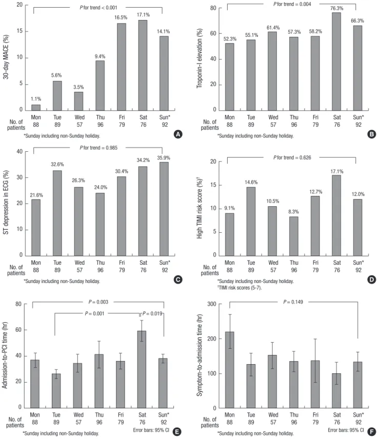
Fig. 3. Outcomes and baseline characteristics according to ER admission day. (A) Thirty-day major adverse cardiac events (MACE) according to ER admission day. The incidence of MACE shows a trend of increase from Monday to Sunday. (B) Baseline Troponin-I elevation according to ER admission day. Distribution of patients with elevated baseline tro- ponin-I shows a trend of gradual increase from Monday to Sunday. (C) Baseline electrocardiographic ST-segment depression according to ER admission day. Distribution of pa- tients with baseline ECG change does not show statistically significant trend. (D) Baseline high TIMI risk score according to ER admission day. Distribution of patients with high TIMI risk score did not get statistically significant trend. (E) Admission to-PCI time according to ER admission day. The admission-to-PCI time has a trend of gradual increase from Monday to Saturday, revealing significantly longer time on Saturday than Tuesday or Sunday. (F) Symptom to-admission time according to ER admission day. The distribu- tion of symptom-to-admission time shows a trend of longest time on Monday and gradual decrease toward the weekend, without statistical significance.
30-day MACE (%)
*Sunday including non-Sunday holiday.
Mon Tue Wed Thu Fri Sat Sun*
88 89 57 96 79 76 92 No. of
patients 20
15
10
5
0
P for trend < 0.001
1.1%
5.6%
3.5%
9.4%
16.5% 17.1%
14.1%
A
Troponin-I elevation (%)
*Sunday including non-Sunday holiday.
Mon Tue Wed Thu Fri Sat Sun*
88 89 57 96 79 76 92 No. of
patients 80
60
40
20
0
P for trend = 0.004
52.3% 55.1%
61.4%
57.3% 58.2%
76.3%
66.3%
B
ST depression in ECG (%)
*Sunday including non-Sunday holiday.
Mon Tue Wed Thu Fri Sat Sun*
88 89 57 96 79 76 92 No. of
patients 40
30
20
10
0
P for trend = 0.985
21.6%
32.6%
26.3%
24.0%
30.4%
34.2% 35.9%
C
High TIMI risk score (%)†
*Sunday including non-Sunday holiday.
†TIMI risk scores (5-7).
Mon Tue Wed Thu Fri Sat Sun*
88 89 57 96 79 76 92 No. of
patients 20
15
10
5
0
P for trend = 0.626
9.1%
14.6%
10.5%
8.3%
12.7%
17.1%
12.0%
D
Admission-to-PCI time (hr)
*Sunday including non-Sunday holiday. Error bars: 95% CI
Mon Tue Wed Thu Fri Sat Sun*
88 89 57 96 79 76 92 No. of
patients 80
60
40
20
0
P = 0.003 P = 0.001
E P = 0.019
Symptom-to-admission time (hr)
*Sunday including non-Sunday holiday. Error bars: 95% CI
Mon Tue Wed Thu Fri Sat Sun*
88 89 57 96 79 76 92 No. of
patients 300
200
100
0
P = 0.149
F
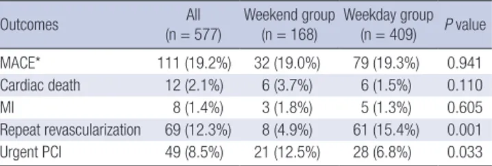
Table 4. Eighteen-month cumulative clinical outcomes
Outcomes All
(n = 577) Weekend group
(n = 168) Weekday group (n = 409) P value
MACE* 111 (19.2%) 32 (19.0%) 79 (19.3%) 0.941
Cardiac death 12 (2.1%) 6 (3.7%) 6 (1.5%) 0.110
MI 8 (1.4%) 3 (1.8%) 5 (1.3%) 0.605
Repeat revascularization 69 (12.3%) 8 (4.9%) 61 (15.4%) 0.001
Urgent PCI 49 (8.5%) 21 (12.5%) 28 (6.8%) 0.033
*MACE (major adverse cardiac event) including cumulative cardiac death, MI, repeat revascularization, and unplanned urgent PCI. MI, myocardial infarction; PCI, percuta- neous coronary intervention.
Clinical outcomes at 30 days of follow-up
The weekend group had higher 30-day MACE rate than the week- day group (15.5% vs. 7.3%, P = 0.005) (Fig. 2). The incidence of cardiac death (3.0% vs. 0.5%, P = 0.025) and that of urgent PCI (12.5% vs. 6.8%, P = 0.033) were also significantly higher in the weekend group (Table 2). Overall cardiac enzyme elevation was more frequent in the weekend group. When the whole patients were subgrouped into NSTEMI (n = 350) and UA (n = 227), there were similar results. The incidence rate of MACE was significant- ly higher in the weekend group in comparison with the week- day group in patients with NSTEMI (16.8% vs. 9.5%, P = 0.047) and in patients with UA (12.2% vs. 4.5%, P = 0.046).
Most MACE occurred within 5 days after PCI in both groups, and the distribution patterns were similar between the two groups (Table 3). Seven patients died of cardiac causes, five in the week- end group and 2 in the weekday group. In the weekend group, four had cardiogenic shocks and one ventricular tachyarrhyth- mia, while in the weekday group one had cardiogenic shock and one ventricular tachyarrhythmia. There was no significant difference in the incidence of MACE among eight PCI opera- tors (P = 0.100) and between four senior operators and four ju- nior operators (7.9% vs. 12.2%, P = 0.082).
Interestingly, there was a trend of gradual increase in MACE from the start of a week toward the weekend with a highest in- cidence on Saturday followed by Friday (Fig. 3A). With regard to the potential factors that can contribute to the occurrence of MACE (Fig. 3B-F), baseline Troponin-I elevation and the ad- mission-to-PCI time showed similar distribution pattern as the MACE distribution with a peak on Saturday, suggesting some causal role in the increase of MACE in the weekend group. The distribution of symptom-to-admission time showed a reverse pattern of MACE, showing a trend of longest time on Monday and gradual decrease toward the weekend, without statistical significance.
However, there was no significant difference in MACE be- tween the weekend group and the weekday group at 18 months (19.0% vs. 19.6%, P = 0.888) (Table 4).
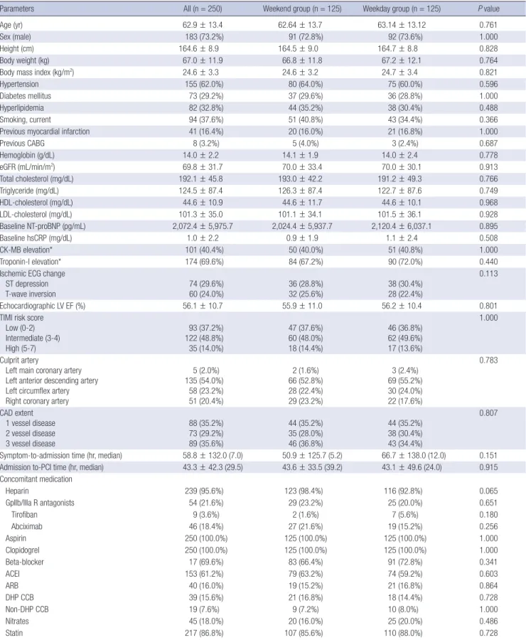
Clinical outcomes in propensity score matched population After propensity score matching, 125 of 168 patients in the week-
end group were successfully matched to an equal number of patients in the weekday group. Baseline characteristics showed no significant differences between the groups after propensity score matching (Table 5). The clinical outcomes of the matched population also showed significantly higher rate of MACE in the weekend group (12.8% vs. 4.8%, P = 0.041) (Table 6).
Survival analysis
The cumulative 30-day MACE-free survival rate was significant- ly lower in the weekend group compared to the weekday group (92.7% vs. 84.5%, P = 0.003) (Fig. 4A), as well as in the propensi- ty-score matched population (87.2% vs. 95.2%, P = 0.026) (Fig.
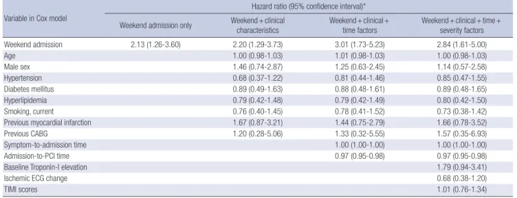
4B). In Cox proportional hazard regression analysis, weekend admission was associated with a 2.1-fold increased hazard for MACE (HR, 2.13; 95% CI, 1.26-3.60, P = 0.005) (Table 7) after sequential adjustment of potential confounding factors includ- ing clinical characteristics, time factors such as symptom-to- admission time and admission-to-PCI, and severity factors such as Troponin-I elevation, ECG change and TIMI score. Interest- ingly, admission-to-PCI time appeared to be a weak, but an in- dependent protective factor of MACE.
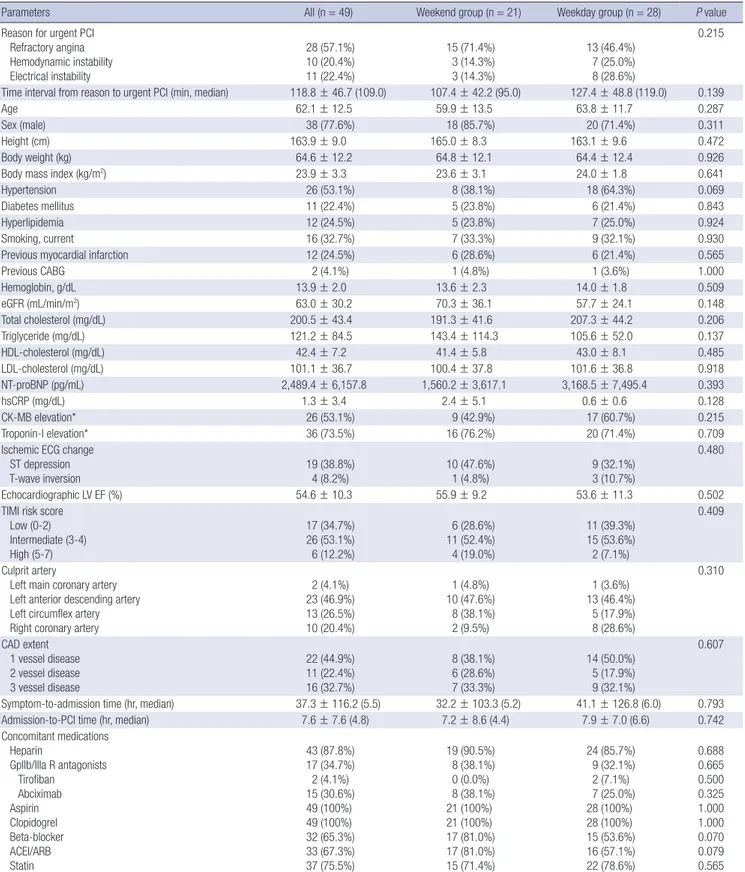
Analysis of urgent PCI subgroup
The weekend group had higher unplanned, urgent PCI rate than the weekday group. Considering the possibility of inappropri- ately performed urgent PCI during the weekend period, we un- derwent further analysis. Of the 49 patients with urgent PCI, 57.1% had refractory angina, 20.4% had hemodynamic instabil- ity, and 22.4% had electrical instability as the reason for urgent PCI (Table 8). There were no significant differences in the rea- sons for urgent PCI (refractory angina, 71.4% vs. 46.4%; hemo- dynamic instability, 14.3% vs. 25.0%; electrical instability, 14.3%
vs. 28.6%, P = 0.215) between the weekend and the weekday group. More importantly, the time interval from unstable clini- cal condition to urgent PCI (107.4 ± 42.2 min vs. 127.4 ± 48.8 min, P = 0.139) were not significantly different between the two groups. In addition, the time interval between unstable clinical condition and urgent PCI did not differ between MACE (+) group (n = 3) and MACE (-) group (n = 46) (104.0 ± 30.3 vs. 120.0 ± 47.7 min, P = 0.661).
DISCUSSION
This study shows that admission on weekends has an adverse influence on clinical outcomes in patients with NSTE-ACS. Week- end admission was associated with a higher incidence of 30- day MACE, which maintained after adjusting the difference in baseline characteristics with propensity score matching and Cox proportional hazard model regression analysis. In addition, there was a trend of gradual increase of MACE from Monday toward the weekend peaking on Saturday with similar patterns
Table 5. Baseline characteristics in propensity-score matched population
Parameters All (n = 250) Weekend group (n = 125) Weekday group (n = 125) P value
Age (yr) 62.9 ± 13.4 62.64 ± 13.7 63.14 ± 13.12 0.761
Sex (male) 183 (73.2%) 91 (72.8%) 92 (73.6%) 1.000
Height (cm) 164.6 ± 8.9 164.5 ± 9.0 164.7 ± 8.8 0.828
Body weight (kg) 67.0 ± 11.9 66.8 ± 11.8 67.2 ± 12.1 0.764
Body mass index (kg/m2) 24.6 ± 3.3 24.6 ± 3.2 24.7 ± 3.4 0.821
Hypertension 155 (62.0%) 80 (64.0%) 75 (60.0%) 0.596
Diabetes mellitus 73 (29.2%) 37 (29.6%) 36 (28.8%) 1.000
Hyperlipidemia 82 (32.8%) 44 (35.2%) 38 (30.4%) 0.488
Smoking, current 94 (37.6%) 51 (40.8%) 43 (34.4%) 0.366
Previous myocardial infarction 41 (16.4%) 20 (16.0%) 21 (16.8%) 1.000
Previous CABG 8 (3.2%) 5 (4.0%) 3 (2.4%) 0.687
Hemoglobin (g/dL) 14.0 ± 2.2 14.1 ± 1.9 14.0 ± 2.4 0.778
eGFR (mL/min/m2) 69.8 ± 31.7 70.0 ± 33.4 70.0 ± 30.1 0.913
Total cholesterol (mg/dL) 192.1 ± 45.8 193.0 ± 42.2 191.2 ± 49.3 0.766
Triglyceride (mg/dL) 124.5 ± 87.4 126.3 ± 87.4 122.7 ± 87.6 0.749
HDL-cholesterol (mg/dL) 44.6 ± 10.9 44.6 ± 11.7 44.6 ± 10.1 0.968
LDL-cholesterol (mg/dL) 101.3 ± 35.0 101.1 ± 34.1 101.5 ± 36.1 0.928
Baseline NT-proBNP (pg/mL) 2,072.4 ± 5,975.7 2,024.4 ± 5,937.7 2,120.4 ± 6,037.1 0.895
Baseline hsCRP (mg/dL) 1.0 ± 2.2 0.9 ± 1.9 1.1 ± 2.4 0.508
CK-MB elevation* 101 (40.4%) 50 (40.0%) 51 (40.8%) 1.000
Troponin-I elevation* 174 (69.6%) 84 (67.2%) 90 (72.0%) 0.440
Ischemic ECG change ST depression
T-wave inversion 74 (29.6%)
60 (24.0%) 36 (28.8%)
32 (25.6%) 38 (30.4%)
28 (22.4%)
0.113
Echocardiographic LV EF (%) 56.1 ± 10.7 55.9 ± 11.0 56.2 ± 10.4 0.801
TIMI risk score Low (0-2) Intermediate (3-4) High (5-7)
93 (37.2%) 122 (48.8%) 35 (14.0%)
47 (37.6%) 60 (48.0%) 18 (14.4%)
46 (36.8%) 62 (49.6%) 17 (13.6%)
1.000
Culprit artery
Left main coronary artery Left anterior descending artery Left circumflex artery Right coronary artery
5 (2.0%) 135 (54.0%)
58 (23.2%) 51 (20.4%)
2 (1.6%) 66 (52.8%) 28 (22.4%) 29 (23.2%)
3 (2.4%) 69 (55.2%) 30 (24.0%) 22 (17.6%)
0.783
CAD extent 1 vessel disease 2 vessel disease 3 vessel disease
88 (35.2%) 73 (29.2%) 89 (35.6%)
44 (35.2%) 35 (28.0%) 46 (36.8%)
44 (35.2%) 38 (30.4%) 43 (34.4%)
0.807
Symptom-to-admission time (hr, median) 58.8 ± 132.0 (7.0) 50.9 ± 125.7 (5.2) 66.7 ± 138.0 (12.0) 0.151 Admission to-PCI time (hr, median) 43.3 ± 42.3 (29.5) 43.6 ± 33.5 (39.2) 43.1 ± 49.6 (24.0) 0.915 Concomitant medication
Heparin 239 (95.6%) 123 (98.4%) 116 (92.8%) 0.065
GpIIb/IIIa R antagonists 54 (21.6%) 29 (23.2%) 25 (20.0%) 0.651
Tirofiban 9 (3.6%) 2 (1.6%) 7 (5.6%) 0.180
Abciximab 46 (18.4%) 27 (21.6%) 19 (15.2%) 0.256
Aspirin 250 (100.0%) 125 (100.0%) 125 (100.0%) 1.000
Clopidogrel 250 (100.0%) 125 (100.0%) 125 (100.0%) 1.000
Beta-blocker 17 (69.6%) 83 (66.4%) 91 (72.8%) 0.341
ACEI 153 (61.2%) 79 (63.2%) 74 (59.2%) 0.603
ARB 40 (16.0%) 19 (15.2%) 21 (16.8%) 0.864
DHP CCB 39 (15.6%) 21 (16.8%) 18 (14.4%) 0.728
Non-DHP CCB 19 (7.6%) 9 (7.2%) 10 (8.0%) 1.000
Nitrates 45 (18.0%) 20 (16.0%) 25 (20.0%) 0.486
Statin 217 (86.8%) 107 (85.6%) 110 (88.0%) 0.728
* > 1 × upper reference limit; CABG, coronary artery bypass grafting; eGFR, estimated glomerular filtration rate; HDL, high-density lipoprotein; LDL, low-density lipoprotein; NT- proBNP, N-terminal-proB-type natriuretic peptide; hsCRP, high-sensitivity C-reactive protein; CK-MB, creatine kinase-myocardial band; ECG, electrocardiography; LV EF, left ventricular ejection fraction; TIMI, thrombolysis in myocardial infarction; CAD, coronary artery disease; PCI, percutaneous coronary intervention; GpIIb/IIIa R, glycoprotein IIb/IIIa receptor; ACEI, angiotensin-converting-enzyme inhibitor; ARB, angiotensin receptor blocker; DHP CCB, dihydropyridine-calcium channel blocker.
Table 6. Thirty-day clinical outcomes in propensity-score matched population
Outcomes All
(n = 250)
Weekend group (n = 125)
Weekday group
(n = 125) P value
MACE 22 (8.8%) 16 (12.8%) 6 (4.8%) 0.041
Cardiac death 1 (0.4%) 1 (0.8%) 0 (0.0%) 1.000
Recurrent MI 2 (0.8%) 2 (1.6%) 0 (0.0%) 0.500
Repeat revascularization 2 (0.8%) 2 (1.6%) 0 (0.0%) 0.500
Urgent PCI 20 (8.0%) 14 (11.2%) 6 (4.8%) 0.064
CK-MB elevation, overall 157 (63.6%) 85 (68.0%) 72 (59.0%) 0.120 Peak CK-MB level 52.0 ± 92.4 62.6 ± 95.1 41.5 ± 88.8 0.072 Troponin-I elevation, overall 213 (86.2%) 109 (87.2%) 104 (52.2%) 0.690 Peak Troponin-I level 17.3 ± 34.5 23.0 ± 43.7 11.6 ± 20.5 0.009 MACE, major adverse cardiac event; MI, myocardial infarction; PCI, percutaneous cor- onary intervention; CK-MB, creatine kinase-myocardial band.
Fig. 4. Thirty-day MACE-free survival. (A) MACE-free survival in all patients. The cumulative 30-day MACE-free survival rate is significantly lower in patients admitted during the weekend (dotted line) compared to patients admitted during the weekday (solid line). Survival curves begin to diverge at day 1 and continue to separate throughout the 30-day follow-up period. (B) MACE-free survival in propensity-matched population. The cumulative 30-day MACE-free survival rate is significantly lower in the weekend group in the propensity score-matched population, too. MACE, major adverse cardiac events.
MACE-free survival (%)
Days
0 10 20 30
409 379 379 379
168 144 143 142
Weekday Weekend 100
90 80 70 60 50
Log-rank P = 0.003
92.7%
84.5%
Weekday Weekend
A
MACE-free survival (%)
Days
0 10 20 30
125 119 119 119
125 110 109 109
Weekday Weekend 100
90 80 70 60 50
Log-rank P = 0.026
95.2%
87.2%
Weekday Weekend
B
Table 7. Adjusted risk of 30-day MACE in sequential Cox models
Variable in Cox model
Hazard ratio (95% confidence interval)*
Weekend admission only Weekend + clinical characteristics
Weekend + clinical + time factors
Weekend + clinical + time + severity factors
Weekend admission 2.13 (1.26-3.60) 2.20 (1.29-3.73) 3.01 (1.73-5.23) 2.84 (1.61-5.00)
Age 1.00 (0.98-1.03) 1.01 (0.98-1.03) 1.00 (0.98-1.03)
Male sex 1.46 (0.74-2.87) 1.25 (0.63-2.45) 1.14 (0.57-2.58)
Hypertension 0.68 (0.37-1.22) 0.81 (0.44-1.46) 0.85 (0.47-1.55)
Diabetes mellitus 0.89 (0.49-1.63) 0.88 (0.48-1.61) 0.89 (0.48-1.65)
Hyperlipidemia 0.79 (0.42-1.48) 0.79 (0.42-1.49) 0.80 (0.42-1.50)
Smoking, current 0.76 (0.40-1.45) 0.78 (0.41-1.52) 0.73 (0.38-1.42)
Previous myocardial infarction 1.67 (0.87-3.21) 1.44 (0.75-2.79) 1.66 (0.78-3.52)
Previous CABG 1.20 (0.28-5.06) 1.33 (0.32-5.55) 1.57 (0.35-6.93)
Symptom-to-admission time 1.00 (1.00-1.00) 1.00 (1.00-1.00)
Admission-to-PCI time 0.97 (0.95-0.98) 0.97 (0.95-0.98)
Baseline Troponin-I elevation 1.79 (0.94-3.41)
Ischemic ECG change 0.68 (0.38-1.20)
TIMI scores 1.01 (0.76-1.34)
*The hazard ratios are derived from sequential Cox proportional-hazard models that included variables that may affect 30-day MACE.
in cardiac biomarker elevation and time delay to PCI. In sub- group of patients with urgent PCI, the causes of and the time
delay to the urgent PCI were not significantly different between the two groups.
In this study, the patients in the weekend group had higher cardiac markers than those in the weekday group indicating they are a “riskier” patients group. Cram et al. (8) showed that patients who were admitted during the weekend were older and had higher in-hospital mortality. Ryan et al. (10) also show- ed that patients admitted during the weekend tended to have high-risk characteristics including older age, more prior CABG and more positive cardiac markers. Consistent with previous studies, our study also showed that patients in the weekend group had more high-risk characteristics.
MACE occurred more often in the weekend group in the pres- ent study. Previously, the MIDAS study showed that patients with acute MI admitted during the weekend had significantly higher 30-day mortality compared to patients admitted during
Table 8. Analysis of subgroup of patients with urgent PCI
Parameters All (n = 49) Weekend group (n = 21) Weekday group (n = 28) P value
Reason for urgent PCI Refractory angina Hemodynamic instability Electrical instability
28 (57.1%) 10 (20.4%) 11 (22.4%)
15 (71.4%) 3 (14.3%) 3 (14.3%)
13 (46.4%) 7 (25.0%) 8 (28.6%)
0.215
Time interval from reason to urgent PCI (min, median) 118.8 ± 46.7 (109.0) 107.4 ± 42.2 (95.0) 127.4 ± 48.8 (119.0) 0.139
Age 62.1 ± 12.5 59.9 ± 13.5 63.8 ± 11.7 0.287
Sex (male) 38 (77.6%) 18 (85.7%) 20 (71.4%) 0.311
Height (cm) 163.9 ± 9.0 165.0 ± 8.3 163.1 ± 9.6 0.472
Body weight (kg) 64.6 ± 12.2 64.8 ± 12.1 64.4 ± 12.4 0.926
Body mass index (kg/m2) 23.9 ± 3.3 23.6 ± 3.1 24.0 ± 1.8 0.641
Hypertension 26 (53.1%) 8 (38.1%) 18 (64.3%) 0.069
Diabetes mellitus 11 (22.4%) 5 (23.8%) 6 (21.4%) 0.843
Hyperlipidemia 12 (24.5%) 5 (23.8%) 7 (25.0%) 0.924
Smoking, current 16 (32.7%) 7 (33.3%) 9 (32.1%) 0.930
Previous myocardial infarction 12 (24.5%) 6 (28.6%) 6 (21.4%) 0.565
Previous CABG 2 (4.1%) 1 (4.8%) 1 (3.6%) 1.000
Hemoglobin, g/dL 13.9 ± 2.0 13.6 ± 2.3 14.0 ± 1.8 0.509
eGFR (mL/min/m2) 63.0 ± 30.2 70.3 ± 36.1 57.7 ± 24.1 0.148
Total cholesterol (mg/dL) 200.5 ± 43.4 191.3 ± 41.6 207.3 ± 44.2 0.206
Triglyceride (mg/dL) 121.2 ± 84.5 143.4 ± 114.3 105.6 ± 52.0 0.137
HDL-cholesterol (mg/dL) 42.4 ± 7.2 41.4 ± 5.8 43.0 ± 8.1 0.485
LDL-cholesterol (mg/dL) 101.1 ± 36.7 100.4 ± 37.8 101.6 ± 36.8 0.918
NT-proBNP (pg/mL) 2,489.4 ± 6,157.8 1,560.2 ± 3,617.1 3,168.5 ± 7,495.4 0.393
hsCRP (mg/dL) 1.3 ± 3.4 2.4 ± 5.1 0.6 ± 0.6 0.128
CK-MB elevation* 26 (53.1%) 9 (42.9%) 17 (60.7%) 0.215
Troponin-I elevation* 36 (73.5%) 16 (76.2%) 20 (71.4%) 0.709
Ischemic ECG change ST depression T-wave inversion
19 (38.8%) 4 (8.2%)
10 (47.6%) 1 (4.8%)
9 (32.1%) 3 (10.7%)
0.480
Echocardiographic LV EF (%) 54.6 ± 10.3 55.9 ± 9.2 53.6 ± 11.3 0.502
TIMI risk score Low (0-2) Intermediate (3-4) High (5-7)
17 (34.7%) 26 (53.1%) 6 (12.2%)
6 (28.6%) 11 (52.4%) 4 (19.0%)
11 (39.3%) 15 (53.6%) 2 (7.1%)
0.409
Culprit artery
Left main coronary artery Left anterior descending artery Left circumflex artery Right coronary artery
2 (4.1%) 23 (46.9%) 13 (26.5%) 10 (20.4%)
1 (4.8%) 10 (47.6%)
8 (38.1%) 2 (9.5%)
1 (3.6%) 13 (46.4%)
5 (17.9%) 8 (28.6%)
0.310
CAD extent 1 vessel disease 2 vessel disease 3 vessel disease
22 (44.9%) 11 (22.4%) 16 (32.7%)
8 (38.1%) 6 (28.6%) 7 (33.3%)
14 (50.0%) 5 (17.9%) 9 (32.1%)
0.607
Symptom-to-admission time (hr, median) 37.3 ± 116.2 (5.5) 32.2 ± 103.3 (5.2) 41.1 ± 126.8 (6.0) 0.793
Admission-to-PCI time (hr, median) 7.6 ± 7.6 (4.8) 7.2 ± 8.6 (4.4) 7.9 ± 7.0 (6.6) 0.742
Concomitant medications Heparin
GpIIb/IIIa R antagonists Tirofiban
Abciximab Aspirin Clopidogrel Beta-blocker ACEI/ARB Statin
43 (87.8%) 17 (34.7%) 2 (4.1%) 15 (30.6%) 49 (100%) 49 (100%) 32 (65.3%) 33 (67.3%) 37 (75.5%)
19 (90.5%) 8 (38.1%) 0 (0.0%) 8 (38.1%) 21 (100%) 21 (100%) 17 (81.0%) 17 (81.0%) 15 (71.4%)
24 (85.7%) 9 (32.1%) 2 (7.1%) 7 (25.0%) 28 (100%) 28 (100%) 15 (53.6%) 16 (57.1%) 22 (78.6%)
0.688 0.665 0.500 0.325 1.000 1.000 0.070 0.079 0.565
* > 1 × upper reference limit; PCI, percutaneous coronary intervention; CABG, coronary artery bypass grafting; eGFR, estimated glomerular filtration rate; HDL, high-density lipo- protein; LDL, low-density lipoprotein; NT-proBNP, N-terminal-proB-type natriuretic peptide; hsCRP, high-sensitivity C-reactive protein; CK-MB, creatine kinase-myocardial band;
ECG, electrocardiography; LV EF, left ventricle ejection fraction; TIMI, thrombolysis in myocardial infarction; CAD, coronary artery disease; GpIIb/IIIa R, glycoprotein IIb/IIIa recep- tor; ACEI, angiotensin-converting-enzyme inhibitor; ARB, angiotensin receptor blocker.
the weekday, supporting the results of our study (5). However, the study population comprised all acute MI patients without differentiating between STEMI and NSTE-ACS. Cram et al. (8) show ed that risk-adjusted mortality was higher in patients with ACS admitted during weekend compared patients admitted during weekday. In contrast, Ryan et al. (10) showed in their registry that weekend admission did not have an adverse effect on clinical outcome in patients with NSTE-ACS. However, in that registry, only high-risk patients who were admitted to the ER within 24 hr of symptoms onset were enrolled. Another trial revealed that next-working day intervention did not worsen car- diac event compared to immediate intervention in patients with NSTE-ACS (17). In the ABOARD trial, immediate intervention with 70 min of delay did not show significant difference in the composite of death, MI, or urgent revascularization at 1-month follow-up in comparison with the next-working day interven- tion with 21 hr of delay. However, there were some differences between the ABOARD trial and our study. First, the two compara- tor groups in our study were determined by the admission date and time of the patients not by the delay in intervention. Sec- ond, the time delays to intervention of both the weekday group (mean admission to PCI time, 35.2 hr) and the weekend group (45.3 hr) in our study were different from those of ABOARD tri- al. Third, the proportion of patients who underwent PCI within 2 hr of admission in our study, who could be classified as im- mediate intervention group in ABOARD trial, was quite low (3.5%) and showed no significant difference between the week- end and the weekday group (3.0% vs. 3.7%, P = 0.680, data not shown). We think that the differences in the group classification and the study population may be responsible for the discrepan- cies between the two studies. In addition, in another study with acute MI patients showing no significant difference between the weekend and weekday group (18), only highly-selected pa- tients with early onset of MI were enrolled. Overall, in previous studies with positive results, the population comprised all acute MI or ACS patients without differentiating between NSTEMI and STEMI, therefore the result is not easily applicable to the current practice guidelines for UA/NSTEMI (19) or STEMI (20).
The present study enrolled NSTE-ACS patients only, which was more consistent with the practice guidelines, and revealed that patients with NSTE-ACS in the weekend had a significantly higher rate of MACE, even after adjustment for differences in baseline characteristics.
In order to find factors contributing to the worse outcome in the weekend group, we first compared the daily-distribution pattern of the most possible contributing factors for MACE. Sec- ond, we conducted Cox proportional hazard analysis by putting possible contributing factors step-by-step. In the analysis of dai- ly-distribution pattern, baseline cardiac enzyme elevation and admission-to-PCI time showed similar pattern with that of MA - CE. However, Cox proportional hazard model analysis showed
that the worse outcome in the weekend group was significant even after adjusting with those two factors. Moreover, it revealed the admission-to-PCI time as an independent predictor of MACE, in contrast to the result of baseline characteristics and daily-dis- tribution pattern. This discrepancy was probably due to the rel- atively short admission-to-PCI time in patients with urgent re- vascularization which was one of the main components of the MACE. Therefore, we can speculate that there might be other, hidden contributing factors which were not measured in the present study. Finally, among other possible contributing fac- tors, time delay from development of patient’s instability to ur- gent PCI was not significantly different in the two groups, either.
One of the reasons why weekend admission was associated with worse outcome is that more severe patients visit hospitals during weekends, because of the difference in accessibility to the hospital between the weekend and the weekdays. Two pre- vious studies showed that patients admitted during the week- end tended to be older and have more positive cardiac markers (8, 10). Another study reported that patient presented with more complex and critical conditions on the weekend than on the weekdays (21). Further prospective study with much detailed variable setting on baseline and therapeutic characteristics would be needed in order to find the hidden contributing factors.
This study has several limitations. The main limitation of this study is the relatively small sample size and enrollment from a single study center during a 4-yr period, although the reason for enrolling from a single center during a relatively short peri- od was to avoid inter-institutional differences in detailed tech- nique of PCI and choices among various anti-thrombotic agents.
Second, the enrollment was confined to patients who under- went PCI, excluding patients treated medically or with CABG.
Although routine invasive treatment is highly recommended for most patients with NSTE-ACS (22-24), this can be an impor- tant limitation of our study. Third, we used composite endpoint of MACE as the primary endpoint. Hence, large portion of the MACE comprised of urgent revascularization, which could be classified as ‘soft’ endpoint. Finally, this study was not a prospec- tive study, rather a retrospective analysis. Therefore, unknown confounding factors could have affected the results. Further large- scale prospective analyses would be required to assess the ef- fect of weekend admission on clinical outcomes in patients with NSTE-ACS.
In conclusion, patients with NSTE-ACS admitted during the weekends have an increased MACE rate. The similarity of daily- distribution pattern of baseline cardiac enzyme elevation and admission-to-PCI with that of MACE suggested those factors as underlying contributing factors to worse outcome with the week- end admission. However, weekend admission still remains an independent risk factor of MACE, suggesting the possibility of hidden, unmeasured contributing factor.
DISCLOSURE
The authors have no conflicts of interest to disclose.
AUTHOR CONTRIBUTION
Guarantor of integrity of the study: Cho YS. Study concepts and coordination: Cho YS. Literature research: Kim HJ, Cho YS. De- sign of ethical issues: Kim HJ, Cho YS. Data acquisition and in- terpretation: Cho YS, Park JJ, Oh IY, Yoon CH, Suh JW, Youn TJ, Chae IH, Choi DJ. Data review and analysis: Kim HJ, Cho YS, Kang J. Manuscript preparation: Kim HJ, Kim KI, Cho YS. Statis- tical analysis: Kim HJ, Cho YS, Kang J, Park JJ. Manuscript edit- ing: Kim HJ, Cho YS, Park JJ. Manuscript revision: Kim HJ, Cho YS, Park JJ. Manuscript approval: all authors.
ORCID
Hyun-Jin Kim http://orcid.org/0000-0002-7885-1695 Jeehoon Kang http://orcid.org/0000-0002-9078-2231 Young-Seok Cho http://orcid.org/0000-0001-9944-9868 Kwang-Il Kim http://orcid.org/0000-0002-6658-047X Jin Joo Park http://orcid.org/0000-0001-9611-1490 Il-Young Oh http://orcid.org/0000-0002-5584-605X Chang-Hwan Yoon http://orcid.org/0000-0001-6305-4442 Jung-Won Suh http://orcid.org/0000-0002-0397-6071 Tae-Jin Youn http://orcid.org/0000-0002-4628-9503 In-Ho Chae http://orcid.org/0000-0003-1644-2105 Dong-Ju Choi http://orcid.org/0000-0003-0146-2189 REFERENCES
1. Angeja BG, Gibson CM, Chin R, Frederick PD, Every NR, Ross AM, Stone GW, Barron HV; Participants in the National Registry of Myocardial In- farction 2-3. Predictors of door-to-balloon delay in primary angioplasty.
Am J Cardiol 2002; 89: 1156-61.
2. Hamm CW, Bassand JP, Agewall S, Bax J, Boersma E, Bueno H, Caso P, Dudek D, Gielen S, Huber K, et al.; ESC Committee for Practice Guide- lines. ESC Guidelines for the management of acute coronary syndromes in patients presenting without persistent ST-segment elevation: the Task Force for the management of acute coronary syndromes (ACS) in pa- tients presenting without persistent ST-segment elevation of the Europe- an Society of Cardiology (ESC). Eur Heart J 2011; 32: 2999-3054.
3. Jneid H, Anderson JL, Wright RS, Adams CD, Bridges CR, Casey DE Jr, Ettinger SM, Fesmire FM, Ganiats TG, Lincoff AM, et al.; American Col- lege of Cardiology Foundation; American Heart Association Task Force on Practice Guidelines. 2012 ACCF/AHA focused update of the guide- line for the management of patients with unstable angina/Non-ST-ele- vation myocardial infarction (updating the 2007 guideline and replac- ing the 2011 focused update): a report of the American College of Cardi- ology Foundation/American Heart Association Task Force on practice guidelines. Circulation 2012; 126: 875-910.
4. Yui Y, Hirayama A, Nonogi H, Kimura K, Kodama K, Hosoda S, Kawai C.
Unstable angina and non-ST elevation acute coronary syndrome: epide- miology and current management in Japan (Japan Multicenter Investi- gation for Cardiovascular Disease-D (JMIC-D) Committee). Circ J 2007;
71: 1335-47.
5. Kostis WJ, Demissie K, Marcella SW, Shao YH, Wilson AC, Moreyra AE;
Myocardial Infarction Data Acquisition System (MIDAS 10) Study Group.
Weekend versus weekday admission and mortality from myocardial in- farction. N Engl J Med 2007; 356: 1099-109.
6. Magid DJ, Wang Y, Herrin J, McNamara RL, Bradley EH, Curtis JP, Pol- lack CV Jr, French WJ, Blaney ME, Krumholz HM. Relationship between time of day, day of week, timeliness of reperfusion, and in-hospital mor- tality for patients with acute ST-segment elevation myocardial infarction.
JAMA 2005; 294: 803-12.
7. Garot P, Juliard JM, Benamer H, Steg PG. Are the results of primary per- cutaneous transluminal coronary angioplasty for acute myocardial in- farction different during the “off” hours? Am J Cardiol 1997; 79: 1527-9.
8. Cram P, Hillis SL, Barnett M, Rosenthal GE. Effects of weekend admis- sion and hospital teaching status on in-hospital mortality. Am J Med 2004; 117: 151-7.
9. Matsui K, Kojima S, Sakamoto T, Ishihara M, Kimura K, Miyazaki S, Ya- magishi M, Tei C, Hiraoka H, Sonoda M, et al. Weekend onset of acute myocardial infarction does not have a negative impact on outcome in Japan. Circ J 2007; 71: 1841-4.
10. Ryan JW, Peterson ED, Chen AY, Roe MT, Ohman EM, Cannon CP, Berg- er PB, Saucedo JF, DeLong ER, Normand SL, et al.; CRUSADE Investi- gators. Optimal timing of intervention in non-ST-segment elevation acute coronary syndromes: insights from the CRUSADE (Can Rapid risk strati- fication of Unstable angina patients Suppress ADverse outcomes with Early implementation of the ACC/AHA guidelines) Registry. Circulation 2005; 112: 3049-57.
11. Hong JS, Kang HC, Lee SH. Comparison of case fatality rates for acute myocardial infarction in weekday vs weekend admissions in South Ko- rea. Circ J 2010; 74: 496-502.
12. Anderson JL, Adams CD, Antman EM, Bridges CR, Califf RM, Casey DE Jr, Chavey WE 2nd, Fesmire FM, Hochman JS, Levin TN, et al. Ameri- can College of Cardiology; American Heart Association Task Force on Practice Guidelines (Writing Committee to Revise the 2002 Guidelines for the Management of Patients With Unstable Angina/Non-ST-Eleva- tion Myocardial Infarction); American College of Emergency Physicians;
Society for Cardiovascular Angiography and Interventions; Society of Thoracic Surgeons; American Association of Cardiovascular and Pul- monary Rehabilitation; Society for Academic Emergency Medicine. ACC/
AHA 2007 guidelines for the management of patients with unstable an- gina/non-ST-Elevation myocardial infarction: a report of the American College of Cardiology/American Heart Association Task Force on Prac- tice Guidelines (Writing Committee to Revise the 2002 Guidelines for the Management of Patients With Unstable Angina/Non-ST-Elevation Myo- cardial Infarction) developed in collaboration with the American Col- lege of Emergency Physicians, the Society for Cardiovascular Angiogra- phy and Interventions, and the Society of Thoracic Surgeons endorsed by the American Association of Cardiovascular and Pulmonary Rehabili- tation and the Society for Academic Emergency Medicine. J Am Coll Car- diol 2007; 50: e1-e157.
13. Thygesen K, Alpert JS, Jaffe AS, Simoons ML, Chaitman BR, White HD,