Received August 28, 2018, Revised October 18, 2018, Accepted for publication December 12, 2018
Corresponding author: Young Min Park, Department of Dermatology, Seoul St. Mary’s Hospital, College of Medicine, The Catholic University of Korea, 222 Banpo-daero, Seocho-gu, Seoul 06591, Korea. Tel: 82-2-2258- 6223, Fax: 82-2-594-3255, E-mail: [email protected]
ORCID: https://orcid.org/0000-0002-3631-0807
This is an Open Access article distributed under the terms of the Creative Commons Attribution Non-Commercial License (http://creativecommons.org/
licenses/by-nc/4.0) which permits unrestricted non-commercial use, distribution, and reproduction in any medium, provided the original work is properly cited.
Copyright © The Korean Dermatological Association and The Korean Society for Investigative Dermatology
https://doi.org/10.5021/ad.2019.31.4.479
Effects of Eupatilin on Insulin-Like Growth Factor 1-Induced Lipogenesis and Inflammation of SZ95 Sebocytes
Ji Hyun Lee, Ye Jin Lee, Ji Young Song1, Yeong Ho Kim, Jun Young Lee, Christos C. Zouboulis2, Young Min Park
Department of Dermatology, Seoul St. Mary’s Hospital, College of Medicine, The Catholic University of Korea, 1Department of Biomedicine & Health Sciences, College of Medicine, The Catholic University of Korea, Seoul, Korea, 2Departments of Dermatology, Venereology, Allergology and Immunology, Dessau Medical Center, Dessau, Germany
Dear Editor:
Acne is inflammatory disorder of the pilosebaceous unit.
Its mechanism is complex, occurring as a result of hyper- keratinization of the pilosebaceous ducts, colonization of Propionibacterium acnes, and perifollicular inflammation with an imbalance of sebum production1. Insulin-like growth factor (IGF)-1 and peroxisome proliferator-acti- vated receptor (PPAR) regulate the growth and differen- tiation of sebocytes and sebum secretion. Hyperglycemic diets induce IGF-1 production and may aggravate acne2. Eupatilin [2-(3,4-dimethoxyphenyl)-5,7-dihydroxy-6-meth- oxychromen-4-one] is the main flavonoid of the Artemisia species. Eupatilin exerts various effects including an- ti-apoptotic, cytoprotective, antioxidant, and anti-inflam- matory nature on many different cell lines3. Eupatilin dem- onstrates antioxidant activity by suppressing reactive oxy- gen species and anti-inflammatory activity by inhibiting 5-lipoxygenase4. In addition, eupatilin downregulates the production of inflammatory cytokines such as tumor necrosis factor (TNF)-α, interleukin (IL)-4, IL-6, and IL-18 through nu- clear factor kappa B (NF-κB) in RBL-2H3 cells3-5. Eupatilin has been used in an attempt to address inflammatory dis- eases, but its therapeutic effect on acne and related mecha- nisms remain unclear. In this study, we aimed to investigate the therapeutic effect of eupatilin on IGF-1-induced in-
flammation and lipogenesis using human sebocytes.
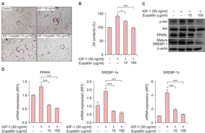
To examine the effects of eupatilin in vitro, we used hu- man SZ95 sebocytes (Supplementary Material). The effects of eupatilin on the proliferation of cultured SZ95 sebo- cytes were determined with various doses of eupatilin. In MTT assay, the proliferation of SZ95 sebocytes was se- quentially decreased according to the concentration (10, 20, 50, or 100 μM of eupatilin) (Supplementary Mater- ial). Thus, we determined experimental concentration as 10- and 100-μM of eupatilin. Next, we stained an intra- cellular lipid droplet formation using Oil Red O to inves- tigate the effects of eupatilin on the lipid synthesis of sebocytes. When SZ95 sebocytes were treated with eupa- tilin, lipid accumulation in the cytoplasm was significantly reduced (Fig. 1A, B). To elucidate how eupatilin sup- pressed IGF-1-induced lipogenesis of SZ95 sebocytes, the effects of eupatilin treatment on the IGF-1-induced ex- pression levels of phosphorylated Akt and lipogenesis-re- lated transcription factors (PPARγ and mature sterol regu- latory element-binding protein [SREBP]-1) were measured.
In comparison with no treatment, 100 μg/ml of eupatilin significantly reduced the protein levels of phosphorylated Akt, PPARγ, and mature SREBP-1 of the sebocytes, which were increased by IGF-1 pretreatment (Fig. 1C). Likewise, eupatilin also significantly downregulated the mRNA ex-
480 Ann Dermatol
Fig. 1. Effects of eupatilin on the intracellular lipid synthesis of SZ95 sebocytes. With the exception of the control group, SZ95 sebocytes were pretreated with 50 ng/ml of insulin-like growth factor (IGF)-1 for 48 hours and then with 10 μg/ml or 100 μg/ml of eupatilin for 48 hours. (A) Intracellular lipid droplets of SZ95 sebocytes treated with eupatilin were detected by Oil Red O staining. Bars=20 μm. (B) Supernatant Oil Red O levels (%) were measured by their optical density at 500 nm. (C) Whole-cell lysates were prepared and analyzed by western blotting. Blots were incubated with antibodies specific for total and phosphorylated forms of Akt, peroxisome proliferator-activated receptor (PPAR)-γ, and mature sterol regulatory element-binding protein (SREBP)-1. (D) Quantitative reverse- transcription polymerase chain reaction of PPARγ, SREBP-1a, and SREBP-1c for the evaluation of mRNA expression was performed.
Data are presented as the mean±standard error of triplicate assay (n=5). Data were analyzed using the Student’s t-test (*p<0.05,
***p<0.001). RFI: relative fold increase.
pression level of PPARγ, SREBP-1a, and SREBP-1c of the sebocytes (Fig. 1D). These results suggest that eupatilin has an inhibitory effect on IGF-I-induced lipogenesis of se- bocytes through suppression of the phosphorylation of Akt, PPARγ, and mature SREBP-1.
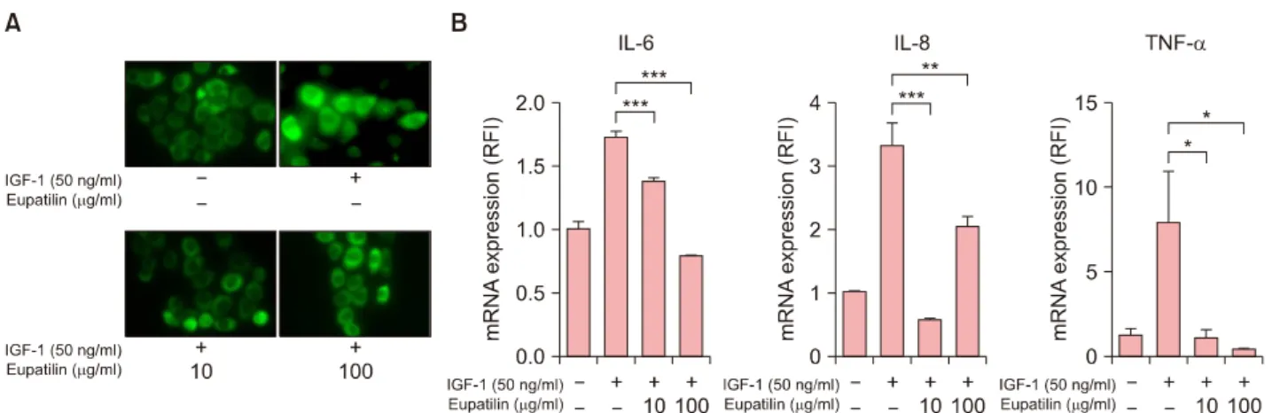
To further investigate the anti-inflammatory effects of eu- patilin on sebocytes, we analyzed the IGF-1-induced pro- inflammatory cytokines of sebocytes. In the immunofluo- rescence study, treatment with eupatilin inhibited the translocation of NF-κB p65 induced by 50 ng/ml of IGF-1 (Fig. 2A). This eupatilin treatment also significantly down- regulated the mRNA expression levels of (pro)inflam- matory cytokines such as TNF-α, IL-6, and IL-8 (Fig. 2B).
In this study, we first showed that the proper concen- trations of eupatilin (10 μg/ml and 100 μg/ml) had po- tent anti-IGF-I effects on SZ95 sebocytes, as follows: 1) eu- patilin suppressed the lipogenesis of sebocytes by interfer- ing with the expression of phosphorylated Akt, PPARγ,
and SREBP-1 induced by IGF-1; and 2) eupatilin reduced the inflammatory response of sebocytes by inhibiting NF-κB activation, with IGF-1 able to induce the inflammatory cy- tokine expression of sebocytes.
SREBP-1 is a major transcription factor that regulates cho- lesterol/fatty acid metabolism6. Smith et al.7 reported that IGF-1 induced SREBP-1. This activation occurred via a phosphoinositide 3-kinase (PI3K)/Akt pathway. They also showed that IGF-I transmits its lipogenic signal in sebo- cytes via an Akt pathway. Meanwhile, PPARγ is a poten- tial modulator of lipid production in human sebocytes8. PPARs are regulators of lipogenesis and differentiation of keratinocytes as well as sebocytes and have three isoforms (i.e., α, δ, and γ). Notably, PPARγ is important for se- baceous gland development and function9. Interestingly, in the present study, the Oil Red O stain showed that eu- patilin suppressed lipid synthesis by sebocytes. Although eupatilin from Artemisia plants has been reported as a se-
Fig. 2. Inhibition of insulin-like growth factor (IGF)-1–induced (pro)inflammatory cytokines of SZ95 sebocytes by eupatilin. With the exception of the control group, SZ95 sebocytes were treated with 50 ng/ml of IGF-1 for 48 hours and then with 10 μg/ml and 100 μg/ml of eupatilin for 48 hours. (A) Cells were incubated with primary antibody (nuclear factor kappa B [NF-κB] p65) and were finally visualized under a fluorescence microscope. (B) Quantitative Reverse-transcription polymerase chain reaction for the evaluation of mRNA expression of tumor necrosis factor (TNF)-α, interleukin (IL)-6, and IL-8 was performed. Data are presented as the mean±standard error of triplicate assay (n=5). Data were analyzed using the Student’s t-test (*p<0.05, **p<0.01, ***p<0.001).
RFI: Relative fold increase.
lective PPARα agonist10, in the present study, the proper concentrations of eupatilin (10 μg/ml and 100 μg/ml) in- hibited the IGF-1-induced SREBP-1 and PPARγ expres- sion of sebocytes. In addition, eupatilin inhibited the phos- phorylation of Akt. This inhibitory effect of eupatilin on IGF-1-induced sebocytes was confirmed by the reduced mRNA expression level of PPARγ, SREBP-1a, and SREPB-1c.
Moreover, we found that eupatiln suppressed the mRNA expression of TNF-α, IL-6, and IL-8 of sebocytes at the transcription level. Likewise, eupatilin also inhibited IGF- 1-induced nuclear translocation of NF-κB p65 of sebocytes.
This study demonstrate that eupatilin strongly downregu- lated IGF-I-induced inflammation and lipogenesis of SZ95 sebocytes. Therefore, our results suggest that eupatilin could be a promising candidate for the management of acne.
ACKNOWLEDGMENT
This work was supported by a National Research Founda- tion of Korea (NRF) grant funded by the Korean govern- ment (MSIP) (no. NRF-2015R1C1A2A01054767 and 2018 R1D1A1B07044100) and by Research Fund of Seoul St.
Mary’s Hospital, The Catholic University of Korea. And This research was supported by a grant from the Korea Health Technology R&D Project through the Korea Health Industry Development Institute (KHIDI) funded by the Ministry of Health & Welfare of the Republic of Korea (no.
HI14C2116).
SUPPLEMENTARY MATERIALS
Supplementary data can be found via http://anndermatol.
org/src/sm/ad-31-479-s001.pdf.
CONFLICTS OF INTEREST
The authors have nothing to disclose.
ORCID
Ji Hyun Lee, https://orcid.org/0000-0002-3671-502X Ye Jin Lee, https://orcid.org/0000-0002-9600-8220 Ji Young Song, https://orcid.org/0000-0002-5749-3032 Yeong Ho Kim, https://orcid.org/0000-0003-4983-4906 Jun Young Lee, https://orcid.org/0000-0002-8650-1759 Christos C. Zouboulis, https://orcid.org/0000-0003-1646-2608 Young Min Park, https://orcid.org/0000-0002-3631-0807
REFERENCES
1. Zouboulis CC. Acne and sebaceous gland function. Clin Dermatol 2004;22:360-366.
2. Melnik BC, Schmitz G. Role of insulin, insulin-like growth factor-1, hyperglycaemic food and milk consumption in the pathogenesis of acne vulgaris. Exp Dermatol 2009;18:833- 841.
3. Lee JH, Lee YJ, Lee JY, Park YM. Topical application of Eupatilin ameliorates atopic dermatitis-like skin lesions in NC/Nga mice. Ann Dermatol 2017;29:61-68.
4. Choi EJ, Oh HM, Na BR, Ramesh TP, Lee HJ, Choi CS, et al.
482 Ann Dermatol
Received September 13, 2018, Revised November 15, 2018, Accepted for publication January 2, 2019
Corresponding author: Young Min Park, Department of Dermatology, Seoul St. Mary’s Hospital, College of Medicine, The Catholic University of Korea, 222 Banpo-daero, Seocho-gu, Seoul 06591, Korea. Tel: 82-2-2258-6223, Fax: 82-2-599-9950, E-mail: [email protected]
ORCID: https://orcid.org/0000-0002-3631-0807
This is an Open Access article distributed under the terms of the Creative Commons Attribution Non-Commercial License (http://creativecommons.org/
licenses/by-nc/4.0) which permits unrestricted non-commercial use, distribution, and reproduction in any medium, provided the original work is properly cited.
Copyright © The Korean Dermatological Association and The Korean Society for Investigative Dermatology Eupatilin protects gastric epithelial cells from oxidative da-
mage and down-regulates genes responsible for the cellular oxidative stress. Pharm Res 2008;25:1355-1364.
5. Lee SH, Bae EA, Park EK, Shin YW, Baek NI, Han EJ, et al.
Inhibitory effect of Eupatilin and jaceosidin isolated from Artemisia princeps in IgE-induced hypersensitivity. Int Im- munopharmacol 2007;7:1678-1684.
6. Engelking LJ, Kuriyama H, Hammer RE, Horton JD, Brown MS, Goldstein JL, et al. Overexpression of Insig-1 in the livers of transgenic mice inhibits SREBP processing and reduces insulin-stimulated lipogenesis. J Clin Invest 2004;
113:1168-1175.
7. Smith TM, Gilliland K, Clawson GA, Thiboutot D. IGF-1 induces SREBP-1 expression and lipogenesis in SEB-1 sebo-
cytes via activation of the phosphoinositide 3-kinase/Akt pathway. J Invest Dermatol 2008;128:1286-1293.
8. Trivedi NR, Cong Z, Nelson AM, Albert AJ, Rosamilia LL, Sivarajah S, et al. Peroxisome proliferator-activated receptors increase human sebum production. J Invest Dermatol 2006;
126:2002-2009.
9. Ferré P. The biology of peroxisome proliferator-activated receptors: relationship with lipid metabolism and insulin sen- sitivity. Diabetes 2004;53 Suppl 1:S43-S50.
10. Choi Y, Jung Y, Kim SN. Identification of Eupatilin from Artemisia argyi as a selective PPARα agonist using affinity selection ultrafiltration LC-MS. Molecules 2015;20:13753- 13763.
https://doi.org/10.5021/ad.2019.31.4.482
Can Body Mass Index and/or Waist Circumference Be the Risk Factors of Chronic Spontaneous Urticaria?:
A Nationwide Population-Based Study
Yoon Seob Kim, Kyungdo Han1, Ji Hyun Lee, Jun Young Lee, Young Min Park
Department of Dermatology, Seoul St. Mary’s Hospital, The Catholic University of Korea, 1Department of Medical Statistics, College of Medicine, The Catholic University of Korea, Seoul, Korea
Dear Editor:
Several studies have suggested an association between chronic spontaneous urticaria (CSU) and body mass index (BMI)1-3 or metabolic syndrome1,4. In contrast, a French study reported that obesity was not associated with severe CSU5. There is little evidence that waist circumference (WC), an- other scale which correlates well with visceral obesity, is associated with CSU. We hypothesized that obesity could be associated with increased CSU risk. The aim of our
study was to investigate the impact of BMI and/or WC on the risk for CSU in an adult Korean population using a na- tionwide database. The study was approved by the Institu- tional Review Board of The Catholic University of Korea (IRB no. KC16EISE0852).
The health check-up database, a sub-dataset of the Korean National Health Insurance Service (NHIS) database (2002∼
2015), was used for data collection. NHIS subscribers are advised to have biannual health check-ups including height,
Ann Dermatol Vol. 31, No. 4, 2019 https://doi.org/10.5021/ad.2019.31.4.479
Supplementary Materials
MATERIALS AND METHODS
Cells and reagents
Immortalized human SZ95 sebocytes (provided by Prof. Christos C. Zouboulis) were maintained in SebomedⓇ basal me- dium (Biochrom GmbH, Berlin, Germany) containing 10% (v/v) fetal bovine serum (Gibco; Invitrogen, Carlsbad, CA, USA), 5 ng/ml of human recombinant epidermal growth factor (Invitrogen, Grand Island, NY, USA), 50 IU/ml of penicillin, and 50 μg/ml of streptomycin (Gibco) in a humidified atmosphere containing 5% CO2 at 37oC (Zouboulis et al., 1999)1. Culture medium was replaced every two days. Eupatilin was provided by Dong-A Pharmaceutical Co., Ltd. (Yongin, Korea) and dissolved in 10% dimethyl sulfoxide. The experimental design is shown in Supplementary Fig. 1.
Determination of cell proliferation by MTT assay
Cell proliferation was analyzed by by 3-(4,5-dimethylthiazol-2-yl)-2,5-diphenyl tetrazolium bromide MTT assay. SZ95 hu- man sebocytes were seeded (1×10³ cells per well) in triplicate into 96-well plates, incubated overnight, and then treated with eupatilin at the concentrations of 10 μg/ml, 20 μg/ml, 50 μg/ml, and 100 μg/ml (Supplementary Fig. 2) in SebomedⓇ media (Biochrom GmbH) without serum at 37oC in 5% CO2. Subsequently, 100 μl of MTT at 5 mg/ml was added to each well and incubation was continued for 4 hours. Supernatants were removed and formazan crystals resulting from mitochondrial enzymatic activity on the MTT substrate were solubilized with dimethyl sulfoxide (Sigma-Aldrich, St.
Louis, MO, USA). The absorbance was measured at 540 nm using an enzyme-linked immunosorbent assay reader (VersaMax; Molecular Devices, Sunnyvale, CA, USA).
Oil Red O staining and lipid detection
SZ95 sebocytes were seeded (1×10⁴ cells/ml) on 12-well culture plates, incubated overnight, and then treated with 50 μg/
ml of IGF-1 and 10 μg/ml or 100 μg/ml of eupatilin for 48 hours. Cells treated with the vehicle served as controls. At the end of the treatment period, cells were washed with phosphate-buffered saline and fixed through incubation in 10% for- malin for one hour at room temperature. Fixed cells were incubated with 60% isopropanol for five minutes and then iso- propanol was completely removed by air-dry. Cells were stained for 10 minutes with filtered Oil Red O working solution, prepared immediately before use by making a 6:4 mixture of stock (0.5% Oil Red O from Sigma-Aldrich in 99% iso- propanol) and dH2O. Supernatant Oil Red O levels were measured by measuring the optical density at 500 nm.
Western blot analysis
Cells were lysed in lysis buffer containing a protease and phosphatase inhibitor cocktail (Thermo Fisher Scientific, Waltham, MA, USA). Equal amounts (30 μg) of extracted protein were resolved using 6% to 12% sodium dodecyl sulfate–
polyacrylamide gel electrophoresis and transferred to polyvinylidene difluoride membranes. Following incubation in blocking solution, the membranes were incubated overnight at 4oC with the appropriate antibodies. Blots were then in- cubated with peroxidase-conjugated secondary antibodies and visualized by enhanced chemiluminescence substrate (Thermo Fisher Scientific). The following primary antibodies were used: phospho-Akt, Akt, peroxisome proliferator-acti- vated receptor (PPAR)γ, mature sterol regulatory element-binding protein (SREBP)-1 (Cell Signaling Technology, Danvers, MA, USA), and actin (Santa Cruz Biotechnologies, Dallas, TX, USA).
Immunofluorescence
SZ95 sebocytes were grown on cell culture slides (SPL Life Sciences, Pocheon, Korea), fixed through incubation with 4%
paraformaldehyde for 20 minutes, and permeabilized through incubation with 0.1% Triton X-100 in phosphate-buffered saline for 10 minutes at room temperature. Cells were then incubated overnight at 4oC with primary antibody (i.e., nuclear factor kappa B [NF-κB] p65) (Santa Cruz Biotechnologies) and then for one hour at room temperature with fluorescein iso- thiocyanate-conjugated secondary antibody (Santa Cruz Biotechnologies), and were finally visualized under a fluorescence
S2 Ann Dermatol
microscope (Olympus, Tokyo, Japan).
Reverse-transcription polymerase chain reaction
To evaluate gene expression, total RNA was isolated using TrizolⓇ Reagent (Invitrogen) according to the manufacturer’s protocols. Equal amounts of RNA (1 μg) were reverse-transcribed into complementary DNA using the Prime ScriptTM RT reagent Kit with gDNA EraserⓇ (Takara Bio, Otsu, Japan). Quantitative reverse-transcription polymerase chain reaction (RT-PCR) was performed using a CFX96TM Real-Time PCR Detection SystemⓇ (Bio-Rad, Hercules, CA, USA) with SYBRⓇ premix EX TaqTM (Takara Bio, Kusatsu, Japan) and specific primers for tumor necrosis factor (TNF)-α, interleukin (IL)-6, IL-8, PPAR-γ, SREBP-1a, and SREBP-1c. The cycling conditions consisted of an initialization step for 10 seconds at 95oC followed by two-step PCR for 40 cycles of 95oC for five seconds (denaturation) and 58oC to 60oC for 30 seconds (annealing/extension). Fluorescence intensity was measured in real time using the optical module. Melt curves were used to determine product specificity. Results were normalized to the level of glyceraldehyde 3-phosphate dehydrogenase gene expression. The analysis of relative gene expression data was conducted using the 2−ΔΔCT method. All experiments were repeated twice. The primers used are as follows : TNF-α, forward 5’-CCCAGGGACCTCTCTCTAATC-3’ and reverse 5’- ATGGGCTACAGGCTTGTCACT-3’; IL-6, forward 5’-ACCCCCAATAAATATAGGACTGGA-3’ and reverse 5’-GAGAAGGC AACTGGACCGAA-3’; IL-8, forward 5’-GGTGCAGTTTTGCCAAGGAG-3’ and reverse 5’-TGGGGTGGAAAGGTTTGGAG- 3’; PPAR-γ, forward 5’-GCCCAGGTTTGCTGAATGTG-3’ and reverse 5’-TGAGGACTCAGGGTGGTTCA-3’; and SREBP- 1a, forward 5’-GCTGCTGACCGACATCGAA-3’, SREBP-1c, forward 5’-GGAGCCATGGATTGCACTTT-3’, and SREBP-1a,c, reverse 5’-TCAAATAGGCCAGGGAAGTCA-3’.
Statistical analysis
All data were expressed as the mean±standard error of the mean. One-way analysis of variance followed by Tukey’s mul- tiple comparison test was used for statistical analysis. The Kruskal–Wallis test was used for comparisons of the four groups.
Statistical significance was set at p<0.05.
REFERENCE
1. Zouboulis CC, Seltmann H, Neitzel H, Orfanos CE. Establishment and characterization of an immortalized human sebaceous gland cell line (SZ95). J Invest Dermatol 1999;113:1011-1020.
Supplementary Fig. 1. Experimen- tal design. IGF-1: insulin-like growth factor-1.
S4 Ann Dermatol
Supplementary Fig. 2. The effects of eupatilin on cell prolifer- ation by MTT assay. The effect of eupatilin on SZ95 sebocyte proliferation was determined by MTT assay. Cells were treated with various concentrations of eupatilin (10∼100 μM). IGF-1:
insulin-like growth factor-1.