© 2012 The Korean Academy of Medical Sciences.
This is an Open Access article distributed under the terms of the Creative Commons Attribution Non-Commercial License (http://creativecommons.org/licenses/by-nc/3.0) which permits unrestricted non-commercial use, distribution, and reproduction in any medium, provided the original work is properly cited.
pISSN 1011-8934 eISSN 1598-6357
Rupture of Right Hepatic Duct into Hydatid Cyst
Echinococcal disease can develop anywhere in the human body. The liver represents its most frequent location. Hepatic hydatid cysts may rupture into the biliary tract, thorax, peritoneum, viscera, digestive tract or skin. We report a rare case with rupture of the right hepatic duct into a hydatid cyst in a woman with known hydatid disease and
choledocholithiasis. The increased intra-luminal pressure in the biliary tree caused the rupture into the adjacent hydatid cyst. The creation of the fistula between the right hepatic duct and the hydatid cyst decompressed the biliary tree, decreased the bilirubin levels and offered a temporary resolution of the obstructive jaundice. Rupture of a hydatid cyst into the biliary tree usually leads to biliary colic, cholangitis and jaundice. However, in case of obstructive jaundice due to choledocholithiasis, it is possible that the cyst may rupture by other way around while offering the patient a temporary relief from his symptoms.
Key Words: Hydatid Disease; Cholelithiasis; Echinococcal Cyst Rupture; Cystobiliary Fistula Nickolaos Michalopoulos, Styliani Laskou,
Theodossis S. Papavramidis, Ioannis Pliakos, Eustathios Kotidis, Isaak Kesisoglou, and
Spiros T. Papavramidis
3rd Department of Surgery, Aristotle University of Thessaloniki, AHEPA University Hospital, Thessaloniki, Greece
Received: 25 February 2012 Accepted: 20 May 2012 Address for Correspondence:
Nickolaos Michalopoulos, MD
3rd Department of Surgery, Aristotle University of Thessaloniki, AHEPA University Hospital, 85 Karakasi St. P.C: 54453, Thessaloniki, Greece
Tel: +00306974092401, Fax: +00302310420293 E-mail: [email protected]
http://dx.doi.org/10.3346/jkms.2012.27.8.953 • J Korean Med Sci 2012; 27: 953-956
CASE REPORT
Infectious Diseases, Microbiology & Parasitology
INTRODUCTION
Hydatid disease, although rare, is still endemic in many coun- tries, representing an important public health problem. It may occur in any organ or tissue. Reviews show that the most fre- quently involved organs are the liver (55%) and lungs (40%).
Rarely, hydatid disease may be found in the spleen (1.8%), kid- neys (1.4%) bones (0.1%) or other sites (1.7%). Multiple simul- taneous locations are observed in 25% of cases (1, 2).
A common complication of hepatic hydatid disease is the rup- ture of the cyst caused by the increased pressure within it (2).
Cysts may rupture into the biliary tree, the peritoneal or pleural cavity, the pericardium, the gastrointestinal tract, or even into blood vessels. Echinococcal cysts may coexist with cholelithia- sis or scarcely with choledocholithiasis (3).
The aim of this study was to present an unusual case of hyda- tid cyst coexisting with choledocholithiasis and obstructive jaun- dice, in which the increased intraluminal pressure in the right hepatic duct led to the decompression of the biliary tree in to the cystic cavity. Diagnostic methods and various treatment modal- ities were also discussed.
CASE DESCRIPTION
A 60-yr-old female, coming from a rural area, was referred in March 2009 to the Emergency Department complaining for right upper quadrant pain and jaundice. She was diagnosed
with echinococcal cyst of the liver and cholelithiasis four years ago. The laboratory examinations on admission were: white blood cell count (WBC): 10,000/μL without eosinophilia; lac- tate dehydrogenase (LDH) 1,209 μ/L; serum glutamic oxalo- acetic transaminase (SGOT) 256 μ/L; serum glutamic pyruvic transaminase (SGPT) 401 μ/L; total bilirubin 5.5 mg/dL with direct bilirubin 3.6 mg/dL. Abdominal ultrasound revealed multiple echinococcal cystic lesions in the right lobe of the liver.
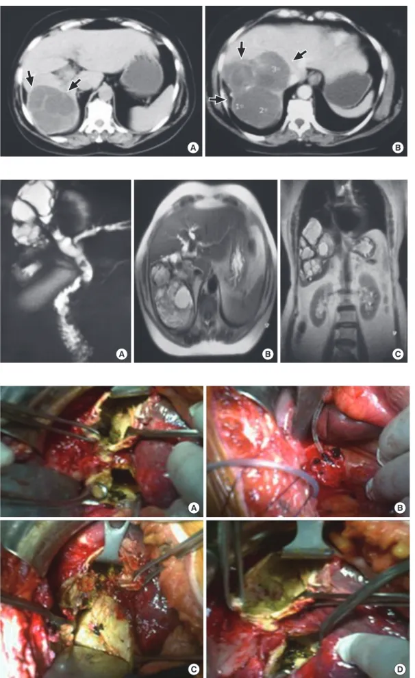
Additionally, the gallbladder was distended, containing multi- ple small stones and mud, while the intrahepatic biliary tree and the common bile duct were dilated. The subsequent com- puted tomography (CT) showed a huge multisegmented hyda- tid cyst of the right lobe occupying the segments V, VI, and VII, The intra- and extra-hepatic biliary trees were dilated (Fig. 1).
She was treated conservatively, her transaminasemia and hyper- bilirubinemia subsided and the patient was discharged seven days later.
Ten days later, she was readmitted with jaundice. Her bio- chemical and hematological tests revealed severe transaminas- emia and hyperbilirubinemia (SGOT 358 μ/L; SGPT 491 μ/L;
LDH 754 μ/L; bilirubin 12.7 mg/dL with direct bilirubin 8.3 mg/
dL). Surprisingly, an acute fall in her bilirubin levels was noticed two days later (LDH 400 μ/L; SGOT 175 μ/L; SGPT 335 μ/L;
gamma-glutamyltransferase (γ-GT) 786 μ/L; alkaline phospha- tase (ALP) 461 μ/L; bilirubin 5.4 mg/dL with direct bilirubin 3,2 mg/dL). Magnetic resonance cholangiopancreatography (MRCP) was performed showing a communication between the echi-
Michalopoulos N, et al. • Rupture of Right Hepatic Duct into Hydatid Cyst
954 http://jkms.org http://dx.doi.org/10.3346/jkms.2012.27.8.953
Fig. 2. MRCP images show- ing communication between the echinococcal cyst and the right hepatic duct. The common hepatic duct and the common bile duct are full with material of unknown origin. (A) Cholangiopancrea- tography. (B) Cross-section imgae. (C) Frontal-section image.
A B C
Fig. 1. CT scan images (A, B) showing a huge multi-locular cyst (arrows) adjacent the right hepatic bile duct.
A B
Fig. 3. Intraoperative images (A-D) showing pigmented gallstones removed from the echinococcal cyst.
A
C
B
D
Michalopoulos N, et al. • Rupture of Right Hepatic Duct into Hydatid Cyst
http://jkms.org 955
http://dx.doi.org/10.3346/jkms.2012.27.8.953
nococcal cyst and the right hepatic duct, while the common he- patic duct and the common bile duct were full with material of unknown origin (Fig. 2).
A surgical intervention was decided upon. An open chole- cystectomy, with exploration of the common bile duct was per- formed. Multiple gallstones were removed from the common bile duct. Additionally, partial cystectomy was performed. Sur- prisingly, pigmented gallstones with daughter cysts were found and removed from the echinococcal cystic cavity (Fig. 3). The intra-operative cholangiogram revealed communication be- tween the echinococcal cyst and the right hepatic duct. The right hepatic duct was ligated. The cystic cavity and the subhepatic space were drained, and a T-tube was placed into the common bile duct. The patient recovered well from the operation and her postoperative course was uneventful. After discharge, albena- zole was administered for 3 month cycles, with 14 day intervals.
Her follow-up included ultrasonography and CT scan 3 months and 12 months after the operation. Two years after the opera- tion the patient was free of disease and symptoms.
DISCUSSION
Hydatid cysts grow at a variable rate. They may stabilize, or be- come calcified, while others may collapse or even completely resolve (4). Becoming symptomatic may be due to pressure ex- ertion of the cyst on the liver parenchyma, or rupture into sur- rounding tissues (3). When referring to biliary tract, the rupture is common in the right and left hepatic duct and rarely may oc- cur into the hepatic duct junction, common bile duct or cystic duct (5). Scarcely, the hydatid cyst could perforate into the gall- bladder (5).
Hydatid cyst rupture has been classified into three types: i) contained when only the endocyst ruptures and the cyst con- tents are confined within the pericyst; ii) communicating when the cyst contents escape via biliary radicles and iii) direct when both the endocyst and the pericyst tear, allowing cyst contents to spill into the pleural or peritoneal spaces (6, 7). In the present case a communicating rupture of the echinococcal cyst occurred causing a fistula between the echinococcal cyst and the right hepatic duct. As a result, the biliary tree was decompressed, se- rum bilirubin levels were decreased and the patient improved.
The communicating type of rupture is the most frequent com- plication, representing approximately 50% of cases on admis- sion (6). The mechanism of intrabiliary rupture seems to be that of entrapment of small bile duct radicles in the pericyst, which due to increased intracystic pressure undergo atrophy resulting in rupture (8). Following cyst enlargement, communication with larger ducts is established (8). Most hydatid cysts of the liver even- tually leak into small bile ducts or perforate into larger ones. In large surgical series, some sort of communication was found in 40%-90% of cysts (9). In our case, the progressive growth of the
cyst led its wall to be adjacent to the right hepatic duct, while choledocholithiasis and subsequent obstruction increased the pressure inside the biliary tree. This combination resulted in the rupture of the right hepatic duct into the hydatid cyst to de- compress the biliary tree and as a result a biliocystic fistula was established.
Intrabiliary rupture can occur with two different clinical set- tings which are followed by certain symptoms. These are occult communication (10%-37%) and frank intrabiliary rupture (3%- 17%) (10). The occult rupture is usually silent and may be accom- panied by suppuration or can evolve towards a frank rupture (9).
In the frank rupture daughter vesicles and fragmented mem- branes escape into the biliary tree causing obstruction, cholan- gitis or septicemia (11). The present case was probably an occult communication, which evolved towards a frank rupture.
The diagnosis of intrabiliary ruptured hydatid cysts has been assisted by imaging tests. Echogenic material, without posterior acoustic shadowing in the extrahepatic ducts, is a finding im- plying the presence of intracystic material (12). Abdominal CT scan may reveal the dilated common bile duct with low attenu- ation intraluminal material, suggesting the presence of hydatid sand and cysts (13). Recently, magnetic resonance imaging (MRI- MRCP) has proven to be a useful noninvasive diagnostic mo- dality in cases of intrabiliary rupture, whereas CT scan and ul- trasound results are inconclusive (13).
Endoscopy is a modality serving both diagnostic and thera- peutic aims. During preoperative endoscopic retrograde chol- angiopancreatography (ERCP), daughter cysts may be seen in the duodenum, impacted in the ampulla of Vater or obstructing any part of the biliary tree (14, 15). Moreover, postoperative ERCP may resolve obstruction or cholangitis due to residual material in biliary ducts, while providing management of postoperative external biliary fistulae (15). Additionally, endoscopic sphinc- terotomy has proved to be an alternative treatment for patients with biliary hydatid disease (12).
In case of cystobiliary communication a surgical intervention is mandatory. Various types of procedures have been proposed such as: partial cystectomy with primary closure, partial cystec- tomy with drainage, cystotomy with drainage, hepatic resection (atypic, segmentary or lobar) and omentoplasty (9). Suturing of the cystobiliary fistula, and if feasible common bile duct explo- ration using intraoperative cholangiography are also required (11). If the biliary tract is cleaned of all cystic content, a T-tube drain is usually sufficient.
In conclusion, rupture of a hepatic hydatid cyst into the biliary tree is the most common complication of hydatid disease. Usu- ally, it leads to biliary colic, cholangitis and jaundice. However, it is possible that the rupture is being done conversely, relieving the patient from the obstructive symptoms. Currently, ERCP is a method of both diagnosis and treatment. Further surgical treat- ment may be required if an obvious communication between
Michalopoulos N, et al. • Rupture of Right Hepatic Duct into Hydatid Cyst
956 http://jkms.org http://dx.doi.org/10.3346/jkms.2012.27.8.953
the biliary tree and the hydatid cyst is displayed.
REFERENCES
1. Dziri C, Haouet K, Fingerhut A, Zaouche A. Management of cystic echi- nococcosis complications and dissemination: where is the evidence?
World J Surg 2009; 33: 1266-73.
2. Prousalidis J, Tzardinoglou E, Sgouradis L, Katsohis C, Aletras H. Un- common sites of hydatid disease. World J Surg 1998; 22: 17-22.
3. Lewall DB, McCorkell SJ. Rupture of echinococcal cysts: diagnosis, clas- sification, and clinical implications. AJR Am J Roentgenol 1986; 146:
391-4.
4. WHO Informal Working Group on Echinococcosis. Guidelines for treat- ment of cystic and alveolar echinococcosis in humans. Bull World Health Organ 1996; 74: 231-42.
5. Manouras A, Genetzakis M, Antonakis T, Lagoudianakis P, Pattas M, Papadima A, Giannopoulos P, Memenakos E. Endoscopic management of a relapsing hepatic hydatid cyst with intrabiliary rupture: a case report and review of the literature. Can J Gastroenterol 2007; 21: 249-53.
6. Marti-Bonmati L, Menor F, Ballesta A. Hydatid cyst of the liver: rupture into the biliary tree. AJR Am J Roentgenol 1988; 150: 1051-3.
7. Papavramidis TS, Pliakos I, Triantafillopoulou K, Michalopoulos N, Polyzonis M, Sapalidis K, Kesisiglou I, Papavramidis ST. Endoscopic removal of biliary tree echinococcal cysts. Ann Gastroenterol 2009; 22:
116-8.
8. Yalin R, Aktan AO, Yeğen C, Döşlüoğlu HH. Significance of intracystic pressure in abdominal hydatid disease. Br J Surg 1992; 79: 1182-3.
9. Prousalidis J, Kosmidis C, Kapoutzis K, Fachantidis E, Harlaftis N, Aletras H. Intrabiliary rupture of hydatid cysts of the liver. Am J Surg 2009; 197:
193-8.
10. Erzurumlu K, Dervisoglu A, Polat C, Senyurek G, Yetim I, Hokelek M.
Intrabiliary rupture: an algorithm in the treatment of controversial com- plication of hepatic hydatidosis. World J Gastroenterol 2005; 11: 2472-6.
11. Spârchez Z, Osian G, Onica A, Bărbânta C, Tantău M, Pascu O. Ruptured hydatid cyst of the liver with biliary obstruction: presentation of a case and review of the literature. Rom J Gastroenterol 2004; 13: 245-50.
12. Mendez Montero JV, Arrazola Garcia J, Lopez Lafuente J, Antela Lopez A, Mendez Fernandez R, Saia Ayala A. Fat-fluid level in hepatic hydatid cyst: a new sign of rupture into the biliary tree? AJR Am J Roentgenol 1996; 167: 91-4.
13. Ozaslan E, Bayraktar Y. Endoscopic therapy in the management of hepa- tobiliary hydatid disease. J Clin Gastroenterol 2002; 35: 160-74.
14. Zargar SA, Khuroo MS, Khan BA, Dar MY, Alai MS, Koul P. Intrabiliary rupture of hepatic hydatid cyst: sonographic and cholangiographic ap- pearances. Gastrointest Radio1 1992; 17: 41-5.
15. Simşek H, Ozaslan E, Sayek I, Savaş C, Abbasoğlu O, Soylu AR, Balaban Y, Tatar G. Diagnostic and therapeutic ERCP in hepatic hydatid disease.
Gastrointest Endosc 2003; 58: 384-9.