• 검색 결과가 없습니다.

Gingival Squamous Carcinoma with Metastatic Lymph Node Involvement of Papillary Thyroid Carcinoma

N/A
N/A
Protected

Academic year: 2021

Share "Gingival Squamous Carcinoma with Metastatic Lymph Node Involvement of Papillary Thyroid Carcinoma"

Copied!
4
0
0

로드 중.... (전체 텍스트 보기)

전체 글

(1)

276

J Korean Assoc Maxillofac Plast Reconstr Surg 2012;34(4):276-279 pISSN:1225-4207 eISSN: 2233-7296

Case Report

RECEIVED May 5, 2012, REVISED June 1, 2012, ACCEPTED June 27, 2012 Correspondence to Jwa-Young Kim

Department of Oral and Maxillofacial Surgery, Hallym University Sacred Heart Hospital 22, Gwanpyeong-ro 170-gil, Dongan-gu, Anyang 431-070, Korea

Tel: 82-31-380-3875, Fax: 82-31-380-3872, E-mail: jwayoung@hanmail.net

CC

This is an open access article distributed under the terms of the Creative Commons Attribution Non-Commercial License (http://creativecommons.org/licenses/

by-nc/3.0) which permits unrestricted non-commercial use, distribution, and reproduction in any medium, provided the original work is properly cited.

Gingival Squamous Carcinoma with Metastatic Lymph Node Involvement of Papillary Thyroid Carcinoma

Ju-Won Kim, Chang-Youn Lee, Seung-Min Oh, Byung-Eun Yang, Jwa-Young Kim, Yun-Jung Song 1 , Kang-Min Ahn 2 , Joo-Yong Park 3

Departments of Oral and Maxillofacial Surgery,

1

Conservative Dentistry, Hallym University College of Medicine,

2

Department of Oral and Maxillofacial Surgery, Seoul Asan Medical Center, College of Medicine, University of Ulsan,

3

Oral Cancer Clinic, Research Institute and Hospital, National Cancer Center

Abstract

The development of multiple primary tumors is a problem leading to the treatment of patients diagnosed with gingival squamous cell carcinoma (SCC). The occurrence of multiple primary cancers in patients with SCC of the head and neck is uncommon.

Thyroid carcinomas have been found incidentally in the cervical lymph nodes after histopathologic examination. A 72-year-old male with SCC of the lower gingiva at the clinical stage T2N0M0 was treated with partial mandibulectomy and selective neck dissection. Histopathologic examination showed the foci of papillary thyroid carcinoma metastasis. The patient subsequently underwent total thyroidectomy. We report a case of papillary thyroid carcinoma associated with SCC of the oral gingiva along with a review of literatures.

Key words: Squamous cell carcinoma, Thyroid cancer, Neck dissection

Introduction

Oral cancer accounts for 3∼4% of total cancer incidences.

More than 90% of oral cancer cases are known as squamous cell carcinoma[1,2]. Therefore, the study of squamous cell carcinoma is very important. The treatment of primary mul- tiple carcinoma is difficult, especially in the patients with squamous cell carcinoma in the head and neck[3]. Primary multiple carcinoma has been reported to have occurred in 20% of the patients with squamous cell carcinoma in the head and neck[4]. Because the respiratory and digestive organs are exposed to the same carcinogen at the same

time, multiple carcinoma, which shows similar histological

patterns, may occur in the esophagus, lung, and head and

neck areas[5]. Unlike with primary multiple carcinoma,

however, detection of thyroid gland carcinoma by chance

is uncommon. In less than 0.5% of the patients with squ-

amous cell carcinoma in the head and neck, thyroid gland

carcinoma was reportedly detected in the lymph node of

the neck by chance during surgery[6]. The authors' depart-

ment reports the case and literature review of the detection

of squamous cell carcinoma in the gingiva in the thyroid

gland and lymph node in the neck of a 72-year-old male.

(2)

Ju-Won Kim: Thyroid Carcinoma Discovered Incidentally in Gingival Squamous Cell Carcinoma Patient 277

Vol. 34 No. 4, July 2012 Fig. 1. Intra oral photograph on first visit. The photograph show

irregular margin and tissue necrosis on gingival.

Fig. 3. Positron emission tomography-computed tomography scan. The photograph show about 3 cm length mass with intense fludeoxyglucose (FDG) uptake in left lower gingiva, mild increased FDG uptake in left submandibular area, and lung nodules with faint FDG uptake in right middle lobe of lung and right lower lobe of lung (circle).

Fig. 2. Histopathologic view of gingival squamous cell carcinoma (H&E, ×200). The photomicrograph show the squamous pearl (white arrow).

Case Report

A 72-year-old male visited the authors' hospital on May 24, 2010 with the symptom of gingival pain in his left lower gum. His gingival pain started in his lower left gum two months earlier, and he was taking the medicine pre- scribed by a clinic doctor, but he came to the authors' hospital due to the worsened pain. His systemic medical history showed hypertension, but no drinking of alcohol and smoking.

In the first medical examination, the white and red col-

ored lesion with the size of 4×3 cm and with an irregular

boundary was observed at the gingiva near the first and

second molars in the lower left gum, and tissue necrosis

was detected in the lower area of the lesion (Fig. 1). Thus,

a biopsy was performed with local anesthesia on the same

day. The biopsy result showed that the patient had squ-

amous cell carcinoma with a moderate level of differ-

entiation (Fig. 2). Additionally, a positron emission tomog-

raphy-computed tomography (PET-CT) scan, a head and

neck magnetic resonance imaging, and a whole body bone

scan were performed on the patient. In PET-CT scan, About

(3)

278 Ju-Won Kim: Thyroid Carcinoma Discovered Incidentally in a Gingival Squamous Cell Carcinoma Patient

J Korean Assoc Maxillofac Plast Reconstr Surg

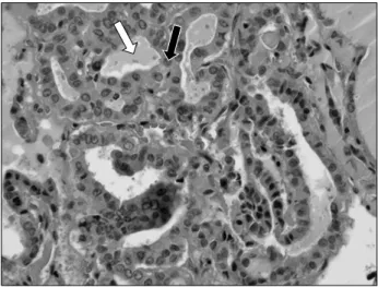
Fig. 5. Histopathologic view of extirpated lymph node at level II, showing papillary thyroid. Carcinoma (H&E staining and Galectin-3 immunostaining, ×200). The photomicrograph show the colloid like material (white arrow) and the intranuclear groove (black arrow).

Fig. 4. Post-operation panoramic view.

3 cm length mass with intense fludeoxyglucose (FDG) up- take in left lower gingiva and mild increased FDG uptake in left submandibular area was found. There was no symp- tom on thyroid (Fig. 3). And partial amputation of the lower jaw, neck dissection (supraomohyoid neck dis- section, including partially level IV), and reconstruction using a left fibula free flap were also performed under systemic anesthesia on June 23, after the patient's hospital- ization on June 21, 2010 (Fig. 4).

The frozen biopsy for safety margin around the primary lesion during the surgery did not show the carcinoma and surgical biopsy for the primary lesion showed squamous cell carcinoma. But metastatic papillary carcinoma was de- tected in the follow-up biopsy on the level II lymph node.

The tumor cell that makes up the gland, the colloid-like material, the intranuclear groove, and the intranuclear in- clusion were observed after H&E staining and Galectin-3 immunostaining of the tissues. The patient was diagnosed as having papillary thyroid carcinoma (Fig. 5). Since he was referred to the Department of Breast and Endocrine Surgery for diagnosis of papillary thyroid carcinoma via ultrasonography and a liquid-based cytology test for the thyroid, thyroidectomy and additional neck dissection (level IV and central) were performed on him under sys- temic anesthesia at the aforementioned department on September 6, 2010. The biopsy results showed metastasis in one of the perithyroid lymph nodes and infiltration of the perithyroid soft tissue. The remnant and metastatic up- take were not observed after the treatment with I

131

in December 2010, after the surgery.

Discussion

Thyroid gland cancer is not commonly detected, unlike

primary multiple carcinoma, in the patients with squamous

cell carcinoma in the head and neck. It can only be de-

tected in the following cases: in the removed thyroid lobe

during a laryngectomy, in case of facilitation in the thyroid,

and in neck dissection[6]. Thyroid papillary cancer is the

most common malignancy in the thyroid, accounting for

50∼90% of thyroid-originating cancers, with frequent

metastasis in the lymph node[7]. Its prognosis is known

as relatively good, however, and metastasis in the lymph

node does not significantly affect the patient's survival

rate[8]. Also, the progress of thyroid papillary cancer is

relatively slow, and its prognosis was recently reported

to be 90∼98% of the 10-year survival rate, due to enhanced

treatment efficacy combined with thyroid-stimulating hor-

mone inhibition therapy[9-11]. Yet due to the occurrence

of local recurrences and remote metastasis in 20∼35% of

cases, follow-up checks are important to find the recur-

rences and metastasis[9,10,12,13]. Thyroid ultrasonography

is mainly used to diagnose a thyroid nodule, and has been

effective as the primary differential test because it can pro-

vide the important information on the shape of the nodule,

the infiltration into the surrounding tissues, and the meta-

stasis in the lymph node in the neck[9,10]. It can also

simultaneously perform the cell inhalation test in cases of

potential metastisis.

(4)

Ju-Won Kim: Thyroid Carcinoma Discovered Incidentally in Gingival Squamous Cell Carcinoma Patient 279

Vol. 34 No. 4, July 2012

Goepfert and Callender[14] reported that the patients di- agnosed as having squamous cell carcinoma in the head and neck have a higher risk of recurrence than the patients with ordinary thyroid papillary cancer if they had thyroid papillary cancer. In addition, Godballe et al .[15] determined the break point of age for the diagnosis of thyroid papillary cancer as 60 years. This is because the cancer shows in- creasing patterns of nuclear dysplasia in the carcinoma, mitosis, remote metastasis, and expansion out of the thy- roid before and after the age of 60 years. Thyroid hormone inhibitory therapy after thyroidectomy, neck dissection, and radioactive iodine treatment are generally performed, and follow-up monitoring with serum thyroglobulin and systemic scanning of the radioactive iodine are used for patients who have a higher risk of recurrence of thyroid papillary cancer[10,12,13,16].

We think that thyroid papillary cancer had been exist with no clinical symptom for a long time and it was found incidentally during neck dissection for gingival squamous cell carcinoma. Although this case is rare, it is important that proper treatment and active management are needed in geriatric patients.

This is the case report of the thyroid papillary cancer detected by chance during surgery on a patient who was diagnosed as having squamous cell carcinoma in the gingi- va of his lower left jaw.

Although it has been reported that the progress of thyroid papillary cancer is slow and its prognosis is good, the literature review revealed that the risk is high in geriatric patients with squamous cell carcinoma in the head and neck.

References

1. Arbes SJ Jr, Olshan AF, Caplan DJ, Schoenbach VJ, Slade GD, Symons MJ. Factors contributing to the poorer survival of black Americans diagnosed with oral cancer (United States). Cancer Causes Control 1999;10:513-23.

2. Shiboski CH, Shiboski SC, Silverman SJ. Trends in oral can-

cer rates in the United States, 1973-1996. Community Dent Oral Epidemiol 2000;28:249-56.

3. Coskun H, Erisen L, Tolunay S, Basut O, Onart S, Tezel I.

Incidental association of thyroid carcinoma and squamous cell carcinoma of head and neck. Am J Otolaryngol 2002;

23:228-32.

4. Larson JT, Adams GL, Fattah HA. Survival statistics for mul- tiple primaries in head and neck cancer. Otolaryngol Head Neck Surg 1990;103:14-24.

5. Jones AS, Morar P, Phillips DE, Field JK, Husband D, Helliwell TR. Second primary tumors in patients with head and neck squamous cell carcinoma. Cancer 1995;75:1343-53.

6. Pitman KT, Johnson JT, Myers EN. Papillary thyroid carcino- ma associated with squamous cell carcinoma of the head and neck: significance and treatment. Am J Otolaryngol 1996;17:190-6.

7. Schlumberger MJ, Filetti S, Hay ID. Nontoxic goiter and thy- roid neoplasia. In: Larsen PR, editor. Williams textbook of endocrinology. 10th ed. Philadelphia: W.B Saunders; 2002.

p.469-73.

8. Shaha AR. Management of the neck in thyroid cancer.

Otolaryngol Clin North Am 1998;31:823-31.

9. Huang SH, Wang PW, Huang YE, et al. Sequential fol- low-up of serum thyroglobulin and whole body scan in thy- roid cancer patients without initial metastasis. Thyroid 2006;16:1273-8.

10. Mazzaferri EL, Kloos RT. Using recombinant human TSH in the management of well-differentiated thyroid cancer: cur- rent strategies and future directions. Thyroid 2000;10:767-78.

11. National Comprehensive Cancer Network. Clinical practice guidelines in oncology (NCCN Guidelines)-Thyroid carci- noma, version 2. 2007 [2011 DEC 19]. Available from:

http//www.nccn.org.

12. Lee E, Lee SJ, Lee CR, et al. FDG-PET as a predictor of re- currence with 131I scan negative differentiated thyroid can- cer: an evaluation compared with pathologic findings. J Korean Soc Endocrinol 1999;14:520-30.

13. Benbassat CA, Mechlis-frish S, Guttmann H, Glaser B, Krausz Y. Current concepts in the follow-up of patients with differentiated thyroid cancer. IAMJ 2007;9:540-5.

14. Goepfert H, Callender DL. Differentiated thyroid cancer-pap- illary and follicular carcinomas. Am J Otolaryngol 1994;

15:167-79.

15. Godballe C, Asschenfeldt P, Sørensen JA, Sørensen MW, Jørgensen K. Papillary thyroid carcinoma: correlations be- tween prognosis, age, and clinicopathological and histo- morphological findings. Laryngoscope 1994;104:747-51.

16. Mansi L, Moncayo R, Cuccurullo V, Dottorini ME, Rambaldi

PF. Nuclear medicine in diagnosis, staging and follow-up of

thyroid cancer. Q J Nucl Med Mol Imaging 2004;48:82-95.

수치

Fig. 3. Positron emission tomography-computed tomography scan. The photograph show about 3 cm length mass with intense  fludeoxyglucose (FDG) uptake in left lower gingiva, mild increased FDG uptake in left submandibular area, and lung nodules with  faint F
Fig. 5. Histopathologic view of extirpated lymph node at level  II, showing papillary thyroid

참조

관련 문서

These results suggest that the bilobalide inhibits the cell proliferation and induces the apoptotic cell death in FaDu human pharyngeal squamous cell carcinoma via both

The expression levels of apoptotic related proteins and caspase dependent proteins by the ostreolysin purified from P.ostreatus on cell viability in FaDu human

Concentration-Dependent inhibition of cell viability by adenosine in human oral fibroblast and FaDu human head and neck squamous cell carcinoma · · ·

It also suggest that cell proliferation inhibits by a novel signal transduction for adenosine effect in human FaDu hypopharynx squamous cell carcinoma cells....

Differential Expression of Desmoglein1, Desmoglein3, Epithelial Membrane Antigen, Ber-EP4 and CD10 in Basal Cell Carcinoma and Squamous Cell Carcinoma

Cooper, M.D.et el Revised American Thyroid Association Management Guidelines for Patients with Thyroid Nodules and Differentiated Thyroid Cancer THYROID Volume

Objective: The purpose of this study was to analyze recent trend in incidence of basal cell carcinoma and squamous cell carcinoma in patients from the Gwangju City

The objective was to access the prevalence of asymptomatic thyroid disease not only thyroid nodules but diffuse thyroid disorders by using ultrasonography