Case Report
원고 접수일 2013년 2월 18일, 원고 수정일 2013년 3월 28일, 게재 확정일 2013년 5월 20일
책임저자 최병준
(130-872) 서울시 동대문구 경희대로 23, 경희대학교 치과병원 구강악안면외과 Tel: 02-958-9440, Fax: 02-966-4572, E-mail: [email protected]
RECEIVED February 18, 2013, REVISED March 28, 2013, ACCEPTED May 20, 2013
Correspondence to Byung-Joon Choi
Department of Oral and Maxillofacial Surgery, Kyung Hee University Dental Hospital
23 Kyungheedae-ro, Dongdaemun-gu, Seoul 130-872, Korea Tel: 82-2-958-9440, Fax: 82-2-966-4572, E-mail: [email protected]
CC This is an open access article distributed under the terms of the Creative Commons Attribution Non-Commercial License (http://creativecommons.org/licenses/
by-nc/3.0) which permits unrestricted non-commercial use, distribution, and reproduction in any medium, provided the original work is properly cited.
관절원판의 유착을 동반한 하악과두의 골연골종의 치료:
증례보고
설동주ㆍ최병준ㆍ김여갑ㆍ이백수ㆍ오주영ㆍ임지민
경희대학교 치의학전문대학원 구강악안면외과학교실
Abstract
Treatment of Osteochondroma of the Mandibular Condyle with Inferior Adhesion of Meniscus: A Case Report
Dong-Ju Seol, Byung-Joon Choi, Yeo-Gab Kim, Baek-Soo Lee, Joo-Young Ohe, Ji-Min Lim Department of Oral and Maxillofacial Surgery, Kyung Hee University School of Dentistry
Osteochondroma is a benign neoplasm, osseous projection surrounded with cartilage, 35.8% of benign osseous tumor, 8.5%
of whole osseous tumor and usually arises from the skeletal bone. Osteochondroma is a cartilaginous derivation and relatively uncommon in the craniofacial bone. Osteochondroma of the mandible has slow growth rates which mainly affect women around forty years of age and it can appear through the coronoid process and mandibular condyle, especially in the medial half. Clinical finding associated with osteochondroma of condyle are primarily a palpable, painless temporomandibular area mass with facial asymmetry, malocclusion and midline deviations. Sometimes pain and dysfunction like trismus often accompany the anatomic derangement. Other features include malocclusion with open-bite on the affected side and cross-bite on the contralateral side. In this study, a 45-years old female patient exhibits pain on the left temporo-mandibular joint area and malocclusion due to loss of the molar region with osteochondroma on the top left of her mandibular condyle head. The patient is able to recover gradually from the symptom through treatments on manipulation, stabilization splint, arthroscopic lavage and surgical excision, thus, this is reported as a clinical case.
Key words: Osteochondroma, Arthroscopic lavage, Surgical excision
서 론
골연골종은 연골로 둘러싸인 골성 융기모양의 양성 신생물이
다. 골성 양성종양의 35.8%, 모든 골성 종양의 8.5%를 차지하며,
주로 골격골에 발생한다. 골연골종은 연골 기원이며, 두개안면
부위에는 비교적 흔하지 않다. 하악골의 골연골종은 성장속도가
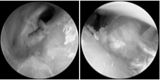
Fig. 2. Wash out excess material
(loose debris and degenerative tissue). Tissue structure seemed relatively normal.Fig. 1. Hard to define the osteochondroma and meniscus.
과두부위의 골연골종에 대한 임상 소견은 이환측에 개방교합을 동반한 부정교합이 나타나며, 비이환측에 반대교합이 나타나는 것이다. 정중선변위, 안면비대칭을 동반하며 종물이 촉진되고 대 부분 동통이 수반되지 않으며, 해부학적 이상소견과 함께 종종 개구제한과 같은 기능이상이 동반된다. 전통적인 치료 방식으로 과두절제술과 보존적인 외과적 절제술이 있으며, 비대칭적 하악 전돌증을 해결해주기 위해 악교정수술이 시행될 수 있다.
본 증례는 45세의 여성환자로 좌측 하악과두 상부에 골연골종 이 관절원판의 하방유착과 동반되어 발생하였고, 좌측 측두하악 관절 부위의 통증과 구치부 상실로 인한 교합이상을 호소하였다.
본 교실에서는 상기 환자에 대해 순차적인 운동요법, 교합안정 장치, 관절강세척술, 악관절경 수술 및 외과적 절제술을 통하여 점차적으로 양호한 결과를 관찰하였기에 이를 보고하는 바이다.
증례보고
환자는 45세 여성으로, 본과로 내원하기 약 2년 전 개인 치과 병원에서 양측성 측두하악관절 장애로 진단받고 물리치료와 장치
양측 측두하악관절의 관절강 내 세척술을 위하여 본과로 의뢰되었 다. 환자는 특이할 만한 전신병력이나 치과병력은 없었다. 환자의 최대 개구량은 약 35 mm로 경도의 개구제한을 보였으며, 양측 측두하악관절 부위에 통증을 호소하였다. 상악 우측 제2소구치와 하악 우측 제2소구치 사이에 조기접촉이 관찰되었으나, 개방교합 과 교차교합은 관찰되지 않았으며, 양측으로의 활주운동에 제한 을 보였다(Fig. 1).
고주파를 이용한 물리치료를 병행하며, 하이알루로닌산과 생 리식염수를 이용한 관절강세척술이 양측 측두하악관절에 각각 3회씩 시행되었다.
약 3개월간의 경과관찰 시기에 우측 측두하악관절 부위의 통증 은 개선되었으나, 좌측 측두하악관절 부위에는 호전을 보이지 않아 전신마취하에 관절경을 이용한 관절강 세척술을 계획하였 다. 관절경하에 일부 조직 잔해들과 퇴행성 조직이 관찰되어 제거 되었고, 전반적으로 정상적인 구조소견을 보였으나 관절돌기 상 부에 약간의 골증식 소견이 관찰되었다(Fig. 2).
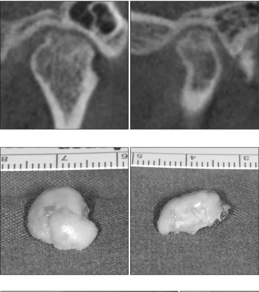
이후 호전양상을 보이지 않았기 때문에 좌측 측두하악관절 부위에 대해 cone-beam computed tomography (CBCT) 촬영 을 시행하였다. CBCT 촬영 결과 관절돌기 상부에 약 10×15 mm 크기의 골표면으로부터 자라나온 불규칙한 형태와 밀도의 골증식 소견이 관찰되었다(Fig. 3). 이 종물에 대해 골연골종으로 잠정진단하고, 술 후 합병증을 최소화하기 위하여 병소 중심의 보존적인 외과적 절제술을 계획하였다. 보존적 술식을 계획하였 으나 병소 위치와 구치부 교합 부재로 인한 수직고경 상실이 예상되었다.
수술은 전신마취하에 좌측 전이개 부위로의 접근을 통하여
이루어졌으며, 좌측 관절돌기 상부의 종물에 대해 외과적 절제술
을 시행하였다. 관절원판과 관절돌기 사이에 유착이 관찰되었으
며, 기구의 접근과 병소 제거 경로의 공간확보는 상관절강을 통하
Fig. 4. Complete removal of the
mass, specimen shows the fusion of disc and condylar head.Fig. 5. The surface is covered by fibrocellular perichondrium (arrow 1) and connective tissue occupies the marrow spaces (arrow 2)
(H&E stain; A: ×40, B: ×200).Fig. 3. Well-defined osteophyte on
left condylar head.Fig. 6. Day 1 post-operation, no retained residual lesion. Premature
contacts between #27 and #37.Fig. 7. Three months post-operation, remodeling is in progress
and no evidence of recurrence. A slight resorption is found on medial and lateral side of left condyle, periosteal new bone formation on anterior area of left condyle with shape of osteophyte.Fig. 8. One year post-operation, increases in over-bite and there
is no pre-mature contacts. Centrally-located condyle noted within articular fossa.Fig. 9. Twenty months post-operation, intra-oral occlusion photo-
graph shows symmetrical and midline corrections on both sides.은 약 10×15 mm의 크기를 보였다(Fig. 4).
술 후 CBCT 촬영상 성공적인 보존적 종물 제거를 확인할
상악 좌측 제2대구치와 하악 좌측 제2대구치 사이에 조기접촉이
임상 및 파노라마 방사선 사진에서 관찰되었다(Fig. 6).
수술 4일 후 교합 회복을 위해 본원 보철과에 의뢰되었다.
상악 좌측 제2대구치와 하악 좌측 제2대구치 사이에 조기접촉, 하악 좌측 제1대구치와 하악 우측 제1, 2대구치의 결손, 하악의 후방전위, 수직고경의 소실, 개구제한 등의 몇 가지 문제점이 지적되었다. 보철계획 수립 시기를 중심위 유도 장치(C.R. splint) 의 적용 후로 정하기로 하였다.
술 후 3개월의 파노라마와 CBCT 촬영 결과 관절돌기의 골개조 가 진행 중임을 관찰할 수 있었으며, 재발 소견은 없었다(Fig. 7).
술 후 4개월에 좌측 측두하악관절에서 clicking sound가 관찰 되었으며 이는 회복가능성을 시사하는 것으로 생각되었다. 지속 적으로 물리치료 및 중심위 유도와 교합안정을 위한 교합장치 치료를 하였다.
술 후 1년 시기에 안정된 악간 관계를 얻었으며, 이를 바탕으로 상실된 구치부의 보철 치료 계획이 수립되었다(Fig. 8).
술 후 1년 8개월 시기의 교합이며, 보철적으로 수복이 완성된 상태이다(Fig. 9).
고 찰
두개안면영역에서 나타나는 골연골종은 두개저, 상악동, 관골 궁, 하악골 등에서 발생할 수 있으며, 하악골에서는 근돌기, 관절 돌기, 하악골 상행지, 몸체, 우각부, 정중부에서 보고된 바 있다[1].
하악 관절돌기 부위에서 발생한 골연골종은 드물며 주로 여성에게 호발하고, 평균 40세 전후로 발생하는 경향을 보인다[2,3]. 또한 좌측의 관절돌기에서 발생률이 좀 더 높다[2,4,5]. 하악 관절돌기 에 발생한 골연골종은 앞서 언급한 대로 특징적인 임상적 양상을 보이며, 이는 방사선 촬영, 특히 컴퓨터단층촬영을 통해서 관찰될 수 있다(Fig. 3)[1,2,6,7]. 조직학적으로 골연골종은 연골성 모자, 연골 내 골화, 피질골 하방 골 내의 연골성 섬 등이 관찰되는 소견을 보인다(Fig. 5)[1,6,7].
골연골종의 발생 기전으로 몇 가지의 가설이 알려져 있다 [1,8,9]. 첫 번째로, 골단부의 골막 결손으로 인한 연골 전구 세포 들의 유출이 골연골종을 형성하게 한다는 가설이다[6,8]. 두 번째 로, 연골모세포와 골모세포를 형성할 수 있는 잠재력을 가진 골막 의 화생적 변화와 관절돌기의 골연골층의 화생적 변화가 골연골종 을 발생시킬 수 있다는 가설이다[2,6,8]. 세 번째는 근육의 인대가 붙는 위치에서 종물이 발생한다는 가설이다[1,2,6,7].
측두하악관절장애와 유사증상을 보이는 경증의 골연골종의 경 우 초기 감별진단을 위하여 plain film 이외의 진단도구인 CBCT 같은 방식을 조기에 사용하는 것이 환자의 시간낭비 및 비용증가 를 막을 수 있는 방법이라 판단된다.
본 증례와 같은 관절돌기 상부의 골연골종은 편측의 관절돌기 과증식과 임상증상이 유사할 수 있다[10,11]. 관절돌기 과증식의 방사선학적 소견은 관절돌기가 전체적으로 확대되어 나타나며,
골연골종의 경우는 정상적인 외형의 관절돌기 표면에서 구상으로 돌출되어 증식된 모양으로 나타나기 때문에 감별진단이 가능하다 [7,10,11]. 조직학적으로 편측의 관절돌기 과증식은 정상적인 연 골을 보이며, 골연골종의 경우에는 비정상적인 연골 증식과 석회 화를 보인다[7,10,12].
골연골종과 감별 진단해야 하는 골관절염은 측두하악관절에서 발생하는 관절염 중의 하나로 발육성인 골연골종과 달리 퇴행성 관절 질환이며, 이러한 퇴행성 변화는 측두하악관절의 과도한 사용이나 전신 질환에 의해서 야기될 수 있다. 골관절염의 방사선 학적 소견은 관절면과 하부 골조직의 파괴, 관절 연골의 소실, 퇴행이 관찰된다. 교합의 변화를 야기시켜 개방교합 등의 증상을 야기하거나 악화시킬 수 있다. 음식을 씹거나 턱을 움직이는 것이 불편하며, 관절이 아프고 때로 관절잡음이 난다. 이 증상이 악화될 경우, 강직으로 인한 개구제한이 생길 수 있으며 그에 따라 음식물 을 섭취하기가 어려워진다.
관절돌기에 발생한 골연골종의 전통적인 치료법은 과두절제술 이지만, 수술 후 교합 변화 등과 같은 합병증과 보존적인 제거술 후의 양호한 결과와 낮은 재발률로 미루어 볼 때 보존적인 외과적 적출술이 권장된다[1,2,8,13,14]. 최근의 후향적 연구에 근거하면 보존적 접근법에 의한 치료의 결과에서도 재발률이 더 높아지지 않았다[13,14]. 이것은 조직학적으로 골연골종은 표면으로부터 자라나온 골증식이며, 침습적이지 않은 종양으로 증식된 부위만 을 절제하는 것으로 충분하다는 연구에서 근거한다[14,15]. 관절 돌기 상부의 골연골종은 보존적인 적출술을 시행하여도 수직고경 변화가 발생할 수 있다. 본 증례에서는 수직고경의 상실 정도를 줄이며, 빠른 기능 회복을 위해 보존적인 외과적 적출술을 시행하 였지만, 수직고경 상실과 더불어 구치부 결손으로 인한 교합상실 이 안정된 하악위를 얻는 데 장애가 되어 수술 후 중심위 유도 교합장치를 통한 장기간의 장치치료를 시행하였다. 또한 관절경 을 통한 접근으로 종양의 일부분을 관찰할 수 있었는데 관절경을 통한 제거술이 불가능하더라도 종양의 존재, 위치 및 형태 등을 직접 볼 수 있다는 점에서 의미있는 술식이라 판단된다.
본 증례를 통하여 관절돌기 상부의 골연골종에도 보존적인 외과적 적출술이 적합한 치료법이며, 술 후 안정된 하악위를 얻기 위해 장치 치료를 한 후 최종 교합회복을 시도하여야 할 것이라 생각한다[1,8].
장기적인 예후평가를 위하여 향후 본 증례의 주기적인 경과관 찰이 필요하다.
References