Early Diagnosis of Burkitt Lymphoma on the Mandible: A Case Report
Miae Kim, Jihyun Park, Yonjoo Mah
Division of Pediatric Dentistry, Department of Dentistry, Ewha Womans University Mokdong Hospital
Burkitt lymphoma (BL) is an aggressive form of non-Hodgkin’s B-cell lymphoma found primarily in the pediatric population. In the oral cavity, this tumor can grow rapidly and often brings about facial swelling or development of an exophytic mass involving the jaws.
A 5-year-old boy was referred for swelling and pain in the left mandibular area. The patient showed diffuse swelling on the left side of the mandible and firm-moderate tenderness upon palpation. An intraoral examination showed moderate mobility and sensitivity to percussion on the left primary first and second molars, without severe caries. A radiographic examination revealed complete loss of the lamina dura on the left primary second molar and permanent first molar. There was a radiolucent osteolytic lesion and destruction of the cortical bone of the left mandibular body. Based on the clinical, radiographic, and immunohistochemical findings, the patient was diagnosed with BL, and was referred to a pediatrician for systemic evaluation and intensive chemotherapy.
Even before the completion of chemotherapy, the swelling resolved and the displaced teeth were relocated to a normal position.
This patient showed a good prognosis due to prompt diagnosis and intensive chemotherapy. Early diagnosis and referral for treatment can prevent the development of BL.
Key words : Burkitt lymphoma, Signs and symptoms, Diagnosis, Chemotherapy, Adjuvant
Abstract
Ⅰ. Introduction
In 1958, Denis Burkitt1) published the first report of Burkitt lymphoma (BL) - derived from facial and ab- dominal lesions in young African children. BL is an ag- gressive form of non-Hodgkin’s lymphoma (NHL), con- sisting of high-grade, diffuse, small, non-cleaved B-lym- phocytes. The affected age group for BL is children and young adults2).
BL may present as one of several types: endemic, spo- radic, or immunodeficiency-associated. The endemic
(African) type almost always occurs in children and in- volves the jaws in 50-70% of cases. Jaw lesions in the endemic type form at the age of 3 years in close to 100%
of known cases. In contrast, jaw lesions are less frequent in the sporadic (American) type and presentation of this type is almost always associated with abdominal compli- cations2). Children diagnosed with sporadic BL are typi- cally older than children suffering from the endemic type. Furthermore, the incidence of BL also increases among patients with various immunodeficiencies, such as HIV infection3).
Corresponding author : Yonjoo Mah
Division of Pediatric Dentistry, Department of Dentistry, Ewha Womans University Mokdong Hospital, 1071, Anyangcheon-ro, Yangcheon-gu, Seoul, 07985, Korea Tel: +82-2-2650-2660 / Fax: +82-2-2650-5764 / E-mail: [email protected]
Received June 8, 2015 / Revised September 22, 2015 / Accepted August 31, 2015
The clinical presentation of BL is characterized by rapid progression with multifactorial extranodular in- volvement. Within the jaws, BL is associated with loos- ening and extrusion of the involved teeth, premature shedding of primary teeth, swelling of alveolar regions, gingival enlargement, pain, and jaw expansion2,4).
The primary treatment of BL is intensive combination chemotherapy. BL is almost always curable via chemotherapy and the prognosis is excellent for those di- agnosed during the early stages of the disease, with sur- vival rates of 85-95%3,5).
This study reports a 5-year-old boy with BL and dis- cusses the clinical, radiological, and immunohistochemi- cal findings of BL. This case report will demonstrate the importance of early diagnosis, prompt referral, and ag- gressive management of BL.
Ⅱ. Case Report
A 5-year-old boy was referred to the department of pe- diatric dentistry with the chief complaint of swelling in the left lower jaw lasting for 7 days. The patient had a history of pain on the same side for 30 days. The patient’s mother reported that the local clinician had at- tributed pain to the eruption of the left mandibular first molar and recommended a wait-and-see approach.
However, sudden and very marked swelling prompted the mother to visit a larger hospital.
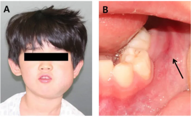
Clinical examination revealed that the boy was in good general health and had no allergic reaction to food or medicine. An extraoral examination showed diffuse swelling in the left mandibular area, from the level of the zygoma to the lower border of the mandible. The surface of the swelling appeared smooth and erythema-
tous (Fig. 1A). The patient showed firm-moderate ten- derness upon palpation, but no discharge was noted.
Also, the left lower primary first and second molars had moderate mobility and sensitivity to percussion. The pa- tient was in a mixed dentition state with the left mandibular permanent first molar partially erupted, but the other first molars had not erupted. The intraoral soft tissue on the left lower posterior area appeared solitary, lobulated, and erythematous, and the swelling mass had an irregular surface (Fig. 1B). The patient had moderate oral hygiene and no severe caries.
The radiographic examination included panoramic and periapical radiographs (Fig. 2). The left lower primary second and permanent first molars showed complete loss of the lamina dura. In the left mandibular area, a radi- olucent osteolytic lesion and destruction of cortical bone were evident. The results of this examination indicated
Fig. 2. Initial radiographs. (A) Panoramic radiograph showing radiolucent osteolytic lesion and the destruction of cortical bone in the left mandibular area.
(B) Periapical radiograph showing complete loss of the lamina dura of the left mandibular primary second molar and permanent first molar.
Fig. 1. Initial photographs. (A) Extraoral photograph showing facial swelling on the left mandibular area from the level of the zygoma to the lower border of the mandible. (B) Intraoral photograph showing prema- ture eruption of the left permanent first molar and a solitary, lobulated, and erythematous swelling in the left lower posterior area.
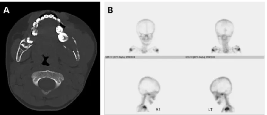
malignancy and further evaluation was necessary to identify the disease. Additional examinations including computed tomography (CT) of the facial bone and bone scintigraphy (Fig. 3). CT showed an osteolytic lesion with cortical bone destruction and regionally enlarged lymph nodes. Bone scintigraphy was performed after in- travenous (IV) injection of 99mTc hydroxyethylene diphos- phonate (HDP) (20 mCi). A photon defect in the mandible was observed and the bone scan ruled out os- teosarcoma.
An incisional biopsy was also performed with the coop- eration of an oral surgeon under IV sedation since the patient was anxious; an electrosurgical generator (Force
FXTM-8C; Valleylab, Boulder, CO) was used for the biop- sy and soft tissue was resected from the left mandibular vestibule (0.8 × 0.8 × 0.4 cm). For the final diagnosis, the specimen was examined by a pathologist, and ana- lyzed chemically (Fig. 4). Routine hematoxylin and eosin stained sections showed diffuse infiltration of atypical medium-sized lymphoid cells into the skeletal muscle (Fig. 5A). Immunohistochemically, the neoplastic cells were positive for LCA (CD45), CD20, Bcl-1, and CD10, and negative for Bcl-2 and terminal deoxynucleotidyl transferase (TdT). The Mib-1 proliferation rate was al- most 100% (Fig. 5B). These features confirmed the presence of high-grade NHL of Burkitt’s type.
Fig. 3. (A) Computed tomography (CT) of the facial bone showing an osteolytic lesion and cortical bone destruction. (B) Bone scintigraphy showing a photon defect in the mandible.
Fig. 4. (A) Incisional biopsy of the left mandibular posterior area. (B) Soft tissue resected by incisional biopsy.
Fig. 5. (A) Histological examination. The diffuse infiltration of atypical medium sized lymphoid cells into the skeletal muscles. (Original magnification × 400) (B) Immunohistochemical stains. Mib-1 staining showing positive in almost 100% of cells. (Original magnification ×400)
The patient was referred to the pediatrics department for further systematic evaluation. A pediatrician per- formed additional examinations including abdominal CT, bone marrow aspiration, chromosomal analysis, and cy- tological examination of cerebrospinal fluid. These analy- ses indicated that the tumor had not spread to other or- gans. After insertion of a Hickman catheter, the patient received intensive chemotherapy based on methotrexate and cyclophosphamide. After 3 months of chemotherapy, the extraoral and intraoral swelling had resolved com- pletely and percussion sensitivity and mobility of the af- fected teeth had decreased (Fig. 6). The radiographic examination showed bone regeneration and recovery of a normal trabecular pattern, and the displaced teeth had relocated to a normal position (Fig. 7). Chemotherapy was planned for 10 months and was finished at the time of writing (Fig. 8).
The patient was recalled at 3-month intervals to eval- uate caries and the eruption of the permanent teeth.
The left mandibular first permanent molar and primary molars had caries limited to the enamel portion and the permanent tooth to erupt was the right and left mandibular first permanent molar. Therefore, topical fluoride application was performed periodically during eruption period. Caries treatment was delayed until the end of chemotherapy.
Ⅲ. Discussion
About 10% of malignant tumors occurring in children younger than 15 years of age are lymphomas, and 60%
of lymphomas developing in children are NHLs. BL is a form of NHL with distinct epidemiological, clinical, pathological, immunological, and molecular cytogenetic characteristics2). The tumor arises through the malignant monoclonal proliferation of early B-lymphocytes, and has a mean potential doubling time of 25.6 h. Thus, BL is known as the most rapidly dividing tumor6).
Fig. 8. Images obtained after 11 months of chemotherapy. (A) Extraoral photograph. (B) Intraoral photograph. (C) Panoramic radiograph. (D) Facial bone CT.
A C D
Fig. 7. Radiographs after 3 months of chemotherapy. (A) Panoramic radiograph showing normal trabecular pattern and teeth relocated to a normal position in the left mandibular area.
(B) CT of facial bone showing cortical bone regeneration in the left mandibular area.
Fig. 6. Photographs after 3 months of chemotherapy. (A) Extraoral photograph showing completely resolved swelling in the left mandibular area (B) Intraoral photo- graph showing resolved gingival swelling.
A A B
Denis Burkitt7)noted the geographic distribution of the tumor and suggested that it may be induced by an in- sect-vectored agent, such as a virus. The discovery of virus particles in cultured lymphoblasts of BL by Epstein8) in 1964 demonstrated that the cause of BL was, in fact, Epstein-Barr virus (EBV). The majority of patients with endemic BL had the EBV genome and a characteristic pattern of viral gene expression9). In African BL, early EBV infection appears to be the initia- tor and malaria the promoter. The prevalence of EBV is also intermediate in non-African BL types. In non- African countries, there is no evidence of malaria, but EBV-positive BL occurs, suggesting that EBV and malarial infection are not necessary and sufficient causes of BL10). Other factors, such as poor nutrition, zinc defi- ciency, and other infectious diseases may be relevant in EBV-positive BL, and EBV has also been identified in 25-40% of immunodeficiency-associated cases2,11). In America, EBV DNA was found in about half of BL cases presenting with AIDS12). Although there are strong rela- tionships between BL and EBV, the exact role of EBV in the pathogenesis of BL has not been clearly demonstrat- ed. EBV is associated with 95% of African BL cases, but only 20% of BL cases in Europe and America. These findings suggest that EBV is not essential for the occur- rence of BL2). Various studies into the etiology of BL have been attempted but have failed to identify clear ge- netic or environmental factors. Arboviruses and plant tumor promoters are other possible local cofactors, and overexpression of the c-myc oncogene and the functional loss of wild-type p53 gene have also been implicated as possible etiologic factors11,13).
There are several differences between endemic (African) and sporadic (non-African or American) BL.
The mean age of occurrence for endemic cases is approx- imately 7 years, while sporadic cases occur in older chil- dren (11-15 years)14). BL is found in almost all organs and the sites primarily involved are the head, neck, and abdominal areas. In sporadic BL, the most common site of presentation is the abdomen, whereas the head and neck are most commonly involved in endemic BL. The percentage of patients with jaw involvement is relatively low in non-endemic BL (12.5-16% of cases), whereas jaw involvement occurs in 60-70% of endemic cases.
Furthermore, the mandible is more commonly involved in sporadic BL, but the maxilla is affected more often in endemic BL14). The sporadic type occurs worldwide, ex- cept in Africa. Therefore, this case could be the sporadic
type based on region, while the patient’s age and posi- tion of occurrence were similar to the endemic type. The characteristics of the tumor cells, their natural history and prognostic features are similar regardless of the type of BL2,15). Therefore, it is difficult to determine whether this case was the endemic or sporadic type.
The clinical signs and symptoms of BL involving the jaws include facial swelling, alveolar and gingival expan- sion, tooth loss, premature eruption of permanent teeth, dental and jaw pain, and paresthesia16). Loss of the lam- ina dura is observed around erupted or developing teeth, as well as widening of the periodontal ligament, enlarge- ment of dental crypts, displacement of the maxillary antrum, and radiolucencies and osteolytic foci of the jaw in the radiographic findings16,17). In this case, clinical signs such as toothache-like pain and facial deformity due to rapid facial swelling and radiographic evidence of floating teeth and loss of the lamina dura were observed.
The clinical presentation of BL is similar to that of many common disorders of the jaws. The differential di- agnosis of BL should include acute dentoalveolar ab- scess, osteomyelitis, rhabdomyosarcoma, periapical le- sions, ameloblastoma, eosinophilic granuloma, multiple myeloma, leukemia, and other fibro-osseous lesions.
Histological features can assist in distinguishing between BL and other oral diseases. Histological sections have a characteristic ‘starry sky’appearance, which is caused by the proliferation of immature, undifferentiated B- lymphocytes with high nuclear to cytoplasmic ratios, in- terspersed with macrophages that contain cellular debris4). A classic immunophenotype is also diagnosed in approximately 94% of BL. The characteristic BL im- munophenotype includes B-cell antigen expression, such as CD20, PAX5, CD19, CD22, and CD79a, with strong CD10 expression, and a very high proliferation rate based on Ki-67 or Mib-1 (cellular markers for prolifera- tion). BL does not express TdT and is usually negative for Bcl-2. This patient had these characteristics and was diagnosed with BL18). Serological evidence of EBV is un- reliable as a diagnostic aid, given that EBV was appar- ent in 6 to 98% of BL patients4).
Dental radiographs can also play an important role in the diagnosis of BL, as changes in radiographs may pre- cede apparent clinical signs. However, some of the changes, such as loss of the lamina dura, widening of the periodontal ligament, and the presence of osteolytic foci are not distinctive characteristics of BL and may ap- pear in other malignant tumors or infections16).
Therefore, diagnosis may be confounded by other dis- eases of the oral cavity, particularly dental infection, which is a common complaint in dental clinics. Saribans et al.19) reported on 16 patients with American-type BL with jaw involvement. Of these, 14 cases were first eval- uated by dentists and 10 were treated with antibiotic therapy or dental extractions for a presumed tooth infec- tion. Symptoms originating from a dental infection re- spond to antibiotic treatment, but those of oncological origin do not improve with antibiotic treatment. As in the case described here, rapid and accurate diagnosis is critical for a favorable prognosis. Thus, it is important for dentists to understand the clinical characteristics of BL of the jaw and include it in their working differential diagnosis.
BL is one of the first human malignancies to be cured by chemotherapy alone. Clinical regression of the tumor is usually achieved within 2 weeks and odontogenesis resumes after successful chemotherapy17). BL treated with intensive chemotherapy has a good prognosis with a 5-year survival rate of 78%14). When the disease is confined to the head and neck, patients can expect tu- mor-free survival for at least 2 years in up to 90% of cases, with current chemotherapy regimens. However, patients with extensive disease, such as involvement of the bone marrow or CNS and those with HIV-associated BL, show lower survival rates17,20). Relapses usually occur within 1 year of diagnosis and patients who survive 2 years without recurrence can be considered cured21). Although 95% of patients achieve an initial complete re- mission following chemotherapy, up to 67% of patients relapse22). Nkrumah et al.23) reported that 52% of pa- tients with initial complete remission relapsed over an observation period ranging from 2 years to over 5 years.
Relapse was common in patients who initially presented with abdominal or CNS involvement, while patients who had facial bone involvement had a much lower recur- rence rate.
Chemotherapy is based on intravenous cyclosporine, vincristine, doxorubicin, methotrexate, and leucovorin;
combination therapy is superior to single-agent chemotherapy. The use of multiple agents maximizes in- tensity while minimizing kinetic failures and may de- crease the chance of drug resistance. Most chemotherapy regimens are cyclophosphamide-based20) and common protocols for BL treatment include the COMP, BMP, LMB86, CODOX-M, and IVAC regimens24). However, chemotherapy has injurious effects on developing teeth.
The worst consequences are seen in the treatment of children younger than 5-6 years; children younger than 12 years also exhibit later changes in tooth development.
The dental abnormalities seen after chemotherapy in- clude hypomineralization of the enamel, arrested root development, microdontia, and aplasia or delayed devel- opment of the permanent teeth25,26). Dental caries are the most commonly reported late dental effects following chemotherapy. Therefore, regular preventive dental care, such as individualized dental procedures and oral hy- giene plans, is needed to improve post-chemotherapy dental outcomes27). This patient underwent regular den- tal check-ups to evaluate the development and eruption of the permanent teeth and caries.
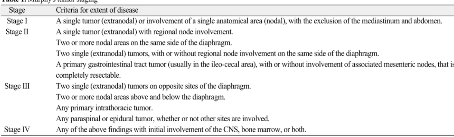
Treatment success depends on the extent of the dis- ease. Otmani et al.28) reported that 54% of BL patients were considered long-term survivors, with survival rates of 100%, 91%, and 27% respectively, for Murphy’s tu- mor stages II, III, and IV (Table 1)29). In another study, the prognosis was excellent for those diagnosed during
Table 1. Murphy's tumor staging29)
Stage Criteria for extent of disease
Stage I A single tumor (extranodal) or involvement of a single anatomical area (nodal), with the exclusion of the mediastinum and abdomen.
Stage II A single tumor (extranodal) with regional node involvement.
Two or more nodal areas on the same side of the diaphragm.
Two single (extranodal) tumors, with or without regional node involvement on the same side of the diaphragm.
A primary gastrointestinal tract tumor (usually in the ileo-cecal area), with or without involvement of associated mesenteric nodes, that is completely resectable.
Stage III Two single (extranodal) tumors on opposite sites of the diaphragm.
Two or more nodal areas above and below the diaphragm.
Any primary intrathoracic tumor.
Any paraspinal or epidural tumor, whether or not other sites are involved.
Stage IV Any of the above findings with initial involvement of the CNS, bone marrow, or both.
the early stages of the disease (I, II), with event-free survival rates of 85-100%. In the advanced stages (III, IV), the survival rate was 75-85%2). Other factors affect- ing prognosis are the patient’s age and the timing of di- agnosis13). Thus, the best prognosis is achieved with BL stage I, and it is important that general dental practi- tioners, who are usually the first point of contact, know the characteristics of this disease and can diagnose it quickly and accurately. Dentists should suspect BL when faced with a patient with unexplained hypermobile teeth, the displacement of unerupted teeth, severe alve- olar bone destruction on radiophotographs, or unex- plained paresthesia. In the maxillofacial region, radi- ographs including both periapical and panoramic views are usually sufficient for the diagnosis of malignant le- sions13). Thus, when a patient with these signs or symp- toms presents, the dentist should refer the patient promptly to a specialist.
Ⅳ. Summary
This case report describes BL in the lower jaw of a 5- year-old male patient. BL is a NHL of B-cell origin with aggressive growth characteristics that requires early di- agnosis and treatment. The clinical presentation of BL in the jaws include the loosening and extrusion of teeth, premature eruption of permanent teeth, gingival en- largement, swelling of facial and alveolar bones, dental pain, and paresthesia. As is typical with BL, the patient showed facial and gingival swelling, dental pain, and premature eruption of a permanent molar. The radi- ographic findings were typical of malignant disease. A biopsy was conducted to determine the diagnosis. This patient was diagnosed with BL, based on the clinical, radiographic, and immunohistochemical analyses, and showed a good prognosis due to prompt diagnosis and intensive chemotherapy. Regular check-ups are also needed to monitor dental developmental abnormalities and dental caries following chemotherapy.
References
1. Burkitt D : A sarcoma involving the jaws in African children. Br J Surg, 46:218-223, 1958.
2. Shapira J, Peylan-Ramu N : Burkitt’s lymphoma.
Oral Oncol, 34:15-23, 1998.
3. Sandlund JT, Downing JR, Crist WM : Non- Hodgkin’s lymphoma in childhood. N Engl J Med,
334:1238-1248, 1996.
4. Ardekian L, Rachmiel A, Rosen D, et al. : Burkitt’s lymphoma of the oral cavity in Israel. J Craniomaxillofac Surg, 27:294-297, 1999.
5. Link MP, Donaldson SS, Berard CW, et al. : Results of treatment of childhood localized non-Hodgkin’s lymphoma with combination chemotherapy with or without radiotherapy. N Engl J Med, 322:1169- 1174, 1990.
6. Iversen OH, Iversen U, Ziegler JL, Bluming AZ : Cell kinetics in Burkitt lymphoma. Eur J Cancer, 10:155-163, 1974.
7. Burkitt D, Wright D : Geographical and tribal distri- bution of the African lymphoma in Uganda. Br Med J, 1:569-573, 1966.
8. Epstein MA, Achong BG, Barr YM : Virus Particles in Cultured Lymphoblasts from Burkitt’s Lymphoma.
Lancet, 1:702-703, 1964.
9. Tao Q, Robertson KD, Manns A, et al. : Epstein- Barr virus (EBV) in endemic Burkitt’s lymphoma:
molecular analysis of primary tumor tissue. Blood, 91:1373-1381, 1998.
10. Evans AS : Epidemiology of Burkitt’s lymphoma:
other risk factors. IARC Sci Publ, 60:197-204, 1985.
11. Pannone G, Zamparese R, Pace M, et al. : The role of EBV in the pathogenesis of Burkitt’s Lymphoma:
an Italian hospital based survey. Infect Agent Cancer, 9:34, 2014.
12. Beral V, Peterman T, Berkelman R, Jaffe H : AIDS- associated non-Hodgkin lymphoma. Lancet, 337:
805-809, 1991.
13. Jan A, Vora K, Sandor GK : Sporadic Burkitt’s lym- phoma of the jaws: the essentials of prompt life-sav- ing referral and management. J Can Dent Assoc, 71:165-168, 2005.
14. Tsui SH, Wong MH, Lam WY : Burkitt’s lymphoma presenting as mandibular swelling--report of a case and review of publications. Br J Oral Maxillofac Surg, 38:8-11, 2000.
15. Lee SE, Sohn MY, Choi SC, et al. : Burkitt’s lym- phoma of the Mandible in a Child. J Korean Acad Pediatr Dent, 39:79-83, 2012.
16. Ardekian L, Peleg M, Samet N, et al. : Burkitt’s lymphoma mimicking an acute dentoalveolar abscess. J Endod, 22:697-698, 1996.
17. Lund DI, Rodd H, Craig GT : Burkitt’s lymphoma presenting with jaw lesions in a young white girl. Br
J Oral Maxillofac Surg, 35:438-441, 1997.
18. O’Malley DP, Auerbach A, Weiss LM : Practical Applications in Immunohistochemistry: Evaluation of Diffuse Large B-Cell Lymphoma and Related Large B-Cell Lymphomas. Arch Pathol Lab Med, 2015.
19. Sariban E, Donahue A, Magrath IT : Jaw involve- ment in American Burkitt’s Lymphoma. Cancer, 53:
1777-1782, 1984.
20. Banthia V, Jen A, Kacker A : Sporadic Burkitt’s lymphoma of the head and neck in the pediatric population. Int J Pediatr Otorhinolaryngol, 67:59- 65, 2003.
21. Ferry JA : Burkitt’s lymphoma: clinicopathologic features and differential diagnosis. Oncologist, 11:
375-383, 2006.
22. Appelbaum FR, Deisseroth AB, Graw RG Jr, et al. : Prolonged complete remission following high dose chemotherapy of Burkitt’s lymphoma in relapse.
Cancer, 41:1059-1063, 1978.
23. Nkrumah FK, Perkins IV : Relapse in Burkitt’s lymphoma. Int J Cancer, 17:455-460, 1976.
24. Philip A. Pizzo DGP : Principle and practice of pedi- atric oncology, 3rd ed. Lippincott-Raven Publishers, 545-587, 1997.
25. Holtta P, Alaluusua S, Saarinen-Pihkala UM, et al.:
Long-term adverse effects on dentition in children with poor-risk neuroblastoma treated with high-dose chemotherapy and autologous stem cell transplanta- tion with or without total body irradiation. Bone Marrow Transplant, 29:121-127, 2002.
26. Kim SK, Kim MJ, Jeong TS, et al. : Management of oral complications in the pediatric population with cancer. J Korean Acad Pediatr Dent, 36:157-167, 2009.
27. Gawade PL, Hudson MM, Kaste SC, et al.: A sys- tematic review of dental late effects in survivors of childhood cancer. Pediatr Blood Cancer, 61:407-416, 2014.
28. Otmani N, Khattab M : Oral Burkitt’s lymphoma in children: the Moroccan experience. Int J Oral Maxillofac Surg, 37:36-40, 2008.
29. Murphy SB, Fairclough DL, Hutchison RE, Berard CW : Non-Hodgkin’s lymphomas of childhood: an analysis of the histology, staging, and response to treatment of 338 cases at a single institution. J Clin Oncol, 7:186-193, 1989.
하악골에서 발생한 Burkitt 림프종의 조기발견과 진단
김미애∙박지현∙마연주
이화여자대학교 의과대학 목동병원 소아치과학교실
Burkitt 림프종은 소아에서 호발하는 악성 종양의 하나로 non-Hodgkin B 림프종의 한 형태이다. 매우 빠르게 증식하는 종양으로 구강 내에서 발생시 안면부 부종 및 이환 치아의 동요도와 변위, 치은의 발적 및 부종 등의 증상을 보인다.
본 증례는 5세 남환으로 왼쪽 하악부의 통증과 안면부 부종으로 개인병원에서 의뢰되었다. 왼쪽 하악골부터 관골까지 퍼진 안면부 부종을 보였으며 좌측 하악 유구치는 심한 충치가 없음에도 불구하고 중등도의 동요도를 보이고 타진에 양성 반응을 보였다. 방사선학적 소견으로는 좌측 하악 제2유구치와 제1영구치의 치조 백선 소실이 관찰 되었으며, 좌측 하악골에는 방사 선 투과성의 골용해성 소견과 피질골의 파괴가 관찰되었다. 환아는 임상적, 방사선학적, 면역조직학적 검사를 바탕으로 Burkitt 림프종으로 진단되었고, 전신적인 검사와 항암 화학요법을 위해 본원 소아과로 의뢰되었다. 화학요법을 시작한 지 3 개월만에 안면부 및 치은의 부종이 소실되었고, 치아의 동요도 및 위치도 정상으로 회복되었다.
Burkitt 림프종으로 진단된 본 환아는 조기 진단과 집중적인 화학요법으로 좋은 예후를 보였기에 이를 보고하는 바이다.
주요어: Burkitt 림프종, 증후와 증상, 진단, 보조 항암화학요법 국문초록